What Is the Best Next Imaging Study After a Positive Radiograph for Diabetic Foot Osteomyelitis?

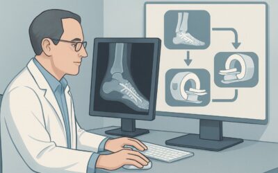
A 64-year-old man with type 2 diabetes presents to your clinic for follow-up on a non-healing ulcer over his first metatarsal head. The area is erythematous and warm, and a plain radiograph you ordered last week is now on your screen. It shows clear cortical erosion and a hazy, disorganized appearance of the trabecular bone, confirming your high suspicion for osteomyelitis. The question is no longer if infection is present, but how far it extends. Deciding between surgical debridement, a specific amputation level, or a prolonged course of IV antibiotics requires a detailed map of the infection’s scope. This article details the American College of Radiology (ACR) Appropriateness Criteria workflow for this exact clinical crossroads: selecting the next imaging study for pretreatment planning when radiographs are already positive for osteomyelitis. For this scenario, an MRI of the foot without and with IV contrast is rated Usually Appropriate.
Who Fits This Clinical Scenario for Pretreatment Planning?
This guidance is specifically for clinicians managing an adult patient with diabetes mellitus where the diagnostic process has already advanced past initial suspicion. The key inclusion criteria are:
- Established Diagnosis: The patient has a clinical picture consistent with a diabetic foot infection (e.g., a deep ulcer, positive probe-to-bone test, localized erythema and warmth).
- Positive Initial Radiographs: An initial X-ray has been performed and demonstrates findings characteristic of osteomyelitis, such as cortical destruction, periosteal reaction, or focal osteopenia.
- Clinical Question: The primary goal of the next imaging study is not to make the initial diagnosis, but to define the precise anatomical extent of the infection to guide treatment decisions—specifically, surgical planning or determining the necessary duration of antimicrobial therapy.
It is crucial to distinguish this situation from similar but distinct clinical presentations that follow different diagnostic pathways. This article does not apply if:
- Initial Radiographs are Negative or Indeterminate: If the X-rays are normal or unclear, the clinical question is different. The goal is to detect occult osteomyelitis, which follows a separate ACR workflow.
- No Initial Imaging Has Been Performed: This guidance is for the second step in imaging, not the first.
- Metallic Hardware is Present: If the patient has surgical screws, plates, or other instrumentation in the area of concern, standard MRI may be severely limited by artifact. This scenario has its own specific recommendations, often involving specialized MRI sequences or alternative modalities like nuclear medicine studies.
What Diagnoses Are You Working Up in This Scenario?
With a positive radiograph, osteomyelitis is the presumptive diagnosis. The purpose of advanced imaging is to confirm its extent and rule out or characterize critical comorbidities that will profoundly impact the treatment plan.
Established Osteomyelitis: The primary goal is to map the infection. Does it involve only the metatarsal head? Has it spread down the shaft (marrow involvement)? Is there a sequestrum (a piece of dead bone) that will act as a nidus for persistent infection if not surgically removed? MRI provides unparalleled detail of bone marrow and soft tissue to answer these questions.
Adjacent Soft Tissue Abscess or Sinus Tract: Osteomyelitis in the diabetic foot is rarely an isolated bone problem. It is almost always associated with overlying soft tissue infection. Advanced imaging must delineate the full extent of any drainable fluid collections (abscesses) or sinus tracts connecting the ulcer to the bone. An un-drained abscess will doom antibiotic therapy to failure.
Charcot Neuroarthropathy: This non-infectious, inflammatory destruction of bone and joints is the great mimicker of osteomyelitis in patients with diabetic neuropathy. The two can look similar on radiographs, showing bone destruction and fragmentation. While less likely to be the sole diagnosis when a deep ulcer is present, Charcot changes can coexist with a superimposed infection. Differentiating the edema of acute Charcot from the enhancement of infection on MRI is a critical, albeit challenging, task for the radiologist.
Septic Arthritis: The infection may not be confined to the bone. If it has spread into an adjacent joint, such as the metatarsophalangeal joint, it becomes septic arthritis. This complication often requires more aggressive surgical intervention, including potential joint resection, and carries a worse prognosis.
Why Is MRI of the Foot the Recommended Next Step for Pretreatment Planning?
The ACR designates MRI foot without and with IV contrast as Usually Appropriate because it directly answers the key questions needed for effective treatment planning. Its superior soft-tissue contrast and high sensitivity for bone marrow pathology make it the ideal tool for defining the three-dimensional extent of the disease.
The rationale for this recommendation includes:
- Anatomical Definition: MRI excels at showing the precise boundaries of the infection. It can distinguish between cellulitis, a drainable abscess, and phlegmon. For the surgeon, this information is invaluable for planning incisions, determining how much bone to debride, or deciding on an amputation level that will result in clean, viable margins.
- Bone Marrow and Soft Tissue Sensitivity: MRI can detect marrow edema and enhancement far more sensitively than CT, providing a clear map of the infection’s spread within the bone. It simultaneously visualizes associated tendon, muscle, and fascial plane involvement.
- Role of IV Contrast: While an MRI without contrast is also rated Usually Appropriate, the addition of a gadolinium-based contrast agent is highly recommended in this pretreatment planning scenario. Contrast helps differentiate viable, inflamed tissue from non-enhancing necrotic tissue or a sequestrum. It also clearly delineates the walls of an abscess and highlights sinus tracts, which can be difficult to see on non-contrast images.
- Safety Profile: MRI uses no ionizing radiation (0 mSv), which is a significant advantage over CT and nuclear medicine studies, especially if repeat imaging may be needed to assess treatment response.
Why are other studies rated lower for this specific task?
- CT foot with IV contrast (May be appropriate): CT is excellent for evaluating cortical bone and identifying sequestra. However, it has significantly lower contrast resolution for soft tissues and bone marrow, making it inferior to MRI for defining the full extent of infection. It remains a valuable alternative if a patient has a contraindication to MRI (e.g., an incompatible implanted device).
- WBC scan foot (May be appropriate): Labeled white blood cell scans are sensitive for detecting infection but provide poor anatomical detail. They can show a “hot spot” of inflammation but cannot tell a surgeon precisely which tissues are involved or where an abscess is located. This lack of spatial resolution makes them suboptimal for surgical planning. These studies also involve a significant radiation dose (☢☢☢☢ 10-30 mSv).
What’s the Next Step After the MRI Results?
The MRI report is not the end of the diagnostic journey; it is the map that guides the next therapeutic action. The downstream workflow depends directly on the findings.
- If MRI confirms localized osteomyelitis with a drainable abscess: The findings provide a clear target for the surgeon. The next step is typically prompt surgical intervention for incision, drainage, and debridement of infected and nonviable bone and soft tissue. Cultures obtained during surgery will guide long-term, pathogen-directed antibiotic therapy.
- If MRI shows extensive, multi-bone osteomyelitis: The report helps the clinical team (e.g., podiatric surgery, orthopedic surgery, vascular surgery, infectious disease) have a realistic discussion with the patient about the likelihood of limb salvage. The imaging may guide the decision toward a more definitive amputation at a level with healthy, well-perfused tissue, rather than multiple failed debridement procedures.
- If MRI findings are indeterminate (e.g., Charcot vs. Osteomyelitis): This is a common and difficult clinical scenario. When MRI cannot definitively distinguish between infection and neuroarthropathy, the next logical step is often an Image-guided biopsy, which the ACR rates as May be appropriate. Obtaining a bone sample for histopathology and microbial culture remains the gold standard for a definitive diagnosis, ensuring the patient receives the correct treatment.
Common Pitfalls to Avoid in This Scenario
Navigating this clinical scenario requires careful integration of imaging findings with the patient’s overall clinical picture. Here are a few common pitfalls to avoid:
- Over-reliance on Imaging Alone: MRI findings must always be correlated with the clinical examination, wound characteristics, and laboratory markers (e.g., ESR, CRP). A classic pitfall is mistaking the intense marrow edema of an acute Charcot foot for osteomyelitis, potentially leading to unnecessary intervention.
- Forgetting to Check Renal Function: Intravenous gadolinium contrast is a key part of the recommended MRI. Always assess the patient’s renal function (eGFR) before ordering a contrast-enhanced study to screen for any contraindications.
- Ignoring the Possibility of Underlying Ischemia: The best surgical plan will fail if there is inadequate blood flow to the foot. Ensure the patient’s vascular status has been assessed. If peripheral artery disease is suspected, a vascular surgery consultation and non-invasive arterial studies may be required before any major surgical debridement or amputation.
- Delaying Action on Critical Findings: If the MRI report describes a large, deep abscess or gas in the soft tissues (suggesting a necrotizing infection), this constitutes a surgical emergency. The workflow should be escalated immediately to the surgical service.
Related ACR Topics and Tools
For a comprehensive overview of all clinical variants and imaging options for this condition, please refer to our parent topic hub article. For additional resources to help you select and understand imaging studies, explore the tools below.
- For breadth across all scenarios in Suspected Osteomyelitis of the Foot in Patients with Diabetes Mellitus, see our parent guide: Suspected Osteomyelitis of the Foot in Patients with Diabetes Mellitus: ACR Appropriateness Decoded.
- ACR Appropriateness Criteria Lookup — for adjacent scenarios
- Imaging Protocol Library — for technique on the recommended study
- Radiation Dose Calculator — for cumulative dose conversations
Frequently Asked Questions
Why is MRI recommended over a CT scan when the X-ray is already positive?
While a CT scan is excellent for showing cortical bone destruction, MRI provides far superior detail of the bone marrow and surrounding soft tissues. For pretreatment planning, the goal is to see the full extent of the infection—including marrow spread, abscesses, and sinus tracts—which MRI visualizes much more clearly than CT. This detailed map is critical for effective surgical planning.
Is intravenous contrast always necessary for the MRI in this scenario?
While the ACR rates MRI both with and without contrast as ‘Usually Appropriate,’ the addition of IV gadolinium contrast is highly recommended for pretreatment planning. Contrast enhances the visibility of abscess walls, sinus tracts, and non-viable tissue (sequestra), providing crucial information for the surgeon that may be missed on non-contrast images.
What if the MRI report is equivocal and cannot distinguish osteomyelitis from Charcot neuroarthropathy?
This is a well-known diagnostic challenge. If the MRI is indeterminate, the next step is often to pursue a bone biopsy for microbiology and histopathology. An image-guided biopsy is rated ‘May be appropriate’ by the ACR and is considered the gold standard for confirming infection and identifying the causative organism to guide antibiotic therapy.
My patient has a pacemaker. Can they still get an MRI for their foot?
It depends on the type of pacemaker. Many modern pacemakers are ‘MRI-conditional,’ meaning a scan can be performed safely under specific protocols. This requires close coordination with the radiology department and cardiology to ensure the device is programmed correctly before and after the scan. If the device is not MRI-compatible, a CT scan or a nuclear medicine study would be the next best alternatives.
How does the presence of a deep ulcer or a positive probe-to-bone test change the need for this MRI?
A deep ulcer and a positive probe-to-bone test are strong clinical indicators of osteomyelitis, which is why the initial radiograph was likely ordered and found to be positive. These clinical findings reinforce the need for advanced imaging (like MRI) to define the extent of the infection. They increase the pre-test probability of osteomyelitis, making the detailed anatomical map provided by MRI even more critical for planning definitive treatment.
Reviewed by Pouyan Golshani, MD, Interventional Radiologist — May 30, 2026