What Is the Best Next Imaging Study for a Suspected Ankle Osteochondral Lesion After Normal Radiographs?
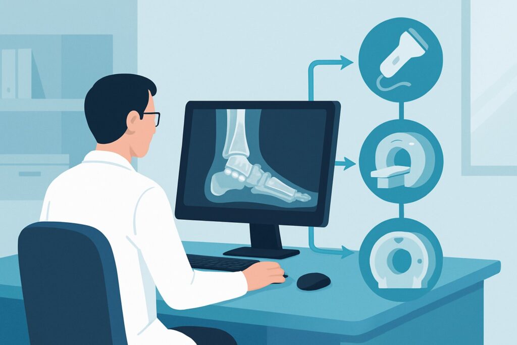
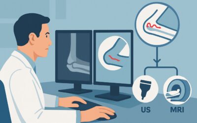
A 28-year-old patient presents with persistent, deep, focal pain in their right ankle, six months after an inversion injury they thought had healed. They are an active runner, but the pain, localized over the talar dome, now limits their activity. You’ve obtained standard ankle radiographs, which show no fracture, dislocation, or significant degenerative change. Given the mechanism and focal nature of the pain, your clinical suspicion is high for an osteochondral lesion of the talus (OLT). The question now is which advanced imaging study will confirm the diagnosis, assess its stability, and guide the orthopedic surgeon’s management plan. For this specific clinical scenario, the American College of Radiology (ACR) Appropriateness Criteria rate **MRI ankle without IV contrast** as *Usually Appropriate*.
Who Fits This Clinical Scenario for Suspected Osteochondral Lesion?
This diagnostic workflow is intended for a specific patient population. The key inclusion criteria are a patient with **chronic ankle pain** (lasting weeks to months) where initial, standard ankle radiographs are normal or non-contributory. The clinical picture, often including a history of trauma like an ankle sprain, points specifically toward an injury of the articular cartilage and underlying bone—an osteochondral lesion. The pain is typically deep, aching, and may be associated with mechanical symptoms like catching or locking.
It is crucial to distinguish this presentation from similar but distinct clinical problems that follow different imaging pathways:
* **Primary Tendon Abnormality:** If the pain is localized along the course of a tendon (e.g., peroneal, posterior tibial) and the primary suspicion is tendinosis or tear, the workup follows the ACR variant for suspected tendon abnormality.
* **Primary Ligamentous Instability:** If the patient’s chief complaint is a feeling of the ankle “giving way” and the examination suggests ligamentous laxity, the workup is guided by the ACR variant for suspected ankle instability.
* **Obvious Radiographic Abnormality:** If initial radiographs clearly show degenerative joint disease, fracture, or another definite cause for the pain, this scenario does not apply. The next steps would be guided by those specific findings.
This guide is for the focused workup of a suspected cartilage and subchondral bone injury in the setting of normal initial X-rays.
What Diagnoses Are You Working Up in This Scenario?
When ordering advanced imaging after normal radiographs for focal chronic ankle pain, you are investigating a specific set of differential diagnoses where cartilage and subchondral bone are the primary structures of interest.
The most common and primary consideration is an **osteochondral lesion of the talus (OLT)**, also known as an osteochondral defect (OCD). This is an injury involving both the articular cartilage and the adjacent subchondral bone of the talar dome. These lesions are most frequently post-traumatic and typically occur in the anterolateral or posteromedial aspects of the talus. The key clinical question is not just presence, but also stability—whether the fragment is loose, which has significant implications for treatment.
Another possibility is a **subchondral insufficiency fracture or stress fracture**. This can present with deep, aching pain and may not be visible on radiographs. It is characterized by bone marrow edema on MRI without a discrete osteochondral fragment. This is an important diagnosis to make, as management is typically conservative with protected weight-bearing.
Less commonly, **avascular necrosis (AVN) of the talus** can cause similar symptoms. While often associated with specific risk factors (e.g., steroid use, significant trauma like a talar neck fracture), it can occur idiopathically. Early AVN is radiographically occult, and MRI is the most sensitive modality for detection.
Finally, an **occult fracture** of the talus or surrounding bones that was not apparent on initial radiographs remains on the differential. MRI can readily identify non-displaced fracture lines and the associated bone marrow edema.
Why Is MRI Ankle without IV Contrast Usually Appropriate for Suspected OLTs?
The ACR designates **MRI ankle without IV contrast** as *Usually Appropriate* because it provides the most comprehensive evaluation for the primary differential diagnoses in this scenario without unnecessary contrast or radiation.
The strength of MRI lies in its superior soft tissue and cartilage contrast. It can directly visualize the articular cartilage, allowing for precise assessment of fibrillation, fissuring, or full-thickness defects. Furthermore, fluid-sensitive sequences (like proton density fat-suppressed or T2-weighted fat-suppressed images) are highly sensitive for detecting the bone marrow edema that accompanies subchondral injury, insufficiency fractures, and early avascular necrosis. For OLTs specifically, MRI is critical for staging, as it can help determine if the lesion is stable (overlying cartilage intact) or unstable (cartilage breached, fluid undermining the fragment). This information is paramount for orthopedic decision-making. Importantly, this study involves no ionizing radiation (adult and pediatric RRL=O 0 mSv).
Alternative studies are rated lower for specific reasons:
* **CT ankle without IV contrast** is rated *May be appropriate*. While CT offers excellent spatial resolution for delineating the bony extent of a lesion, subchondral cysts, or loose bodies, it cannot directly visualize articular cartilage or bone marrow edema. It is a viable alternative if MRI is contraindicated (e.g., incompatible hardware, severe claustrophobia), but it is less comprehensive for the primary diagnosis and involves a small amount of ionizing radiation (adult RRL=☢ <0.1 mSv).
* **MRI ankle without and with IV contrast** is rated *Usually not appropriate*. For the initial diagnosis of a native (non-operated) osteochondral lesion, intravenous gadolinium contrast adds little diagnostic information. The key features of cartilage integrity and subchondral stability can be assessed on a standard non-contrast protocol. Contrast is typically reserved for post-operative imaging to assess the vascularity and integration of a graft.
* **Ultrasound (US) ankle** is rated *Usually not appropriate*. Ultrasound cannot adequately penetrate bone to visualize the majority of the talar dome’s articular surface, making it unsuitable for evaluating or ruling out an OLT.
What’s Next After MRI? Downstream Workflow
The results of the ankle MRI will directly guide the subsequent clinical pathway, which almost always involves consultation with an orthopedic surgeon specializing in foot and ankle conditions.
* **Positive for a Stable OLT:** If the MRI confirms an osteochondral lesion but the overlying cartilage is intact and there is no fluid undermining the fragment, the lesion is considered stable. The next step is typically a trial of conservative management, which may include activity modification, immobilization in a boot, and physical therapy. Follow-up imaging may be considered if symptoms persist.
* **Positive for an Unstable OLT:** If the MRI shows a breach in the articular cartilage with joint fluid tracking beneath the osteochondral fragment, or if a loose body is identified, the lesion is unstable. This finding generally necessitates surgical intervention. The orthopedic surgeon will use the MRI findings to plan the procedure, which could range from microfracture to osteochondral autograft or allograft transplantation.
* **Negative for OLT, Positive for Other Cause:** If the MRI is negative for an OLT but reveals another cause for the pain, such as a stress fracture, sinus tarsi syndrome, or significant tendinosis, management is directed at that specific diagnosis. A stress fracture, for example, would be managed with a period of non-weight-bearing or protected weight-bearing.
* **Negative or Nonspecific MRI:** If the MRI is entirely normal or shows only nonspecific findings, the focus shifts. The next step may involve re-evaluating for other causes of pain, such as nerve entrapment or early complex regional pain syndrome. If clinical suspicion for an intra-articular process remains high despite the negative MRI, the workup may proceed to the ACR variant for chronic ankle pain of uncertain etiology, where diagnostic injection or arthroscopy could be considered.
Pitfalls to Avoid (and When to Get Help)
When working up a suspected osteochondral lesion, several common pitfalls can delay diagnosis or lead to suboptimal management.
First, avoid the pitfall of **prolonged conservative therapy without a definitive diagnosis**. If a patient with a history of trauma has persistent, deep ankle pain for months despite normal radiographs, advanced imaging is warranted. Delaying an MRI can lead to the progression of an unstable lesion.
Second, be cautious of **misinterpreting the clinical signs**. While OLT is a primary concern, remember to thoroughly examine for associated ligamentous instability or tendon pathology, as these can coexist and may also require treatment.
Third, ensure the **MRI protocol is appropriate**. A general “ankle MRI” order is usually sufficient, but the interpreting radiologist should be aware of the specific clinical question to ensure high-resolution, cartilage-sensitive sequences are performed through the talar dome.
If the diagnosis remains elusive after a high-quality MRI or if the patient’s symptoms are out of proportion to the imaging findings, it is time to escalate. This typically involves a referral to a foot and ankle orthopedic specialist for further evaluation, which may include diagnostic arthroscopy.
Related ACR Topics and Tools
This article focuses on a single, specific clinical scenario. For a comprehensive overview of all imaging pathways related to chronic ankle pain, from initial workup to other specific suspected etiologies, refer to our parent guide.
* For breadth across all scenarios in Chronic Ankle Pain, see our parent guide: Chronic Ankle Pain: ACR Appropriateness Decoded.
For clinicians ordering or interpreting imaging, the following GigHz resources can help streamline workflows and improve communication:
- ACR Appropriateness Criteria Lookup — for quickly checking recommended studies for adjacent or different clinical scenarios.
- Imaging Protocol Library — for understanding the technical details of the recommended MRI study.
- Radiation Dose Calculator — for discussing cumulative radiation exposure with patients, particularly when considering CT as an alternative.
Frequently Asked Questions
Why is a non-contrast MRI preferred over an MR arthrogram for an initial OLT workup?
For the initial diagnosis of a native (unoperated) osteochondral lesion, a high-quality non-contrast MRI is typically sufficient. The joint’s natural synovial fluid provides enough contrast to outline cartilage defects and assess for fluid undermining an unstable fragment on T2-weighted images. An MR arthrogram, which involves injecting contrast directly into the joint, is rated ‘May be appropriate’ and is generally reserved for cases where the non-contrast study is equivocal or for some post-operative assessments.
If my patient has a pacemaker and cannot get an MRI, is CT arthrography a good alternative?
Yes. In cases where MRI is contraindicated, CT is the next best modality. A standard non-contrast CT is rated ‘May be appropriate’ and is excellent for evaluating the bony detail of the lesion. A CT arthrogram, also rated ‘May be appropriate,’ adds value by using intra-articular contrast to delineate the cartilage surface, making it the best non-MRI alternative for assessing the integrity of the cartilage cap over the lesion.
How does the location of the suspected OLT (e.g., anterolateral vs. posteromedial) affect the imaging choice?
The choice of imaging modality (MRI) does not change based on the suspected location. However, the location is clinically important. Anterolateral lesions are almost always associated with trauma (inversion injuries), while posteromedial lesions can be traumatic or atraumatic. MRI is equally effective at visualizing lesions in both common locations.
What specific information should I include in the imaging order for a suspected OLT?
To ensure the radiologist performs the optimal protocol, the order should include: 1) The specific indication, ‘Chronic ankle pain with normal radiographs, rule out osteochondral lesion of the talus.’ 2) The chronicity of symptoms and any history of trauma. 3) The precise location of the patient’s pain on physical exam. This context helps the radiologist focus the evaluation and tailor the imaging sequences if necessary.
Are radiographs ever useful for follow-up of a known OLT?
Radiographs have a limited role in the follow-up of most OLTs, as they cannot assess cartilage healing or changes in bone marrow edema. However, they may be used in the long-term to monitor for secondary osteoarthritis or to assess the healing of a large bony fragment after surgical fixation. For assessing the healing and integration of the lesion itself, MRI remains the study of choice.
Reviewed by Pouyan Golshani, MD, Interventional Radiologist — May 21, 2026