What Is the Next Imaging Step for a Child with an Equivocal Appendix Ultrasound?
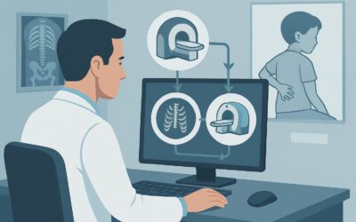
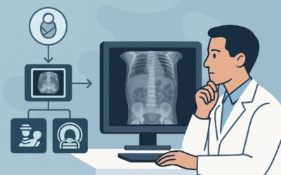
It’s 9 p.m. in the emergency department, and you are evaluating a 10-year-old with a day-long history of migrating right lower quadrant (RLQ) pain, anorexia, and a low-grade fever. The clinical picture is concerning for acute appendicitis, so you ordered the standard initial imaging study: a focused RLQ ultrasound. The preliminary report is unsatisfying: “Appendix not clearly visualized due to body habitus and overlying bowel gas. No clear secondary signs of inflammation.” The child’s pain is persisting, and the surgical team is asking for more definitive imaging before deciding on an operative plan. You are now faced with a critical decision: what is the appropriate next study to order? This article provides a detailed clinical workflow for this exact scenario, guiding you through the differential diagnosis, imaging rationale, and downstream management based on the American College of Radiology (ACR) Appropriateness Criteria. For this specific presentation, the ACR rates MRI abdomen and pelvis without and with IV contrast as Usually Appropriate.
Who Fits This Clinical Scenario?
This guidance is specifically for a pediatric patient where there is a continued clinical suspicion for acute appendicitis after an initial right lower quadrant (RLQ) ultrasound has been performed and yielded equivocal, nondiagnostic, or technically limited results. This is a common and challenging situation in clinical practice.
Inclusion criteria for this workflow:
- The patient is a child (infant to adolescent).
- Clinical signs and symptoms are suggestive of acute appendicitis (e.g., RLQ pain, fever, elevated inflammatory markers).
- A prior focused RLQ ultrasound was performed but failed to definitively confirm or exclude the diagnosis. This includes reports stating the appendix was not visualized, incompletely visualized, or findings were ambiguous.
It is crucial to distinguish this scenario from others. This workflow does not apply to:
- Initial imaging for a low-risk child: For patients with a low pre-test probability of appendicitis, the initial step might be observation or ultrasound, not an immediate second-line study.
- A clearly positive or negative ultrasound: If the initial ultrasound confidently identifies a normal or an inflamed appendix, this workflow is unnecessary, and management proceeds accordingly.
- High clinical suspicion with signs of perforation: In a child with a rigid abdomen, high fever, and hemodynamic instability, imaging should not delay an urgent surgical consultation, as they may proceed directly to the operating room.
What Diagnoses Are You Working Up in This Scenario?
While acute appendicitis is the primary concern, an equivocal ultrasound necessitates broadening the differential diagnosis. The next imaging study must be capable of not only identifying an inflamed appendix but also diagnosing common mimics. The key considerations in this pediatric population include:
Acute Appendicitis
This remains the leading diagnosis to exclude. The goal of the next imaging study is to visualize the appendix directly and assess for signs of inflammation, such as a diameter greater than 6 mm, wall thickening, hyperenhancement, and surrounding fat stranding or fluid. Complications like perforation or abscess formation must also be evaluated.
Mesenteric Adenitis
A frequent mimic of appendicitis in children, this condition involves inflammation and enlargement of the mesenteric lymph nodes, often secondary to a viral or bacterial infection. A definitive imaging study should be able to demonstrate prominent lymph nodes in the absence of an abnormal appendix, redirecting care toward supportive management.
Terminal Ileitis
Inflammation of the terminal ileum, most commonly from infectious enterocolitis or as an initial presentation of Crohn’s disease, can produce symptoms identical to appendicitis. The ideal imaging modality will clearly visualize the terminal ileum to assess for bowel wall thickening, mucosal enhancement, and adjacent inflammation.
Ovarian Pathology (in female patients)
In adolescent and pre-adolescent females, ovarian torsion, a ruptured or hemorrhagic ovarian cyst, or pelvic inflammatory disease can present with acute RLQ pain. The initial ultrasound may have been too focused on the appendix to fully evaluate the adnexa. The subsequent study must provide a comprehensive view of the pelvis to rule out these time-sensitive gynecologic emergencies.
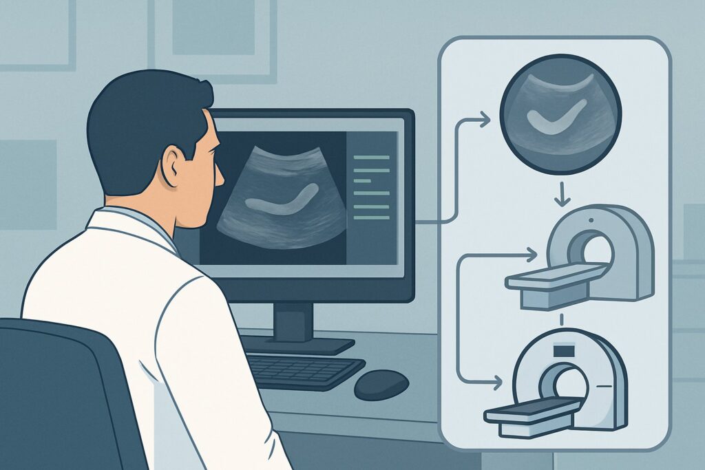
Why Is MRI of the Abdomen and Pelvis the Recommended Study for This Presentation?
When an ultrasound is inconclusive, the ACR designates MRI abdomen and pelvis without and with IV contrast as a Usually Appropriate next step. This recommendation is driven by MRI’s high diagnostic accuracy combined with its superior safety profile in children.
The primary rationale for choosing MRI is its ability to provide a definitive diagnosis without using ionizing radiation. MRI has demonstrated sensitivity and specificity for acute appendicitis that is comparable to CT. It excels at soft-tissue contrast, allowing for clear visualization of the appendix and surrounding structures. Furthermore, it is highly effective at diagnosing the alternative conditions on the differential, such as mesenteric adenitis, inflammatory bowel disease, and ovarian pathology.
Let’s compare this to the other options rated by the ACR for this scenario:
- CT abdomen and pelvis with IV contrast is also rated Usually Appropriate. It is fast, widely available, and highly accurate. However, its significant drawback is the use of ionizing radiation (ped_rrl=☢☢☢☢ 3-10 mSv). Given the heightened sensitivity of pediatric tissues to radiation and the cumulative lifetime risk, the ACR supports an “as low as reasonably achievable” (ALARA) principle, making radiation-free MRI the preferred choice when it can be performed in a timely manner.
- A repeat US abdomen RLQ is rated as May be Appropriate. If the initial study was limited by an inexperienced operator or suboptimal equipment, a repeat scan by a senior sonographer may be successful. However, if the limitation was patient-dependent (e.g., body habitus, bowel gas), a second ultrasound is likely to be inconclusive as well, leading to further delays in diagnosis.
- CT abdomen and pelvis without IV contrast is rated May be Appropriate (Disagreement). While it avoids the risks of IV contrast, its diagnostic accuracy for appendicitis and its mimics is lower than a contrast-enhanced study, particularly in thin children with little intra-abdominal fat.
The use of IV contrast with MRI enhances the visualization of inflammation, increasing diagnostic confidence for both appendicitis and its alternatives. Therefore, for a child with an equivocal ultrasound, MRI provides the most diagnostic information with the least potential for harm.
What’s Next After MRI? Downstream Workflow
The results of the abdominal and pelvic MRI will guide the subsequent clinical pathway. The clarity provided by MRI typically allows for a confident decision, minimizing unnecessary hospital stays or operations.
If the MRI is positive for acute appendicitis:
A definitive diagnosis of uncomplicated or complicated appendicitis warrants an immediate surgical consultation for appendectomy. The detailed imaging can also help surgeons anticipate challenges, such as a retrocecal position or the presence of an abscess that may require preoperative percutaneous drainage.
If the MRI is negative for appendicitis but identifies an alternative diagnosis:
The workflow shifts to address the specific finding.
- A diagnosis of mesenteric adenitis or infectious enterocolitis leads to supportive care, hydration, and pain management, allowing for discharge home when clinically stable.
- Findings suggestive of Crohn’s disease (e.g., terminal ileitis) require a consultation with pediatric gastroenterology for further evaluation and management.
- An ovarian cyst or torsion in a female patient necessitates an urgent gynecology consultation.
If the MRI is entirely negative:
When the MRI shows a normal appendix and no other cause for the patient’s symptoms, it provides strong evidence against an acute intra-abdominal surgical emergency. The clinical team can confidently pursue a course of observation, pain control, and consideration of non-gastrointestinal causes of pain. This often prevents a non-therapeutic appendectomy.
Pitfalls to Avoid (and When to Get Help)
Navigating this clinical scenario requires careful judgment to avoid common missteps. Be mindful of these potential pitfalls:
- Excessive Delay: While MRI is preferred, prolonged delays (e.g., waiting until the next day if the patient is worsening) may be inappropriate. If MRI is not readily available and the clinical suspicion is high or rising, a low-dose protocol CT may be the most pragmatic choice.
- Ignoring the Radiation Risk: Defaulting to CT without first considering the availability and feasibility of MRI is a significant pitfall in pediatric care. Always weigh the radiation burden against the clinical urgency.
- Patient Motion on MRI: Younger children may require sedation to obtain a diagnostic-quality MRI. This requires coordination with anesthesiology and should be factored into the decision-making and timing.
- Over-reliance on a Single Finding: Do not anchor on the initial equivocal ultrasound. Treat it as a non-diagnostic data point and move forward with a more definitive study based on the persistent clinical concern.
If the patient develops signs of peritonitis, sepsis, or hemodynamic instability at any point, escalate immediately to a pediatric surgeon. Definitive imaging should never delay life-saving intervention.
Related ACR Topics and Tools
This article focuses on a single, common clinical decision point. For a comprehensive overview of all pediatric scenarios related to suspected appendicitis, and for tools to help with imaging decisions and patient communication, please refer to the following resources.
- For breadth across all scenarios in Suspected Appendicitis-Child, see our parent guide: Suspected Appendicitis-Child: ACR Appropriateness Decoded.
- ACR Appropriateness Criteria Lookup — for adjacent scenarios
- Imaging Protocol Library — for technique on the recommended study
- Radiation Dose Calculator — for cumulative dose conversations
Frequently Asked Questions
Why not just order a CT scan if it’s faster and also rated ‘Usually Appropriate’?
While CT is fast and accurate, the primary reason to prefer MRI in children is to avoid ionizing radiation. A pediatric CT of the abdomen and pelvis delivers a dose of 3-10 mSv, which carries a small but real lifetime attributable risk of cancer. Since MRI offers comparable diagnostic accuracy without any radiation, it is the preferred next step whenever it can be performed in a clinically appropriate timeframe.
What should I do if MRI is not available 24/7 at my hospital?
This is a practical challenge. If a patient is clinically stable, they can be admitted for observation with a plan for an MRI the following morning. However, if the patient’s condition is worsening or clinical suspicion remains high overnight, a discussion with the radiologist and surgical team is warranted. In this case, a low-dose protocol CT scan is a reasonable and appropriate alternative to avoid a dangerous delay in diagnosis.
Is intravenous contrast always necessary for the MRI?
The ACR lists both ‘MRI abdomen and pelvis without and with IV contrast’ and ‘MRI abdomen and pelvis without IV contrast’ as ‘Usually Appropriate’. While non-contrast protocols can be effective, IV contrast often increases diagnostic confidence by highlighting inflammation in the appendiceal wall and surrounding tissues. It is particularly helpful for identifying abscesses and evaluating for alternative diagnoses like inflammatory bowel disease. The decision should be made in consultation with the on-call radiologist.
How does the workflow change for an adolescent female patient?
The workflow is largely the same, but the index of suspicion for gynecologic pathology (ovarian torsion, hemorrhagic cyst, pelvic inflammatory disease) is higher. This makes MRI an even more valuable tool, as it provides superior evaluation of the ovaries and fallopian tubes compared to CT. The initial ultrasound in a female patient should always include a pelvic component for this reason.
What if the child is too young or anxious to hold still for an MRI?
Patient motion can significantly degrade MRI quality. For young children (typically under 7-8 years old) or those with severe pain or anxiety, sedation or general anesthesia may be required to complete the scan. This requires coordination with the pediatric anesthesia team and should be part of the initial planning when ordering the MRI. The risks and benefits of sedation must be weighed against the alternatives, such as proceeding with a faster CT scan.
Reviewed by Pouyan Golshani, MD, Interventional Radiologist — May 29, 2026