What Is the Next Imaging Step for a Suspicious Axillary Node Found on CT or MRI?

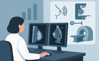
A 58-year-old female undergoes a CT angiogram of the chest for shortness of breath, which rules out a pulmonary embolism. However, the radiologist notes an incidental 1.5 cm left axillary lymph node with rounded morphology and loss of its fatty hilum, flagging it as suspicious. The emergency department physician now faces a common clinical question: what is the appropriate next step to evaluate this finding? This is not a palpable lump, nor is it from a screening mammogram. This article provides a focused, evidence-based workflow for this specific scenario, guiding the choice of the next imaging study. According to the American College of Radiology (ACR) Appropriateness Criteria, for a suspicious axillary node incidentally found on a modality like CT or MRI, dedicated US axilla is rated Usually Appropriate.
## Who Fits This Clinical Scenario?
This guidance is specifically for a female patient who has a suspicious-appearing axillary lymph node discovered incidentally on an imaging study that was not a mammogram or breast ultrasound. The initial finding is typically from a cross-sectional study like a Computed Tomography (CT) scan (of the chest, abdomen, or neck), a Magnetic Resonance Imaging (MRI) scan performed for a non-breast indication, or a PET/CT scan.
This workflow is designed for when the axillary finding is the primary question to be answered. It is crucial to distinguish this situation from several related, but distinct, clinical presentations that follow different diagnostic pathways:
- New Palpable Axillary Lump: If the patient presents with a new lump they can feel in their axilla, the initial imaging approach is different. This scenario is addressed in a separate ACR variant.
- Known Breast Cancer Diagnosis: If the patient has a newly diagnosed breast cancer, axillary imaging is performed for staging purposes, and the workup is integrated into their overall cancer care plan.
- Finding on Mammogram or Breast US: If the suspicious node was first identified on a screening or diagnostic mammogram or a dedicated breast ultrasound, the workup continues within the breast imaging pathway, often involving immediate further characterization or biopsy.
This article focuses solely on the “incidental” axillary node, where the immediate goal is to determine its significance and rule out an underlying, previously unknown malignancy.
## What Diagnoses Are You Working Up in This Scenario?
When a suspicious axillary node is found incidentally, the differential diagnosis is broad, but the primary concern is ruling out malignancy. The imaging workup is designed to characterize the node and search for a potential primary source.
The most critical diagnosis to exclude is occult breast cancer. Axillary lymph node metastasis can be the very first clinical sign of an otherwise undetectable breast primary. The term “occult” refers to a cancer that is not palpable and not visible on initial mammography. The axilla is the most common site of regional lymph node drainage from the breast, making this the leading concern in this scenario.
Another important consideration is lymphoma. Both Hodgkin and non-Hodgkin lymphoma can present with axillary lymphadenopathy. While often associated with more widespread or systemic symptoms (fever, night sweats, weight loss), isolated axillary involvement can occur. Ultrasound can help characterize the nodes, but a definitive diagnosis requires tissue sampling.
Metastasis from a non-breast primary cancer is also on the differential. Malignancies such as melanoma, lung cancer, thyroid cancer, or gastrointestinal cancers can metastasize to the axillary nodes. The patient’s history, risk factors, and the appearance of the node on the initial study can provide clues.
Finally, benign reactive or inflammatory adenopathy is a common cause of enlarged lymph nodes. This can be due to localized infection (e.g., from the arm or hand), systemic infection (e.g., mononucleosis), autoimmune conditions (e.g., rheumatoid arthritis, lupus), or recent vaccination. A detailed history, including recent immunizations and their location, is essential, as post-vaccination adenopathy can mimic malignancy.
## Why Is Axillary Ultrasound the Recommended First Step?
For a suspicious axillary node incidentally discovered on CT or MRI, the ACR identifies both US axilla and diagnostic mammography (including digital breast tomosynthesis) as Usually Appropriate. These studies are complementary and often performed together to provide a comprehensive evaluation.
The primary role of US axilla is to directly characterize the lymph node in question. Ultrasound offers superior spatial resolution compared to CT or MRI for superficial structures. It allows a radiologist to meticulously evaluate key morphological features that suggest malignancy, such as:
- Cortical thickening (focal or diffuse)
- Loss or compression of the fatty hilum
- Rounded (rather than oval) shape
- Non-hilar cortical blood flow on color Doppler
Because ultrasound provides real-time imaging without using ionizing radiation (0 mSv), it is the ideal modality for confirming the suspicious finding and guiding a potential biopsy.
Concurrently, diagnostic mammography or digital breast tomosynthesis (DBT) is also Usually Appropriate because it directly addresses the most common underlying cause: occult breast cancer. The goal is to scrutinize the ipsilateral breast for any subtle architectural distortion, suspicious calcifications, or a mass that may not have been visible on the initial CT or MRI.
Other procedures are rated lower for this initial workup step. US-guided fine needle aspiration (FNA) or core biopsy of the axillary node are rated May be appropriate. This rating reflects that tissue sampling is a downstream action, not the initial diagnostic imaging step. Imaging characterization with ultrasound and mammography should occur first to confirm there is a valid target and to search for a primary breast lesion. Proceeding directly to biopsy without this preliminary workup is generally not the standard of care.
## What’s Next After the Initial Workup? Downstream Workflow
The results of the axillary ultrasound and diagnostic mammogram will guide the subsequent steps in the patient’s care. The workflow branches based on these findings.
- If US confirms a suspicious node AND/OR mammography is positive: The patient should be referred to a breast specialist. The next step is almost always a US-guided core needle biopsy of the most suspicious axillary lymph node. A core biopsy is generally preferred over FNA because it provides tissue architecture and allows for immunohistochemical studies (e.g., hormone receptor status) if breast cancer is found. If the mammogram identifies a suspicious breast lesion, that will also be biopsied.
- If US confirms a suspicious node but mammography is negative: A US-guided core biopsy of the lymph node is still indicated. If the biopsy confirms metastatic carcinoma (typically adenocarcinoma), and the mammogram is negative, the patient is diagnosed with occult primary breast cancer (axillary presentation). Further evaluation with breast MRI is often recommended to search for the primary lesion.
- If US shows only benign-appearing nodes AND mammography is negative: If the ultrasound demonstrates that the node has a normal, benign appearance (e.g., a preserved fatty hilum and thin cortex), and the mammogram is clear, the workup may be complete. This suggests the node was likely reactive. Short-term follow-up ultrasound in 3-6 months may be considered to ensure stability, depending on the degree of suspicion on the initial CT or MRI.
- If the workup is indeterminate: In cases where findings are equivocal, a multidisciplinary discussion involving the radiologist, surgeon, and oncologist is invaluable. Further imaging with breast MRI or even a PET/CT might be considered to resolve the diagnostic uncertainty.
## Pitfalls to Avoid (and When to Get Help)
Navigating this clinical scenario requires careful integration of imaging findings and clinical context. Several common pitfalls can delay diagnosis or lead to unnecessary procedures.
1. Ignoring the Incidental Finding: Do not dismiss a radiologist’s comment about a “suspicious” axillary node on a CT or MRI. While many are benign, they can be the first sign of a serious underlying condition and warrant a dedicated workup.
2. Forgetting the Breast: The most frequent error is focusing solely on the axillary node without evaluating the breast. Always order a diagnostic mammogram/tomosynthesis along with the axillary ultrasound.
3. Overlooking Vaccination History: In the era of frequent vaccinations (e.g., for COVID-19, influenza, shingles), reactive axillary adenopathy is common. Always ask the patient about recent vaccinations, including the date and which arm, as this can explain the finding and may warrant a short-term follow-up instead of an immediate biopsy.
4. Premature Biopsy: Avoid referring for a biopsy based on the CT/MRI report alone. Dedicated axillary ultrasound is necessary first to confirm the finding, characterize the node, and provide a target for a potential procedure.
If the dedicated ultrasound and mammogram are both negative but the node appeared highly suspicious on the initial study (e.g., intensely FDG-avid on a PET/CT), it is crucial to escalate. This case should be discussed with a breast imaging specialist or presented at a multidisciplinary tumor board to determine the next best step, which could include breast MRI or excisional biopsy.
## Related ACR Topics and Tools
This article covers one specific variant within the broader topic of axillary imaging. For a complete overview of all clinical scenarios, from palpable lumps to post-treatment follow-up, please see our comprehensive parent guide.
- For breadth across all scenarios in Imaging of the Axilla, see our parent guide: Imaging of the Axilla: ACR Appropriateness Decoded.
To explore adjacent clinical questions or refine your understanding of imaging techniques, the following GigHz resources are available:
- ACR Appropriateness Criteria Lookup — for adjacent scenarios
- Imaging Protocol Library — for technique on the recommended study
- Radiation Dose Calculator — for cumulative dose conversations
Frequently Asked Questions
Why not just order a PET/CT scan to evaluate the suspicious axillary node?
While a PET/CT is excellent for staging known cancer, it is not the recommended initial study for an isolated, incidentally found axillary node. It involves significant radiation exposure and has lower spatial resolution than ultrasound for evaluating lymph node morphology. Furthermore, both malignant and inflammatory nodes can be FDG-avid, leading to false positives. The standard, ACR-recommended approach is to start with targeted ultrasound and diagnostic mammography.
The CT report mentioned multiple enlarged nodes. Does that change the recommendation for axillary ultrasound?
No, the recommendation remains the same. Axillary ultrasound is even more valuable in this situation. It allows the radiologist to survey the entire axilla, identify the most suspicious-looking node based on morphology (not just size), and select the best target for a potential biopsy. Diagnostic mammography is still essential to evaluate the ipsilateral breast.
What if the patient is male? Does this workflow still apply?
This specific ACR variant is written for a female patient, primarily because the risk of occult breast cancer is the main driver of the workup. While male breast cancer is rare, it can also present with axillary metastasis. For a male patient with a suspicious axillary node, axillary ultrasound is still the best initial imaging test. Diagnostic mammography of the male breast would also be considered, along with a broader differential that includes lymphoma and other metastatic diseases.
How long after a vaccine can axillary lymph nodes remain enlarged?
Post-vaccination reactive lymphadenopathy is common and can persist for several weeks. Most guidelines suggest that if a patient is asymptomatic and has a clear vaccination history in the ipsilateral arm, a short-term follow-up ultrasound in 6-12 weeks is a reasonable alternative to immediate biopsy to ensure resolution or stability.
If the axillary ultrasound and mammogram are negative, is a breast MRI always necessary?
Not always. If the initial finding on CT/MRI was only mildly suspicious and the dedicated ultrasound shows a clearly benign-appearing node, the workup may be complete. However, if the node remains suspicious on ultrasound despite a negative mammogram, or if it was very concerning on the initial study (e.g., highly FDG-avid on PET), then a breast MRI is often the next step to search for a mammographically occult primary cancer before proceeding to an excisional biopsy of the node.
Reviewed by Pouyan Golshani, MD, Interventional Radiologist — May 29, 2026