What Is the Next Imaging Study for a Child with a Suspected Vascular Malformation on Ultrasound?
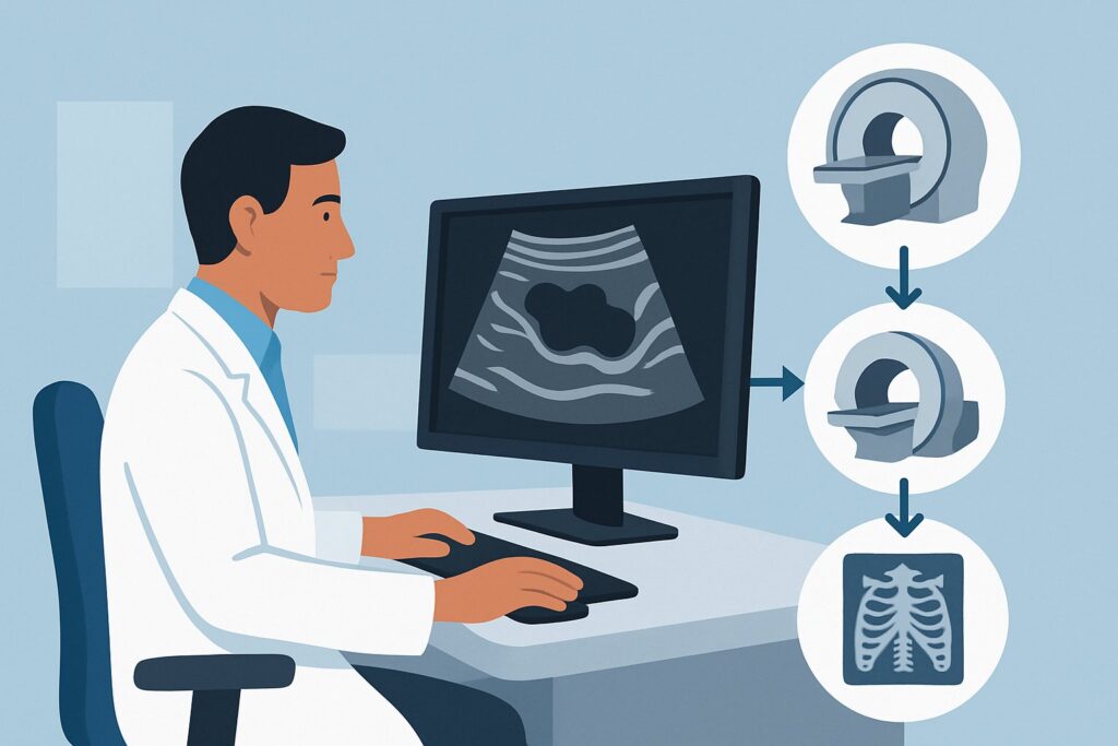
A 7-year-old presents to your pediatric clinic with a soft, compressible, non-pulsatile mass on her calf that has grown slowly over the past year. An initial ultrasound performed by a colleague shows a collection of hypoechoic, serpiginous channels with minimal flow on Doppler, raising suspicion for a vascular malformation. You agree with the preliminary assessment, but the ultrasound cannot fully delineate the extent of the lesion or its relationship to the deep fascia and neurovascular structures. To guide referral to a specialist and plan potential treatment, you need a more definitive imaging study. According to the American College of Radiology (ACR), the next step in this clinical workflow is clear: MRA and MRV area of interest without and with IV contrast is rated Usually appropriate.
Who Fits This Clinical Scenario?
This guidance applies specifically to a child (i.e., not an infant in the first few months of life) who has already undergone an initial ultrasound for a soft tissue anomaly, and the sonographic features are suspicious for a vascular malformation. The key inclusion criteria are:
- The patient is a child.
- An initial ultrasound has been performed.
- Ultrasound findings suggest a vascular malformation (e.g., cystic or tubular structures, abnormal flow on Doppler).
It is crucial to distinguish this situation from similar but distinct clinical presentations that follow different diagnostic pathways. This article does not apply if:
- The patient is an infant with a classic “strawberry” mark. A bright red, rapidly growing lesion in an infant is highly suggestive of an infantile hemangioma. The workup for this is covered in a separate ACR scenario, which often does not require advanced imaging unless there are specific concerns.
- The patient has a known vascular malformation with new symptoms. If a diagnosis has already been established with prior imaging and the child presents with new pain, bleeding, or functional impairment, the imaging choice is guided by the need to evaluate for complications like thrombosis or hemorrhage.
- This is the initial presentation without any prior imaging. If you are evaluating a child with clinical signs of a vascular anomaly for the first time, the decision is about which initial imaging modality to choose, not the next step after an inconclusive ultrasound.
What Diagnoses Are You Working Up in This Scenario?
After an initial ultrasound suggests a vascular malformation, the primary goal of the next imaging study is to precisely classify the lesion, define its anatomical extent, and rule out mimickers. The differential diagnosis is centered on the International Society for the Study of Vascular Anomalies (ISSVA) classification.
Venous Malformation (VM): This is the most common type of symptomatic vascular malformation. These are low-flow lesions composed of dysplastic venous channels. On ultrasound, they appear as compressible, hypoechoic spaces with slow or undetectable flow. MRI is essential to confirm the diagnosis and map the full extent of the malformation, which is often larger than clinically apparent.
Lymphatic Malformation (LM): These are congenital anomalies of the lymphatic system, categorized as macrocystic, microcystic, or mixed. They typically present as soft, non-compressible masses. While ultrasound can identify the cystic nature, MRI provides superior detail regarding the lesion’s composition and its infiltration into surrounding tissues, which is critical for planning sclerotherapy or surgical excision.
Arteriovenous Malformation (AVM): A less common but more consequential diagnosis, AVMs are high-flow lesions with direct connections between arteries and veins without an intervening capillary bed. An initial ultrasound may have shown high-velocity flow and arteriovenous shunting. MRA is critical to map the feeding arteries, the central nidus, and the draining veins, which is prerequisite knowledge for any endovascular treatment.
Combined or Complex Malformations: Some patients have malformations with combined elements (e.g., venolymphatic malformations) or are associated with syndromes like Klippel-Trenaunay syndrome (which involves capillary, venous, and lymphatic malformations with limb overgrowth). MRI is unparalleled in characterizing these complex anomalies.
Soft Tissue Sarcoma: Although rare, a malignant soft tissue tumor can mimic a vascular anomaly. Certain sarcomas can be highly vascular. MRI with contrast helps differentiate the infiltrative, solid-enhancing components of a tumor from the characteristic appearance of a malformation, making it a critical tool for avoiding misdiagnosis.
Why Is MRA and MRV the Recommended Next Study for a Suspected Vascular Malformation?
The ACR designates MRA and MRV area of interest without and with IV contrast as Usually appropriate because it provides the most comprehensive, non-invasive assessment for this specific clinical question. The combination of magnetic resonance imaging (MRI), angiography (MRA), and venography (MRV) offers a complete picture of the lesion’s morphology, flow dynamics, and anatomical relationships, all without the use of ionizing radiation.
The rationale for this recommendation includes:
- Superior Soft Tissue Characterization: Standard MRI sequences (like T1, T2, and fat-suppressed T2) excel at defining the lesion’s boundaries and its relationship to adjacent muscles, nerves, bones, and vessels. This is crucial for determining resectability or planning percutaneous access for sclerotherapy.
- Flow Dynamics Assessment: The addition of MRA and MRV techniques allows for the differentiation between low-flow (venous, lymphatic) and high-flow (arteriovenous) malformations. This distinction is the most critical branch point in management. Contrast-enhanced dynamic sequences can visualize feeding arteries and draining veins, which is essential for AVMs.
- Safety in a Pediatric Population: With a radiation dose of 0 mSv, MRI avoids the risks of ionizing radiation associated with CT. This is a paramount consideration in children, who are more sensitive to the long-term effects of radiation.
Alternative studies are rated lower for specific reasons. For instance, CTA and CTV area of interest with IV contrast is rated May be appropriate. While it provides excellent vascular mapping and is faster than MRI, it involves a significant radiation dose (rated as ‘Varies’) and offers inferior soft tissue contrast. It is typically reserved for cases where MRI is contraindicated or unavailable. Similarly, MRI area of interest without IV contrast is rated Usually not appropriate. Omitting intravenous contrast severely limits the ability to assess vascularity, enhancement patterns, and flow dynamics, which are essential for accurate classification and for distinguishing malformations from neoplastic processes.
Once you’ve decided on MRA and MRV, our protocol guide covers key principles for technique and interpretation. While the linked example is for the brain, the fundamental principles of MRA/MRV sequencing and contrast administration are applicable to other body parts: MRA Brain Without Contrast (3D TOF).
What’s Next After MRA and MRV? Downstream Workflow
The results of the MRA/MRV study will guide the subsequent management, which almost always involves a multidisciplinary team.
- If the study confirms a low-flow malformation (VM or LM): The next step is referral to a vascular anomalies center. The team, often including an interventional radiologist, pediatric surgeon, and dermatologist, will use the MRI as a roadmap. The primary treatment for many symptomatic VMs and LMs is percutaneous sclerotherapy, and the detailed anatomy provided by the MRI is essential for planning a safe and effective procedure.
- If the study confirms a high-flow malformation (AVM): This finding necessitates a more urgent referral to a specialized center. The MRA findings are used to plan treatment, which typically involves endovascular embolization by an interventional radiologist or neurointerventionalist to block the abnormal arteriovenous connections. Catheter-based digital subtraction angiography, which is Usually not appropriate for initial diagnosis, becomes the next step for therapeutic intervention.
- If the findings are indeterminate or suspicious for a tumor: The MRI will be used to guide a biopsy. The radiologist can identify the safest path to the most suspicious-looking solid component of the mass, avoiding major vessels. The case should be discussed with a pediatric oncologist and surgeon.
- If the study is negative: If the MRA/MRV is normal and does not identify a vascular anomaly, the clinical picture should be reassessed. Other causes of a soft tissue mass, such as a post-traumatic hematoma, a benign non-vascular tumor (e.g., lipoma), or an abscess, should be considered. Clinical follow-up is the most common next step.
Pitfalls to Avoid (and When to Get Help)
Navigating the workup of a suspected vascular malformation requires careful attention to detail to avoid common errors.
- Pitfall 1: Ordering a non-contrast MRI. As noted, contrast is critical for characterizing flow and ruling out neoplasm. A non-contrast study is often non-diagnostic and will likely need to be repeated.
- Pitfall 2: Accepting an incomplete study. Ensure the ordering protocol specifies both MRA and MRV sequences, along with standard anatomic imaging. Simply ordering “MRI of the leg” may not provide the necessary vascular information.
- Pitfall 3: Defaulting to CT. Unless MRI is absolutely contraindicated, the radiation exposure from CT makes it a suboptimal choice for the initial characterization of these benign conditions in children.
If the clinical signs suggest a rapidly expanding, painful, or ulcerating lesion, or if an AVM is confirmed on imaging, escalate care by making an urgent referral to a multidisciplinary vascular anomalies team.
Related ACR Topics and Tools
This article focuses on a single, common clinical decision point. For a comprehensive overview of imaging for all related pediatric vascular anomalies, consult our parent guide. For help with adjacent scenarios or technical details, the following resources are available.
- For breadth across all scenarios in Soft Tissue Vascular Anomalies: Vascular Malformations and Infantile Vascular Tumors (Non-CNS)-Child, see our parent guide: Soft Tissue Vascular Anomalies: Vascular Malformations and Infantile Vascular Tumors (Non-CNS)-Child: ACR Appropriateness Decoded.
- ACR Appropriateness Criteria Lookup — for adjacent scenarios
- Imaging Protocol Library — for technique on the recommended study
- Radiation Dose Calculator — for cumulative dose conversations
Frequently Asked Questions
Why is MRA/MRV preferred over a standard MRI with and without contrast for this scenario?
While a standard MRI with and without contrast is also rated ‘Usually appropriate,’ explicitly ordering MRA (Magnetic Resonance Angiography) and MRV (Magnetic Resonance Venography) ensures the protocol includes specific sequences designed to visualize blood flow and vessel anatomy. This provides a more dynamic and comprehensive assessment of the lesion’s vascularity, which is crucial for distinguishing high-flow from low-flow malformations—the most important factor for determining management and urgency.
Is sedation required for an MRA/MRV in a child?
Often, yes. MRI scans can take 45-60 minutes or longer, and the child must remain perfectly still to acquire high-quality images. Most young children (typically under age 8) require sedation or general anesthesia to complete the study successfully. This should be discussed with the parents and the radiology department when scheduling the exam.
What if the ultrasound showed a classic infantile hemangioma, not a malformation?
That is a different clinical scenario. Most infantile hemangiomas are diagnosed clinically and do not require imaging. Imaging is reserved for cases with diagnostic uncertainty, deep lesions, or when there is concern for complications or associated syndromes (e.g., PHACE syndrome). The ACR has separate guidelines for that specific presentation.
Can I order a CT scan if my facility has a long wait time for pediatric MRI?
CTA/CTV is rated ‘May be appropriate’ and can be considered if MRI is not readily available and a diagnosis is needed to guide urgent management. However, this decision should be made in consultation with a radiologist, weighing the diagnostic need against the significant drawback of ionizing radiation exposure in a child. For most stable, slow-growing lesions, waiting for an MRI is the preferred course.
What specific information should I include in the order for the MRA/MRV?
In your order, be as specific as possible. Include the patient’s age, the exact location and size of the lesion, relevant clinical history (e.g., ‘slow-growing, compressible mass’), and the findings from the prior ultrasound (‘suspicion for low-flow vascular malformation’). This context helps the radiologist tailor the MRI protocol to best answer the clinical question.
Reviewed by Pouyan Golshani, MD, Interventional Radiologist — May 30, 2026