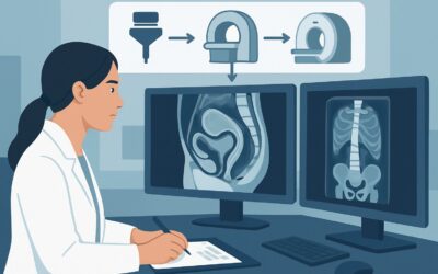
What Is the Next Imaging Study for Indeterminate Postmenopausal Pelvic Pain?

A 62-year-old female returns to your clinic for follow-up on her persistent, dull pelvic ache. The pain has been present for three months, unresponsive to over-the-counter analgesics. A transvaginal ultrasound performed last week was inconclusive; the report notes a “complex-appearing left adnexal structure” but cites poor visualization due to body habitus. The clinical question is clear and urgent: what is the most appropriate next imaging study to characterize this finding and rule out a serious underlying pathology?
This article provides a detailed clinical workflow for this specific scenario, guided by the American College of Radiology (ACR) Appropriateness Criteria. For a postmenopausal patient with subacute or chronic pelvic pain of suspected gynecologic origin and an indeterminate ultrasound, the ACR rates MRI pelvis without and with IV contrast as Usually Appropriate. We will explore the rationale for this recommendation, the differential diagnoses in play, and the downstream clinical pathway based on the MRI results.
Who Fits This Clinical Scenario?
This guidance is specifically for a postmenopausal female presenting with subacute or chronic pelvic pain (lasting weeks to months) where the initial clinical suspicion points toward a gynecologic etiology. The crucial element of this scenario is that a first-line imaging study—typically a transvaginal and/or transabdominal pelvic ultrasound—has already been performed and returned an indeterminate, equivocal, or technically limited result.
This workflow applies when you need to better characterize a finding seen on ultrasound or when the ultrasound was negative but clinical suspicion for a gynecologic source of pain remains high.
This guidance does not apply to several similar-but-distinct clinical situations:
- Initial Imaging: If you are ordering the very first imaging study for this patient’s symptoms, that constitutes a different clinical scenario with its own set of recommendations. This article is strictly for the next step after an inconclusive ultrasound.
- Acute Pelvic Pain: Patients presenting with acute, severe pain, especially if accompanied by fever, leukocytosis, or signs of peritonitis, require a more urgent workup. The differential in acute pain includes ovarian torsion, tubo-ovarian abscess, or a ruptured ectopic pregnancy (rare postmenopause, but possible), which may alter the choice and timing of imaging.
- Clear Non-Gynecologic Suspicion: If the clinical picture strongly suggests a gastrointestinal (e.g., diverticulitis, colon cancer) or urologic (e.g., ureteral stone, bladder mass) source, the imaging workup should be tailored to that system, often starting with a CT scan.
What Diagnoses Are You Working Up in This Scenario?
When an ultrasound is indeterminate in a postmenopausal woman with pelvic pain, the primary goal of subsequent imaging is to differentiate benign from potentially malignant conditions. The differential is broad, but MRI is particularly well-suited to evaluate the following possibilities.
The most consequential diagnosis to exclude is an ovarian or other adnexal malignancy. In a postmenopausal patient, any new, complex, or solid adnexal mass is considered malignant until proven otherwise. Ultrasound may struggle to fully characterize internal architecture, septations, or solid components. MRI provides superior tissue characterization to identify features that raise or lower the suspicion of cancer, guiding the need for referral to a gynecologic oncologist.
Benign uterine pathology, such as a degenerating leiomyoma (fibroid), is a common cause of pain. While fibroids are often asymptomatic, they can outgrow their blood supply and undergo necrosis, causing significant pain. A large or atypically located fibroid can also cause mass effect. MRI can definitively diagnose fibroids, assess for features of degeneration, and precisely map their location relative to other pelvic structures.
While less common after menopause, adenomyosis—the presence of endometrial glands and stroma within the myometrium—can persist and cause chronic pain and uterine enlargement. Ultrasound findings can be subtle or non-specific, whereas MRI is the non-invasive gold standard for diagnosis, typically showing a thickened junctional zone.
Finally, imaging helps evaluate for less common but important etiologies like hydrosalpinx (a fluid-filled, blocked fallopian tube) or rare uterine sarcomas. MRI can also identify non-gynecologic causes of pain that may mimic a gynecologic source, such as pelvic floor disorders, sacral nerve pathology, or occult musculoskeletal issues.
Why Is MRI Pelvis with Contrast the Recommended Next Study?
The ACR designates MRI pelvis without and with IV contrast as a Usually Appropriate study in this scenario due to its unparalleled soft-tissue resolution and safety profile. It directly addresses the diagnostic uncertainty left by the initial ultrasound.
The fundamental advantage of MRI is its superior ability to differentiate between various pelvic soft tissues. It can distinguish the uterine zonal anatomy (endometrium, junctional zone, myometrium), characterize the contents of an ovarian cyst (simple fluid, blood, or fat), and identify solid, enhancing tissue within a mass that is suspicious for malignancy. This level of detail is critical for problem-solving after an equivocal ultrasound.
The addition of intravenous gadolinium-based contrast is key for assessing tissue vascularity. Malignant tumors often demonstrate robust and early enhancement compared to benign tissues. The pattern and degree of enhancement within a complex adnexal mass or uterine lesion provide crucial data points for risk stratification.
Let’s compare this to the other rated procedures:
- CT Pelvis with IV Contrast: While also rated Usually Appropriate, CT is generally considered a secondary option for this specific clinical question. Its soft-tissue contrast is significantly inferior to MRI for characterizing uterine and ovarian pathology. CT is excellent for detecting calcifications, gross metastatic disease, or bowel obstruction, but it cannot provide the detailed tissue analysis needed to evaluate an indeterminate adnexal mass. It also involves ionizing radiation (ACR RRL ☢☢☢, 1-10 mSv).
- CT Pelvis without and with IV Contrast: This combined study is rated Usually not appropriate. It delivers a high radiation dose (ACR RRL ☢☢☢☢, 10-30 mSv) without offering a significant diagnostic advantage over a contrast-enhanced MRI or even a single-phase CT for this indication. The non-contrast portion adds little information for the primary workup of a suspected gynecologic mass.
Crucially, MRI involves no ionizing radiation (ACR RRL O, 0 mSv). This is an important consideration for any patient, ensuring that the diagnostic workup does not contribute to cumulative radiation exposure.
What’s Next After MRI Pelvis with Contrast? Downstream Workflow
The results of the pelvic MRI will guide the subsequent clinical pathway, transforming diagnostic uncertainty into an actionable plan.
If the MRI is positive for suspected malignancy: Findings such as a solid, enhancing adnexal mass, thickened irregular endometrium with myometrial invasion, or evidence of peritoneal disease are highly concerning. The immediate next step is an urgent referral to a gynecologic oncologist. This specialist will typically proceed with surgical staging and treatment, which may involve a total hysterectomy, bilateral salpingo-oophorectomy, and lymph node assessment.
If the MRI confirms a benign gynecologic cause: When the MRI identifies a clear benign source for the pain, such as a degenerating fibroid, adenomyosis, or a benign ovarian cyst (e.g., a simple cyst or endometrioma), the workflow shifts from oncologic workup to symptom management. Treatment options may include medical management (e.g., hormonal therapy, analgesics), referral for a minimally invasive procedure like uterine artery embolization for fibroids, or consultation with a general gynecologist for potential surgical intervention if symptoms are refractory.
If the MRI is negative or non-specific: A negative MRI provides strong evidence against a significant gynecologic cause for the patient’s pain. In this case, the differential diagnosis must be broadened to include non-gynecologic etiologies. The next steps may involve referrals to other specialists, such as Gastroenterology for a colonoscopy, Urology for a cystoscopy, or Physical Medicine and Rehabilitation to evaluate for musculoskeletal or pelvic floor dysfunction.
Pitfalls to Avoid (and When to Get Help)
Navigating the workup for an indeterminate pelvic finding requires careful consideration to avoid common missteps.
- Pitfall: Defaulting to CT. In many clinical settings, CT is the go-to cross-sectional imaging modality. However, for characterizing a suspected gynecologic mass, MRI is diagnostically superior. Resist the reflex to order a CT unless MRI is contraindicated or unavailable.
- Pitfall: Omitting IV contrast. While a non-contrast MRI is also rated Usually Appropriate, it can be insufficient. For evaluating a complex or solid mass where malignancy is a concern, the enhancement pattern seen with IV contrast is a critical piece of the diagnostic puzzle. Always specify “without and with IV contrast” unless there is a specific contraindication.
- Pitfall: Providing insufficient clinical history. The value of a radiology report is maximized when the radiologist understands the specific clinical question. Be sure to include the patient’s menopausal status, the specific findings of the prior ultrasound, and the primary question (e.g., “characterize indeterminate left adnexal mass, rule out malignancy”).
If the patient’s chronic pain suddenly becomes acute and severe, or if she develops fever or hemodynamic instability while awaiting an outpatient MRI, this constitutes a clinical emergency. She should be directed to the emergency department for immediate evaluation to rule out a superimposed acute event like ovarian torsion or hemorrhage into a mass.
Related ACR Topics and Tools
For a comprehensive overview of all clinical variants related to this topic, please consult our parent guide. For further exploration of appropriateness criteria, imaging protocols, and radiation dose, the following GigHz resources are available.
- For breadth across all scenarios in Postmenopausal Subacute or Chronic Pelvic Pain, see our parent guide: Postmenopausal Subacute or Chronic Pelvic Pain: ACR Appropriateness Decoded.
- ACR Appropriateness Criteria Lookup — for adjacent scenarios
- Imaging Protocol Library — for technique on the recommended study
- Radiation Dose Calculator — for cumulative dose conversations
Frequently Asked Questions
What if my patient has a contraindication to MRI, like a non-compatible pacemaker?
In cases where MRI is contraindicated, `CT pelvis with IV contrast` is the best alternative and is also rated *Usually Appropriate* by the ACR. While its soft-tissue resolution is lower than MRI’s, it can still provide crucial information regarding mass size, the presence of solid components, calcifications, and any evidence of metastatic spread to lymph nodes or other organs.
Is a non-contrast MRI ever sufficient for this scenario?
The ACR rates `MRI pelvis without IV contrast` as *Usually Appropriate*. It can be sufficient for characterizing certain lesions with classic signal characteristics, such as a fat-containing dermoid cyst (teratoma) or a hemorrhagic endometrioma. However, for an indeterminate solid or complex cystic-solid mass where malignancy is the primary concern, intravenous contrast is strongly recommended to assess tissue vascularity, which is a key feature in differentiating benign from malignant processes.
The ultrasound report was very vague. Should I repeat the ultrasound before ordering an MRI?
If the initial ultrasound was of poor technical quality (e.g., due to severe obesity or bowel gas), a repeat ultrasound at a specialized imaging center could be considered. However, if the study was technically adequate but the findings themselves were inherently indeterminate (e.g., a complex cyst with septations), proceeding directly to MRI is the more efficient and diagnostically powerful next step, as recommended by the ACR.
Does my patient need any special preparation for a pelvic MRI with contrast?
Yes, most imaging centers will have a standard preparation protocol. Patients are typically asked to be NPO (nothing by mouth) for 4-6 hours before the scan to reduce bowel motion artifacts. They will also need a recent creatinine level to ensure adequate kidney function for the administration of gadolinium-based contrast. Some protocols may also involve the administration of an anti-peristaltic agent like glucagon to further minimize bowel motion.
What if the MRI shows a likely benign fibroid, but the patient’s pain is severe?
Even if the MRI confirms a benign diagnosis like a leiomyoma, this can still be the source of significant pain, especially if it is large or degenerating. The MRI result in this case is still highly valuable, as it confidently rules out malignancy and allows the clinical focus to shift to symptom management. The next step would be a discussion with the patient and her gynecologist about treatment options, which could range from medical therapy to procedures like uterine artery embolization or surgical myomectomy/hysterectomy.
Reviewed by Pouyan Golshani, MD, Interventional Radiologist — May 30, 2026