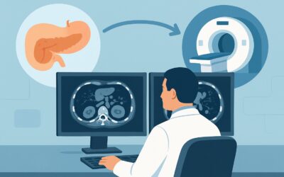
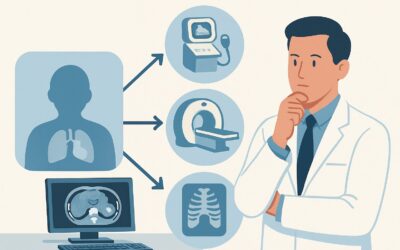
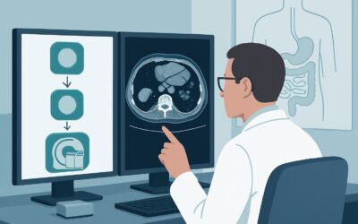
What Is the Right First Imaging Study for Suspected Acute Pancreatitis?

A 45-year-old male presents to the emergency department with a 24-hour history of severe, constant epigastric pain radiating to his back, accompanied by nausea. He is hemodynamically stable. His lab work returns with a lipase level more than three times the upper limit of normal, confirming a diagnosis of acute pancreatitis. This is his first documented episode. As the admitting physician, you initiate fluid resuscitation and pain control, but the key question remains: what is the etiology, and what imaging, if any, is needed in this initial phase? This article provides a focused, evidence-based workflow for this specific clinical scenario, explaining why the American College of Radiology (ACR) rates US abdomen as Usually appropriate for the initial evaluation.
Who Fits This Clinical Scenario for Suspected Acute Pancreatitis?
This guidance applies to a well-defined patient population: those with a suspected first episode of acute pancreatitis where the diagnosis is already strongly supported by clinical and laboratory findings.
Inclusion criteria for this workflow:
- First-time presentation: The patient has no prior history of pancreatitis.
- Classic signs and symptoms: The presentation is typical, primarily severe epigastric pain.
- Confirmed biochemical diagnosis: Amylase and/or lipase levels are significantly elevated (typically >3x the upper limit of normal).
- Early in the clinical course: The patient presents less than 48 to 72 hours after symptom onset.
It is critical to distinguish this straightforward presentation from more complex situations that require a different imaging strategy. This workflow does not apply if:
- The diagnosis is uncertain: If the patient presents with atypical symptoms or has equivocal lab results, the diagnostic goal of imaging is different. This falls under the ACR variant for atypical presentations.
- The patient is critically ill: A patient with signs of Systemic Inflammatory Response Syndrome (SIRS), organ failure, or severe clinical scores (e.g., APACHE II) needs imaging to assess for complications like necrosis, not just etiology.
- The patient fails to improve or deteriorates: If a patient’s condition worsens or does not improve with initial supportive care after several days, the imaging focus shifts to identifying developing complications, such as fluid collections or necrosis.
Applying the wrong imaging strategy in these distinct scenarios can lead to delayed diagnosis of complications or unnecessary radiation exposure.
What Diagnoses Are You Working Up with Initial Imaging?
In a patient with a clear clinical and lab-based diagnosis of acute pancreatitis, the primary goal of initial imaging is not to re-confirm the diagnosis, but to determine the underlying cause. Identifying the etiology is paramount as it directly influences subsequent management.
The key differential diagnosis to investigate is gallstone pancreatitis. This is the most common cause of acute pancreatitis in many populations. An imaging study that can reliably detect gallstones (cholelithiasis) or evidence of a stone passing through the common bile duct (choledocholithiasis) is essential. Finding a biliary cause may trigger a consultation for endoscopic retrograde cholangiopancreatography (ERCP) or cholecystectomy.
While the main target is identifying a biliary etiology, initial imaging can also provide supportive evidence for the primary diagnosis by showing pancreatic inflammation or edema. It may also reveal less common causes, such as a pancreatic mass, though this is not the primary objective.
Finally, imaging helps exclude other serious causes of acute epigastric pain that can mimic pancreatitis, such as acute cholecystitis, a perforated peptic ulcer, or mesenteric ischemia. Although less likely when lipase is markedly elevated, these conditions remain on the differential, and a well-chosen initial study can help rule them out.
Why Is Abdominal Ultrasound the Recommended First Study for This Presentation?
For a patient with a first episode of biochemically confirmed acute pancreatitis within the first 72 hours, the ACR designates US abdomen as Usually appropriate. The rationale is directly tied to the clinical questions that need to be answered at this early stage.
The primary objective is to identify a biliary etiology. Abdominal ultrasound is an excellent modality for this purpose, offering high sensitivity and specificity for detecting gallstones within the gallbladder. It is non-invasive, widely available, cost-effective, and, critically, involves no ionizing radiation (0 mSv). This makes it the ideal screening tool to answer the most urgent etiological question.
In contrast, other powerful imaging modalities are rated lower for this specific initial workup because they represent a mismatch of tool to task.
- CT abdomen and pelvis with IV contrast is rated May be appropriate. While CT is the gold standard for evaluating pancreatic necrosis and peripancreatic fluid collections, these complications often take 48 to 72 hours or longer to become radiographically apparent. Performing a CT scan too early can underestimate the severity of the disease, providing false reassurance while exposing the patient to significant ionizing radiation (adult RRL ☢☢☢ 1-10 mSv). It is less sensitive than ultrasound for detecting small gallstones. Therefore, in an uncomplicated, early presentation, the risks and costs of CT outweigh the benefits.
- CT abdomen and pelvis without IV contrast is rated Usually not appropriate. A non-contrast CT offers little diagnostic advantage over ultrasound for the primary question of gallstones and is inferior to a contrast-enhanced study for evaluating the pancreatic parenchyma, vasculature, and potential complications. It exposes the patient to radiation without providing the necessary diagnostic detail for assessing severity.
The logical, evidence-based approach is to start with the safest, most direct test to rule in or rule out the most common and actionable cause. That test is an abdominal ultrasound.
What’s Next After an Abdominal Ultrasound? Downstream Workflow
The results of the initial abdominal ultrasound guide the subsequent management and imaging pathway. The workflow branches based on the findings.
- If the US is positive for gallstones or biliary ductal dilation: This strongly suggests gallstone pancreatitis. The immediate next step is a surgical consultation to plan for cholecystectomy, typically during the same hospital admission, to prevent recurrence. If there is evidence of a persistent stone in the common bile duct (choledocholithiasis) or concurrent cholangitis, the patient may require an MRCP or an urgent ERCP for stone extraction.
- If the US is negative and the pancreas is well-visualized: With a biliary cause less likely, the focus shifts to investigating other etiologies. This involves a thorough history review for alcohol use and obtaining a triglyceride level. If the patient improves clinically with supportive care, no further imaging may be necessary during the acute phase.
- If the US is indeterminate or non-diagnostic: A common limitation of ultrasound is obscuration of the pancreas and distal common bile duct by overlying bowel gas. If the study is technically limited but clinical suspicion for a biliary cause remains high, or if the patient is not improving as expected, the next logical step is MRI abdomen without and with IV contrast with MRCP (May be appropriate). MRCP is highly sensitive for detecting common bile duct stones and provides excellent detail of the pancreatic and biliary systems without using ionizing radiation.
- If the patient’s clinical status deteriorates: Should the patient develop signs of sepsis, organ failure, or SIRS, they transition to a different clinical scenario. The imaging priority shifts from etiology to assessing for complications. At this point, a CT abdomen and pelvis with IV contrast becomes the study of choice to evaluate for pancreatic necrosis and fluid collections.
Common Pitfalls to Avoid in Early Pancreatitis Imaging
Navigating the initial workup requires avoiding several common missteps that can lead to diagnostic delays or unnecessary procedures.
- Ordering a CT scan too early. The most frequent pitfall is ordering a contrast-enhanced CT within the first 24-48 hours for an uncomplicated case. Necrosis may not be visible this early, leading to an underestimation of severity. Reserve CT for patients who are critically ill on presentation or who fail to improve after 48-72 hours.
- Stopping the workup after a “negative” ultrasound. A negative ultrasound for gallstones does not definitively rule out a biliary cause. A small stone may have already passed. If no other cause (like alcohol or hypertriglyceridemia) is found, it is often presumed to be of biliary origin (“biliary sludge” or microlithiasis).
- Forgetting to check triglycerides. Severe hypertriglyceridemia (typically >1000 mg/dL) is a crucial, non-biliary cause of acute pancreatitis. This simple lab test should be part of the initial workup for any patient with pancreatitis of unknown etiology.
If a patient shows signs of hemodynamic instability, worsening organ dysfunction, or an abrupt drop in hemoglobin, escalate immediately. This suggests a severe complication like hemorrhagic pancreatitis or infected necrosis, warranting urgent ICU consultation and likely a contrast-enhanced CT scan.
Related ACR Topics and Tools
For a comprehensive overview of imaging recommendations across all clinical variants of acute pancreatitis, from atypical presentations to chronic complications, please see our parent guide. Further resources for selecting appropriate imaging and understanding protocols are also available.
- For breadth across all scenarios in Acute Pancreatitis, see our parent guide: Acute Pancreatitis: ACR Appropriateness Decoded.
- Imaging Appropriateness Selector — for adjacent scenarios
- Imaging Protocol Library — for technique on the recommended study
- Radiation Dose Calculator — for cumulative dose conversations
Frequently Asked Questions
If the diagnosis of acute pancreatitis is already confirmed by labs, why is any imaging needed at all?
In this scenario, the primary goal of imaging is not to confirm pancreatitis but to determine its cause (etiology). Identifying gallstones as the cause is critical because it changes management, often leading to surgery (cholecystectomy) to prevent recurrence. Ultrasound is the safest and most effective first step for this specific question.
My patient’s ultrasound was negative for gallstones. Should I order a CT scan next?
Not necessarily. If the patient is clinically improving and the ultrasound was of good quality, the next step is to investigate other causes like alcohol use and hypertriglyceridemia. A CT scan is generally reserved for patients who are critically ill or fail to improve after 48-72 hours, as its main role is to detect complications like necrosis, not to find the initial cause.
The ultrasound report says the pancreas was ‘obscured by bowel gas.’ What should I do?
This is a common limitation. If your clinical suspicion for a biliary cause remains high (e.g., elevated liver enzymes), the next best test is an MRI with MRCP. It provides excellent, non-invasive visualization of the bile ducts and pancreas without the radiation of a CT scan. If the patient is stable, this is preferred over an early CT.
Is there any role for a plain abdominal X-ray in this initial presentation?
Generally, no. Plain radiographs have a very low yield for diagnosing acute pancreatitis or its common causes. They are not recommended by the ACR for this clinical scenario. Their primary role would be to look for free air in the rare case of a perforated ulcer mimicking pancreatitis, but CT is far more sensitive for this.
When does CT become the right first choice for suspected pancreatitis?
CT with IV contrast becomes the appropriate initial study when the patient is critically ill on presentation (e.g., in shock, with organ failure, or high severity scores) or if the diagnosis is uncertain and other emergent conditions like mesenteric ischemia or bowel perforation are high on the differential. In the stable, first-time presentation described in this article, ultrasound remains the correct first step.
Reviewed by Pouyan Golshani, MD, Interventional Radiologist — May 26, 2026