What Is the Right Imaging for Posttreatment Colon Cancer Evaluation? An ACR Workflow
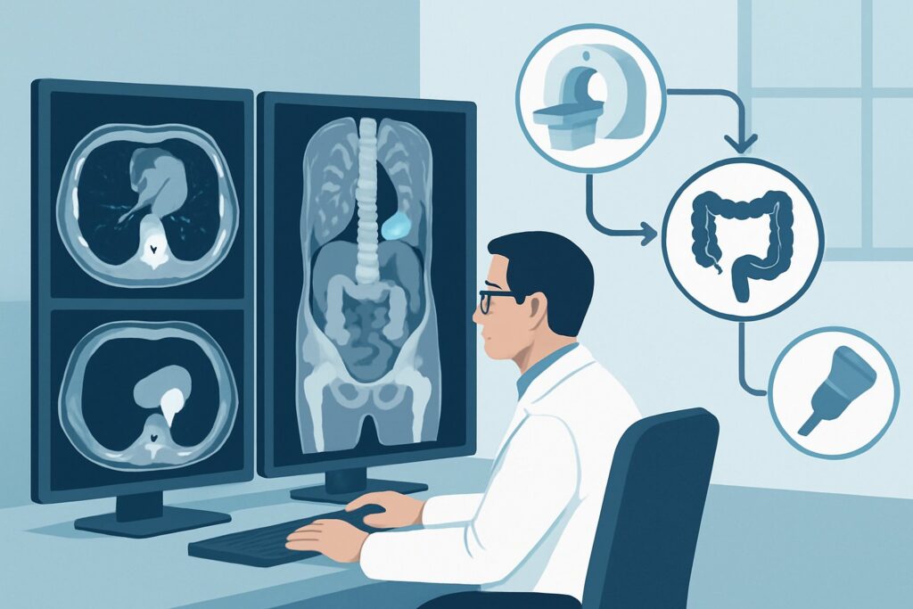
A 62-year-old man is in your clinic for his 18-month follow-up after a right hemicolectomy and adjuvant chemotherapy for Stage III colon adenocarcinoma. He feels well, and his physical exam is unremarkable. His carcinoembryonic antigen (CEA) level is stable and within the normal range. According to surveillance guidelines, it is time for his routine imaging to assess for disease recurrence. You need to decide which study provides the most reliable and comprehensive evaluation without exposing him to unnecessary procedures or radiation. This article details the American College of Radiology (ACR) evidence-based workflow for this specific clinical scenario: posttreatment evaluation of an adult with colon cancer. For this presentation, the ACR rates `CT abdomen and pelvis with IV contrast` as ‘Usually Appropriate’.
Who Fits This Clinical Scenario for Colon Cancer Surveillance?
This guidance applies specifically to adult patients who have completed definitive treatment for colon cancer and are now undergoing routine, scheduled surveillance for disease recurrence or metachronous primary tumors. The patient is typically asymptomatic or has non-specific symptoms, and the imaging is part of a planned follow-up strategy, often performed at predefined intervals (e.g., annually for several years post-treatment).
This workflow is not intended for patients who fit into closely related but distinct clinical situations. You should seek different guidance for:
- Initial Staging of Colon Cancer: Patients with a new colonoscopy-proven diagnosis of colon cancer who have not yet undergone treatment require initial staging to determine the extent of disease. This is a separate ACR variant with its own imaging recommendations.
- Evaluation of Appendiceal Cancer: While anatomically related, appendiceal neoplasms have different patterns of spread (e.g., pseudomyxoma peritonei) and distinct imaging protocols for monitoring.
- Symptomatic Patients with Suspected Recurrence: A patient presenting with acute symptoms like bowel obstruction, new-onset abdominal pain, or a rapidly rising CEA level may require a more urgent or problem-focused imaging approach rather than routine surveillance.
Correctly identifying your patient’s clinical context ensures the chosen imaging study aligns with the diagnostic question at hand, optimizing diagnostic yield and patient care.
What Diagnoses Are You Working Up in Posttreatment Evaluation?
Surveillance imaging after colon cancer treatment is primarily focused on detecting recurrent or new disease at its earliest, most treatable stage. The differential diagnosis you are evaluating includes several key possibilities.
Locoregional Recurrence
This is a primary concern and refers to the return of cancer at or near the original tumor site. It most commonly occurs at the surgical anastomosis, the point where the bowel was reconnected, or in the adjacent mesenteric lymph nodes. These recurrences can be subtle and may present as soft tissue thickening or new, enlarging lymph nodes.
Distant Metastases
The most common and life-threatening form of recurrence is the development of distant metastases. For colon cancer, the liver is the most frequent site, followed by the lungs. Peritoneal carcinomatosis, the spread of tumor nodules along the lining of the abdominal cavity, is another critical finding to assess for. Less common sites include bone and brain, which are not routinely screened for in asymptomatic patients.
Metachronous Primary Cancer
Patients with a history of colon cancer are at an increased risk of developing a new, independent primary tumor elsewhere in the colon. While colonoscopy is the primary tool for detecting these, cross-sectional imaging can sometimes identify a new, unsuspected mass.
Benign Post-Surgical Changes
Not every new finding is cancer. It is essential to differentiate true recurrence from benign postoperative changes, such as scar tissue (fibrosis), inflammatory granulomas at the site of surgical clips, or stable fluid collections (seromas). These can mimic malignancy, making high-quality imaging and careful comparison to prior studies crucial.
Why Is CT with IV Contrast the Recommended Study for Colon Cancer Surveillance?
For routine posttreatment evaluation of colon cancer, the ACR designates both `CT abdomen and pelvis with IV contrast` and `CT chest with IV contrast` as ‘Usually Appropriate’. In practice, these are typically combined into a single comprehensive examination to assess the most common sites of recurrence.
The rationale for this recommendation is based on the modality’s high diagnostic accuracy, wide availability, and speed. Intravenous contrast is critical because it enhances the visibility of tumor deposits relative to normal background tissue. In the liver, colon cancer metastases are typically hypovascular and appear as dark, hypoenhancing lesions against the brightly enhanced normal liver parenchyma during the portal venous phase. Similarly, IV contrast helps delineate enlarged or necrotic lymph nodes and subtle peritoneal nodules that would be invisible on non-contrast imaging. Therefore, ordering a `CT abdomen and pelvis without IV contrast` is ‘Usually not appropriate’ for this indication, as it severely limits the detection of metastatic disease.
While other modalities exist, they are rated lower for routine surveillance:
- MRI abdomen and pelvis without and with IV contrast is rated ‘May be appropriate’. MRI offers superior soft tissue contrast and is excellent for characterizing indeterminate liver lesions found on CT. However, it is more expensive, takes longer to perform, and is less effective than CT for evaluating the lungs. It is best used as a problem-solving tool rather than a first-line surveillance method.
- FDG-PET/CT skull base to mid-thigh is also rated ‘May be appropriate’. PET/CT is highly sensitive for detecting metabolically active cancer cells. Its primary role is not in routine surveillance but rather in specific scenarios, such as investigating a rising CEA level when conventional CT imaging is negative, or to clarify equivocal findings from other studies. Its higher radiation dose (☢☢☢☢ 10-30 mSv) and lower specificity (inflammation can be PET-avid) make it less suitable for routine annual screening.
The radiation dose for a contrast-enhanced CT of the chest, abdomen, and pelvis is a valid consideration (each component is ☢☢☢ 1-10 mSv), especially given the need for repeat scans over several years. However, the benefit of detecting early, potentially curable recurrence is generally considered to outweigh the risk associated with this radiation exposure. Once you’ve decided on the top procedure, our protocol guide covers the technique, contrast, and reading principles: CT Chest/Abdomen/Pelvis with IV Contrast.
What’s Next After CT? Downstream Clinical Workflow
The results of the surveillance CT scan will guide your next steps, creating a clear decision tree for patient management.
If the study is clearly positive for recurrence:
If the CT identifies unequivocal evidence of locoregional recurrence or distant metastases (e.g., new liver lesions with typical features, enlarging retroperitoneal nodes), the next step is typically a biopsy to confirm the diagnosis histologically. This is often performed via interventional radiology (e.g., CT-guided or ultrasound-guided biopsy). The patient should also be referred back to a multidisciplinary tumor board, including medical oncology, surgical oncology, and radiation oncology, to determine the optimal treatment strategy, which could include systemic chemotherapy, targeted therapy, surgery (metastasectomy), or radiation.
If the study is negative:
A negative or stable surveillance CT is reassuring. The patient continues with the established follow-up schedule, which typically includes regular clinical visits, CEA monitoring, and subsequent imaging at the next planned interval (e.g., in one year). No immediate further action is needed.
If the study is indeterminate or equivocal:
Often, imaging reveals findings that are not clearly benign but also not definitively malignant, such as a new, sub-centimeter liver or lung nodule. In this situation, the downstream workflow depends on the specific finding and level of suspicion. Options include:
- A short-interval follow-up CT (e.g., in 3-6 months) to assess for stability or growth.
- A problem-solving study, such as an MRI of the liver to better characterize an indeterminate liver lesion.
- A PET/CT scan, particularly if the CEA level is also rising, to assess for metabolic activity in the equivocal finding.
This approach avoids invasive procedures for what may be benign findings while maintaining vigilance for early recurrence.
Pitfalls to Avoid in Colon Cancer Surveillance Imaging
When ordering and interpreting surveillance imaging for colon cancer, several common pitfalls can compromise diagnostic accuracy and patient care. Be mindful of the following:
- Ordering Without IV Contrast: The most critical error is ordering a non-contrast CT. This drastically reduces sensitivity for detecting liver, nodal, and peritoneal metastases, potentially missing an opportunity for early intervention.
- Neglecting Comparison to Priors: Post-surgical anatomy can be complex. Always ensure the radiologist has access to prior imaging studies, especially the first postoperative baseline scan. This is essential for differentiating stable post-surgical changes from new, evolving disease.
- Over-reliance on CEA Alone: While a rising CEA is a strong indicator of recurrence, a significant minority of colon cancer recurrences are not CEA-producing. Do not defer scheduled imaging solely because the CEA level is normal.
- Misinterpreting Inflammatory Changes: Post-surgical or post-radiation inflammation can mimic tumor recurrence. Be cautious about indeterminate soft tissue thickening at the anastomosis, especially in the first year after treatment, and consider short-term follow-up imaging.
If you encounter a complex or equivocal case, especially one with discordant imaging and tumor marker results, escalation to a multidisciplinary tumor board is the most appropriate next step.
Related ACR Topics and Tools
This article provides a deep dive into one specific clinical scenario. For a broader view of related imaging decisions or to explore the technical aspects of the recommended studies, the following resources are valuable.
- For breadth across all scenarios in this topic, see our parent guide: Staging and Disease Monitoring of Colon Cancer and Appendiceal Cancer: ACR Appropriateness Decoded.
- To review other evidence-based imaging guidelines, use the ACR Appropriateness Criteria Lookup.
- For detailed technical specifications on imaging studies, consult the Imaging Protocol Library.
- To discuss cumulative radiation exposure with your patients, the Radiation Dose Calculator can help frame the conversation.
Frequently Asked Questions
How often should surveillance CT scans be performed after colon cancer treatment?
The frequency of surveillance imaging depends on the initial stage of the cancer and national guidelines (e.g., NCCN, ASCO). For patients with Stage II or III disease, annual CT scans of the chest, abdomen, and pelvis are typically recommended for up to 5 years post-treatment, as this is the period of highest risk for recurrence.
What if my patient has a severe allergy to iodinated contrast or has renal insufficiency?
For patients with a contraindication to iodinated contrast, MRI of the abdomen and pelvis with a gadolinium-based contrast agent is an excellent alternative and is rated ‘May be appropriate’ by the ACR. For the chest, a non-contrast CT may be performed to screen for lung metastases, though it is less sensitive for mediastinal disease. For patients with severe renal insufficiency, a non-contrast CT or MRI without contrast may be considered, but the limitations in detecting metastases must be acknowledged.
Is there a role for PET/CT in routine, scheduled surveillance for an asymptomatic patient?
No, for routine surveillance in an asymptomatic patient with a normal CEA, PET/CT is generally not recommended as the primary imaging tool. The ACR rates it ‘May be appropriate’ but not ‘Usually appropriate’. Its main utility is as a second-line, problem-solving tool for cases with a rising CEA and negative conventional imaging or to evaluate an equivocal finding on CT or MRI.
Should oral contrast be used for these surveillance CT scans?
The use of oral contrast varies by institutional protocol. While it can help delineate the bowel loops and identify anastomotic recurrence, many modern protocols rely on neutral oral contrast agents (like water) or no oral contrast, using the excellent soft tissue resolution of multidetector CT to evaluate the bowel. IV contrast remains the most critical component for detecting distant metastases.
If a small, indeterminate lung nodule is found, what is the next step?
The management of an incidental lung nodule on a surveillance CT follows established guidelines, such as the Fleischner Society criteria. For a small (e.g., <6 mm) solid nodule in a high-risk patient, a follow-up CT chest in 6-12 months is often recommended to ensure stability. The goal is to confirm it is a benign granuloma or scar without subjecting the patient to unnecessary invasive procedures.
Reviewed by Pouyan Golshani, MD, Interventional Radiologist — May 30, 2026