What Is the Right Initial Imaging for a Traumatic Visual Defect and Suspected Orbital Injury?
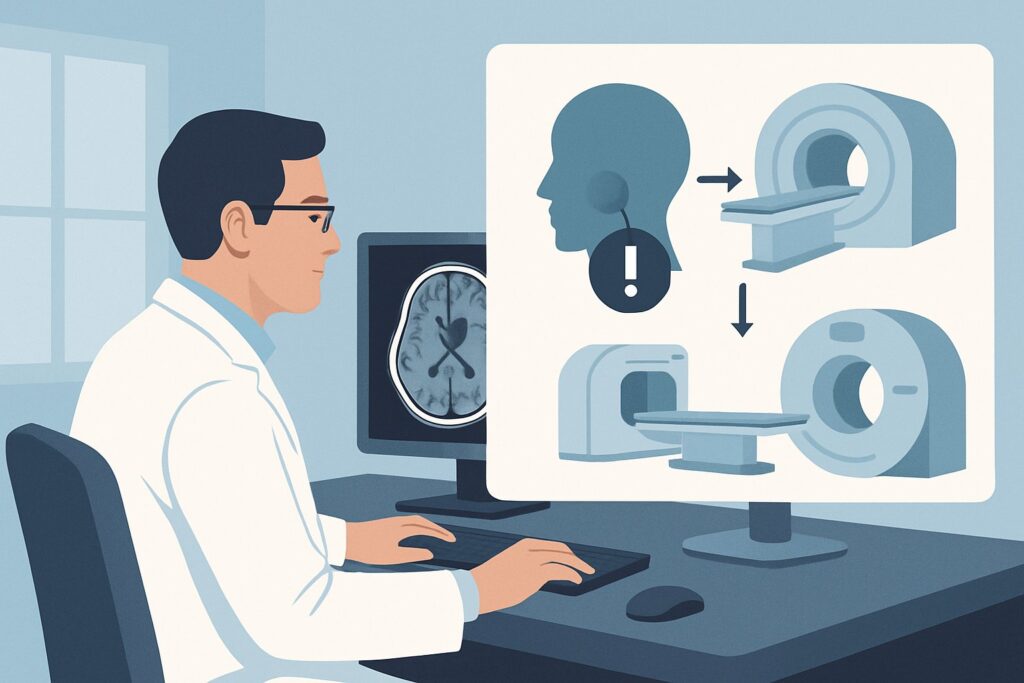
A 34-year-old man presents to the emergency department after being struck in the face during a basketball game. He complains of blurry vision and pain in his right eye. On examination, you note periorbital ecchymosis and swelling, with restricted upward gaze. You suspect an orbital injury, possibly a blowout fracture with muscle entrapment, but you need to rule out more severe, vision-threatening pathology like a retrobulbar hematoma. The immediate clinical question is which imaging study will provide the fastest, most accurate diagnosis to guide emergent consultation and treatment. For this specific scenario, the American College of Radiology (ACR) rates CT head without IV contrast as Usually appropriate, making it the primary recommended study.
Who Fits This Clinical Scenario?
This guidance applies to patients presenting with an acute visual deficit following direct or indirect trauma to the orbit or head. The key inclusion criteria are a clear history of trauma and a new, subjective, or objective change in vision. This could manifest as diplopia, blurred vision, decreased visual acuity, or a visual field cut. The clinical suspicion for an underlying orbital injury—such as a fracture, hemorrhage, or globe injury—is high.
This workflow is distinct from several related but different clinical presentations. It does not apply to patients with:
- Nontraumatic orbital asymmetry or exophthalmos. A patient with slowly progressive, painless proptosis likely has an underlying process like thyroid eye disease or an orbital mass, which requires a different imaging workup, often starting with MRI.
- Suspected orbital cellulitis. If the primary concern is infection, characterized by fever, erythema, and pain with eye movements without a clear traumatic trigger, the imaging choice may differ, often involving IV contrast to assess for abscess formation.
- Isolated superficial trauma. A patient with only a periorbital contusion (“black eye”) but with a completely normal ophthalmologic exam, including full extraocular movements and intact visual acuity, may not require any imaging.
This article focuses exclusively on the initial imaging decision for a new visual defect in the immediate post-traumatic setting.
What Diagnoses Are You Working Up in This Scenario?
When ordering imaging for traumatic visual loss, you are primarily investigating for injuries that require urgent intervention to preserve sight. The differential diagnosis is focused on structural damage to the orbit and its contents.
A primary concern is an orbital fracture, most commonly a “blowout” fracture of the orbital floor or medial wall. These can lead to entrapment of the inferior or medial rectus muscles, causing diplopia and restricted gaze. CT is exceptionally sensitive for identifying these bony disruptions and their effect on adjacent soft tissues.
Another critical, time-sensitive diagnosis is a retrobulbar hematoma. Bleeding behind the globe can rapidly increase intraorbital pressure, leading to orbital compartment syndrome. This compresses the optic nerve and retinal artery, causing ischemic optic neuropathy and irreversible vision loss if not treated with emergent lateral canthotomy. Non-contrast CT readily identifies this acute, high-density hemorrhage.
The imaging study also assesses for globe injury, such as rupture, lens dislocation, or an intraocular foreign body. While CT has limitations in visualizing the globe’s internal architecture compared to ultrasound, it can detect gross deformities like the “flat tire” sign of globe rupture or locate radiopaque foreign bodies.
Finally, the workup considers direct optic nerve injury. This can include nerve transection, avulsion, or an optic nerve sheath hematoma. While subtle nerve injuries are better characterized by MRI, significant hematomas or bony fragments impinging on the nerve can be identified on the initial CT scan.
Why Is CT Head Without IV Contrast the Recommended Study for This Presentation?
In the acute trauma setting, the choice of imaging must balance diagnostic accuracy with speed, availability, and safety. The ACR designates CT head without IV contrast and the more focused CT orbits without IV contrast as Usually appropriate because they optimally meet these needs for evaluating a suspected orbital injury.
The primary strength of non-contrast CT is its unparalleled ability to visualize bone and acute hemorrhage. It is the gold standard for detecting orbital fractures, identifying the location and displacement of fracture fragments, and assessing for related complications like muscle entrapment. It is also highly sensitive for identifying acute retrobulbar hematomas, which appear as high-density collections that are the key finding for orbital compartment syndrome. The speed of a non-contrast CT—typically completed in minutes—is critical when vision is at stake.
In contrast, other modalities are rated lower for this initial workup:
- Radiography orbit is rated Usually not appropriate. Plain X-rays have poor sensitivity for non-displaced fractures and provide almost no information about the soft tissues, globe, or optic nerve. They have been largely replaced by CT for this indication.
- MRI orbits without and with IV contrast is rated May be appropriate or May be appropriate (Disagreement). While MRI offers superior soft-tissue contrast and is excellent for evaluating the optic nerve, it is slower, less available in an emergency, and susceptible to motion artifact. It is also less sensitive than CT for detecting acute fractures. MRI is often a valuable second-line study if the initial CT is negative but a high suspicion for optic nerve or subtle soft-tissue injury remains.
The use of IV contrast is generally not necessary for the initial evaluation. Fractures and acute blood are best seen on non-contrast images. Contrast administration adds time, carries a risk of allergic reaction and post-contrast acute kidney injury, and increases the radiation dose. The radiation dose for a non-contrast head or orbits CT is moderate (adult RRL ☢☢☢, 1-10 mSv).
Once you’ve decided on the top procedure, our protocol guide covers the technique, contrast, and reading principles: CT Brain Without Contrast.
What’s Next After CT Head Without Contrast? Downstream Workflow
The results of the initial CT scan will dictate the immediate next steps in patient management. The workflow branches based on whether a critical, vision-threatening finding is identified.
If the CT is positive for a significant injury:
- Retrobulbar hematoma with signs of orbital compartment syndrome: This is a surgical emergency. The imaging result confirms the diagnosis, and the immediate next step is an emergent ophthalmology consultation for consideration of lateral canthotomy and cantholysis to decompress the orbit. Imaging should never delay this procedure if clinical signs are present.
- Orbital fracture with muscle entrapment: This requires urgent consultation with ophthalmology and potentially oral and maxillofacial surgery (OMFS) or plastic surgery. While surgery is not always as emergent as for a hematoma, it is often required within days to weeks to prevent permanent muscle fibrosis.
- Globe rupture: This is another ophthalmologic emergency requiring immediate consultation for surgical exploration and repair to salvage the eye.
If the CT is negative but symptoms persist:
If the non-contrast CT shows no fracture, hemorrhage, or other clear cause for the patient’s visual deficit, the workup must continue. The next step is a thorough ophthalmologic examination to look for pathology not visible on CT, such as traumatic iritis, retinal detachment, or commotio retinae. If suspicion for an optic nerve injury or a carotid-cavernous fistula remains high, a follow-up MRI/MRA of the orbits and head (May be appropriate) may be warranted to better visualize the nerve, soft tissues, and vasculature.
Pitfalls to Avoid (and When to Get Help)
Navigating the workup for traumatic orbital injury requires vigilance to avoid common diagnostic and management errors.
- Not specifying orbital views: A standard “CT head” protocol may use thick slices (e.g., 5 mm) that can miss subtle orbital floor fractures. If your clinical suspicion is high, explicitly request thin-slice (1-2 mm) images with coronal and sagittal reformats through the orbits or order a dedicated “CT orbits.”
- Delaying intervention for imaging: If a patient has classic clinical signs of orbital compartment syndrome (rock-hard proptotic eye, afferent pupillary defect, markedly elevated intraocular pressure), do not wait for CT results to perform a lateral canthotomy. This is a clinical diagnosis, and imaging serves to confirm it and rule out other injuries.
- Overlooking associated injuries: Orbital trauma rarely occurs in isolation. Always maintain a high index of suspicion for concomitant intracranial injuries (e.g., epidural or subdural hematoma), facial fractures, and cervical spine injuries.
- Missing non-radiopaque foreign bodies: While CT is excellent for metal or glass, organic materials like wood or plastic can be isodense to soft tissue and easily missed. If the history suggests such a foreign body, MRI may be a better subsequent test (after ensuring no metallic components are present).
If you identify a globe rupture, retrobulbar hematoma, or evidence of optic nerve compression, escalate immediately to the on-call ophthalmologist.
Related ACR Topics and Tools
This article covers one specific variant within the broader topic of orbital imaging. For a comprehensive overview of all related clinical scenarios, from nontraumatic proptosis to suspected optic neuritis, please see our parent guide.
- For breadth across all scenarios in Orbits, Vision, and Visual Loss, see our parent guide: Orbits, Vision, and Visual Loss: ACR Appropriateness Decoded.
- To explore other ACR Appropriateness Criteria topics, use the ACR Appropriateness Criteria Lookup.
- To review detailed imaging techniques for hundreds of studies, visit the Imaging Protocol Library.
- To discuss radiation exposure with patients, consult the Radiation Dose Calculator.
Frequently Asked Questions
Why not start with an MRI for a traumatic eye injury?
While MRI provides excellent soft tissue detail, it is not the preferred initial study in acute trauma. CT is much faster, more widely available, superior for detecting fractures, and excellent for identifying acute hemorrhage. MRI is often used as a second-line test if the CT is inconclusive or if there is high suspicion for optic nerve injury without a fracture.
Is a ‘CT orbits’ different from a ‘CT head’?
Yes. While a standard CT head includes the orbits, a dedicated CT orbits protocol uses much thinner image slices (typically 1-1.5 mm vs. 5 mm) and includes specific coronal and sagittal reconstructions. This provides far greater detail of the orbital bones, extraocular muscles, and globe, making it superior for detecting subtle fractures and their complications. If orbital injury is your primary concern, ordering a dedicated CT orbits is best practice.
When should I add IV contrast to the CT scan in orbital trauma?
IV contrast is generally not needed for the initial evaluation of fractures or hemorrhage. However, it should be considered if there is a specific concern for a vascular injury, such as a carotid-cavernous fistula, which can be a rare complication of trauma. In that case, a CTA (Computed Tomography Angiography) would be the appropriate study. Contrast may also be used if there’s a superimposed concern for infection or abscess, though that represents a different clinical scenario.
What if the patient with orbital trauma is pregnant?
Imaging a pregnant patient requires a careful risk-benefit discussion. The radiation dose from a head/orbits CT is low, and the fetus can be shielded. If there is a vision-threatening injury like a retrobulbar hematoma or globe rupture, the benefit of an accurate and rapid diagnosis with CT almost always outweighs the minimal fetal radiation risk. MRI, which does not use ionizing radiation, is an alternative but may not be practical or sufficient in the acute setting.
Are plain X-rays of the orbits ever useful anymore?
For the evaluation of a traumatic visual defect, the ACR rates orbit radiography as ‘Usually not appropriate.’ It has been almost entirely superseded by CT, which is vastly superior for detecting fractures and evaluating soft-tissue structures. The only potential, albeit rare, use for plain films might be in a resource-limited setting to screen for a large, radiopaque foreign body.
Reviewed by Pouyan Golshani, MD, Interventional Radiologist — May 29, 2026