Should You Order Radiographs or MRI for Thoracic Pain with Fracture Risk?
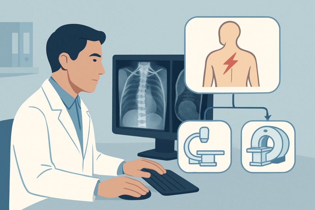
An 82-year-old woman with a history of polymyalgia rheumatica on long-term prednisone presents to your clinic. She stumbled yesterday while gardening—not a full fall, just a jolt—and now has sharp, 7/10 mid-back pain. On exam, she has exquisite point tenderness over the T8 spinous process but no neurologic deficits. You are concerned about a potential fracture given her age and steroid use. What is the right initial imaging study to order? This article provides a step-by-step clinical workflow for this exact scenario, guiding you through the differential, imaging rationale, and downstream decision-making. For this patient, the American College of Radiology (ACR) rates Radiography thoracic spine as Usually appropriate.
Who Fits This Clinical Scenario?
This guidance applies to a specific subset of adult patients presenting with thoracic back pain. The key inclusion criteria are the presence of the back pain (with or without associated myelopathy or radiculopathy) combined with one or more of the following risk factors for vertebral fracture:
- Low-velocity trauma (e.g., a fall from standing height, a sudden twist, or even a forceful sneeze)
- Known diagnosis of osteoporosis
- Elderly individual
- Chronic systemic steroid use
This workflow is designed for the initial imaging decision in these at-risk patients. It is crucial to distinguish this presentation from others that require a different approach. For example, this guidance does not apply to:
- Young, healthy adults with acute pain and no red flags: A patient in their 30s with new mid-back pain after starting a new weightlifting routine would follow a different ACR variant, often involving a trial of conservative management before imaging.
- High-energy trauma: A patient involved in a motor vehicle collision or a fall from a significant height requires evaluation under established trauma protocols, which typically involve CT imaging.
- Patients with “red flag” symptoms suggesting cancer or infection: If the back pain is accompanied by unexplained weight loss, fever, night sweats, or a history of cancer, the workup shifts to a scenario focused on malignancy or infection, which may prioritize MRI with contrast.
What Diagnoses Are You Working Up in This At-Risk Population?
In this clinical context, the differential diagnosis is focused on conditions where bone integrity is compromised. The primary goal of initial imaging is to identify or exclude an acute fracture, which is the most common cause of severe, new-onset pain in this population.
Vertebral Compression Fracture (VCF): This is the leading diagnosis to consider. In patients with osteoporosis, on chronic steroids, or of advanced age, the vertebral bodies can fracture with minimal or no trauma. The pain is typically acute, localized, and exacerbated by movement. Identifying a VCF is critical as it confirms the cause of pain and can trigger a necessary workup for underlying osteoporosis.
Pathologic Fracture from Malignancy: While less common than osteoporotic fractures, a pathologic fracture due to an underlying tumor must be excluded. Metastatic disease (from breast, lung, prostate, or kidney cancer) and multiple myeloma are the most frequent culprits. Imaging features like pedicle destruction, a convex posterior vertebral body wall, or an associated soft tissue mass raise suspicion for malignancy over a benign compression fracture.
Severe Degenerative Disease: Thoracic spondylosis and degenerative disc disease are ubiquitous in older adults and can be a source of chronic pain. While radiographs can show these changes (e.g., disc space narrowing, osteophytes), they are often incidental. The key is to determine if an acute process, like a fracture, is superimposed on this chronic background.
Infection (Discitis/Osteomyelitis): This is a less common but consequential consideration, particularly in patients who may be immunocompromised from chronic steroid use. While radiographs are insensitive in the early stages of infection, they may show endplate erosion or disc space narrowing later in the course. If clinical suspicion for infection is high (e.g., fever, elevated inflammatory markers), more advanced imaging is warranted.
Why Are Thoracic Spine Radiographs the Recommended First Step?
The ACR designates three different imaging modalities as Usually appropriate for this scenario, but thoracic spine radiography is often the most logical and efficient starting point. The rationale is based on its ability to effectively answer the most pressing clinical question: is there a fracture?
Anteroposterior (AP) and lateral radiographs of the thoracic spine are highly effective for detecting vertebral body height loss, the hallmark of a compression fracture. They are widely available, inexpensive, and can be performed quickly. This modality provides a rapid confirmation or exclusion of the most likely diagnosis, guiding immediate management. Radiographs can also reveal signs of other pathologies, such as lytic or blastic lesions concerning for malignancy or advanced degenerative changes.
While radiographs are the typical first step, it’s important to understand the roles of the other Usually appropriate studies:
- MRI thoracic spine without IV contrast: This study provides superior evaluation of the spinal cord, nerve roots, and bone marrow. It is the best test for detecting an “occult” fracture (one not visible on radiographs) and is essential if the patient has myelopathy or radiculopathy. However, it is more costly, takes longer to perform, and is less accessible than radiography. It is often used as the next step if radiographs are negative but clinical suspicion for fracture remains high or if neurologic symptoms are present.
- CT thoracic spine without IV contrast: CT offers exquisite detail of the bony anatomy and is superior to radiography for characterizing complex fracture patterns, assessing for fracture line extension into the posterior elements, and evaluating spinal canal compromise. It is often used after a fracture is identified on a radiograph to guide surgical planning or if radiography is negative in a patient with persistent, high suspicion for a bony injury.
The choice between these appropriate studies depends on the specific clinical presentation. For a neurologically intact patient where the primary question is fracture, radiography is the most resource-conscious and effective initial test. For a patient presenting with acute myelopathy, proceeding directly to MRI may be more appropriate. Both radiography and CT involve ionizing radiation (adult relative radiation level of ☢☢☢, 1-10 mSv), while MRI does not (O, 0 mSv).
What’s the Next Step After Thoracic Spine Radiographs?
The results of the initial radiograph create clear branching points in the clinical workflow. Your subsequent actions depend directly on the findings.
If the radiograph is positive for a simple compression fracture: The diagnosis is established. The next steps are primarily management-focused: pain control, consideration for bracing, and initiating or optimizing treatment for osteoporosis. Further advanced imaging is typically not required unless there are atypical features suggesting malignancy (e.g., pedicle destruction) or if neurologic symptoms develop.
If the radiograph is negative but clinical suspicion remains high: This is a critical juncture. In an at-risk patient with persistent, severe, and localized pain despite a normal radiograph, an occult fracture is a strong possibility. The appropriate next step is an MRI thoracic spine without IV contrast. The MRI can reveal bone marrow edema indicative of an acute, non-displaced fracture that was not visible on the plain film.
If the radiograph is equivocal or shows findings suspicious for malignancy: If the fracture has atypical features, or if a destructive lesion is seen, further characterization is mandatory. An MRI thoracic spine without and with IV contrast, which is rated May be appropriate, is often the best next test. The contrast helps delineate any associated soft tissue mass and assess the pattern of enhancement, which can help differentiate tumor from a benign fracture or infection. A CT scan can also be valuable for assessing the pattern of bone destruction.
If the radiograph shows only degenerative changes: If no acute fracture or aggressive lesion is identified, the pain may be attributable to the degenerative process. The next step is typically a trial of conservative management. If the patient has persistent radicular symptoms, an MRI may be considered later to evaluate for nerve root compression from a herniated disc or foraminal stenosis.
Common Pitfalls to Avoid in This Thoracic Pain Workup
Navigating this scenario requires vigilance to avoid common diagnostic errors. Be mindful of these potential pitfalls:
- Stopping the workup at a negative radiograph: In a high-risk patient with persistent focal pain or any neurologic signs, a normal plain film is not sufficient to rule out pathology. Maintain a low threshold to proceed to MRI to evaluate for an occult fracture or cord compression.
- Misinterpreting an old fracture as acute: Radiographs may show a chronic, healed compression fracture. Correlating the imaging with the acute, focal tenderness on physical exam is key. If there is ambiguity, an MRI can differentiate an acute (edematous) from a chronic fracture.
- Overlooking the thoracolumbar junction: The T11-L2 region is a biomechanical transition zone and a very common location for compression fractures. Ensure the imaging requested and obtained provides clear visualization of this area.
If a patient presents with rapidly progressing neurologic deficits, such as leg weakness, sensory level, or bowel/bladder dysfunction, this constitutes a neurologic emergency. This requires urgent escalation for an immediate MRI and consultation with a spine surgeon or neurologist, bypassing the standard outpatient workflow.
Related ACR Topics and Tools
For a broader overview of imaging for all thoracic back pain scenarios, from acute uncomplicated pain to post-operative evaluation, please see our comprehensive parent guide. For further exploration of the modalities discussed here, the following GigHz resources are available:
- For breadth across all scenarios in Thoracic Back Pain, see our parent guide: Thoracic Back Pain: ACR Appropriateness Decoded.
- To look up appropriateness criteria for adjacent or alternative clinical scenarios, use the ACR Appropriateness Criteria Lookup.
- For detailed technical parameters of the recommended studies, consult the Imaging Protocol Library.
- To discuss radiation exposure with your patients, the Radiation Dose Calculator can help frame the conversation.
Frequently Asked Questions
Why not start with an MRI for an elderly patient with back pain and fracture risk factors?
While MRI is also rated ‘Usually appropriate’, radiography is often preferred as the initial step because it is faster, more widely available, less expensive, and highly effective for diagnosing the most common cause in this scenario: a vertebral compression fracture. MRI is typically reserved as the next step if radiographs are negative but clinical suspicion remains high, or if the patient presents with significant neurologic symptoms like myelopathy.
If the patient has radiculopathy, shouldn’t I order an MRI first?
This is a key clinical decision point. In this specific scenario—an at-risk patient for fracture—the ACR considers both radiography and non-contrast MRI to be ‘Usually Appropriate’. If the radiculopathy is the dominant feature and your suspicion for a soft tissue cause (like a disc herniation) is higher than for a fracture, starting with MRI is a very reasonable choice. However, starting with a radiograph is also appropriate to first rule out a bony abnormality compressing the nerve root.
Does a fall from standing height change the recommendation from simple back pain?
No, in fact, it solidifies it. A fall from standing height is a classic example of the ‘low-velocity trauma’ that places a patient squarely in this clinical scenario. This history significantly increases the pre-test probability of a fracture and reinforces the recommendation to start with radiography to assess for bony injury.
The patient is on chronic steroids. Does this change which contrast I should use if I order an MRI or CT?
Chronic steroid use increases the risk for both osteoporotic fracture and infection. The decision for contrast depends on the specific clinical question. For the initial evaluation of a suspected fracture, non-contrast imaging (radiograph, CT, or MRI) is sufficient. If, however, there are clinical or laboratory signs concerning for infection (e.g., fever, elevated ESR/CRP) or malignancy, then a contrast-enhanced MRI would be the preferred advanced imaging study to evaluate for abscess formation or tumor enhancement.
My patient has osteoporosis but is only 55. Do they still fit this scenario?
Yes, absolutely. The criteria for this scenario are the presence of one or more risk factors, not all of them. A 55-year-old with a known diagnosis of osteoporosis who presents with new thoracic back pain fits this workflow perfectly, even if they are not considered ‘elderly’ and have no history of trauma or steroid use.
Reviewed by Pouyan Golshani, MD, Interventional Radiologist — May 30, 2026