What’s the Best Initial Imaging for Postmenopausal Pelvic Pain with Gynecologic Suspicion?

A 62-year-old female presents to your clinic with four months of a vague, deep pelvic ache. The pain is intermittent, non-radiating, and not associated with gastrointestinal or urinary symptoms. Her pelvic exam reveals some mild adnexal fullness, but is otherwise limited by body habitus. You suspect a gynecologic etiology for her subacute pain and need to decide on the most appropriate initial imaging study. This clinical scenario—a postmenopausal female with subacute or chronic pelvic pain and suspected gynecologic causes—is a common diagnostic challenge. This article provides a focused workflow based on the American College of Radiology (ACR) Appropriateness Criteria, explaining why specific ultrasound examinations are rated Usually Appropriate as the first-line investigation.
Who Fits This Clinical Scenario for Postmenopausal Pelvic Pain?
This guidance is specifically for postmenopausal adult females presenting with subacute (lasting weeks) or chronic (lasting months) pelvic pain where the clinical suspicion points toward a gynecologic origin. This includes pain localized by the patient to the lower abdomen or pelvis, or findings on physical exam such as adnexal fullness, a palpable mass, or uterine tenderness that suggest the reproductive organs are the source.
This workflow does not apply to several similar-but-distinct clinical situations:
- Acute Pelvic Pain: Patients with sudden, severe pain require an emergent workup for conditions like ovarian torsion, ruptured ectopic pregnancy (rare but possible), or acute appendicitis, which may alter the initial choice of imaging.
- Suspected Non-Gynecologic Cause: If the history and exam strongly suggest a gastrointestinal (e.g., diverticulitis), urologic (e.g., nephrolithiasis), or musculoskeletal source, the imaging pathway will differ.
- Known Malignancy: Patients with a known gynecologic cancer presenting with new pain are evaluated under surveillance or staging protocols, not initial diagnostic guidelines.
- Indeterminate Ultrasound: This article covers the initial imaging choice. If an ultrasound has already been performed and the results are inconclusive, the next step in imaging is covered by a different ACR variant.
What Diagnoses Are You Working Up in This Scenario?
In a postmenopausal woman with chronic pelvic pain, the differential diagnosis is broad, but the initial imaging is tailored to identify or exclude several key gynecologic pathologies. Any new adnexal or uterine finding in this population warrants careful evaluation.
Ovarian or Adnexal Mass: This is a primary concern. While many adnexal masses are benign (e.g., cystadenomas, hydrosalpinx), the risk of ovarian malignancy increases with age. Ultrasound is highly effective at characterizing adnexal structures, identifying solid components, septations, or vascularity that raise suspicion for cancer.
Endometrial Pathology: Endometrial carcinoma is the most common gynecologic malignancy, and while it classically presents with postmenopausal bleeding, it can manifest as pain, especially with uterine distention from hematometra or pyometra. Ultrasound can accurately measure endometrial thickness—a key screening parameter—and identify focal masses like polyps.
Uterine Leiomyomas (Fibroids): Though fibroids are estrogen-dependent and typically shrink after menopause, they can persist and cause chronic pain, particularly if they are large, pedunculated, or undergoing degenerative changes. Ultrasound is the primary modality for identifying and mapping uterine fibroids.
Less Common Gynecologic Causes: Other potential etiologies include chronic tubo-ovarian abscesses, pelvic adhesions from prior surgeries or infections, or pelvic organ prolapse. While not always directly visualized, ultrasound can reveal secondary signs of these conditions, such as a fixed, retroverted uterus or a hydrosalpinx.
Why Is Pelvic Ultrasound the Recommended First Step for This Presentation?
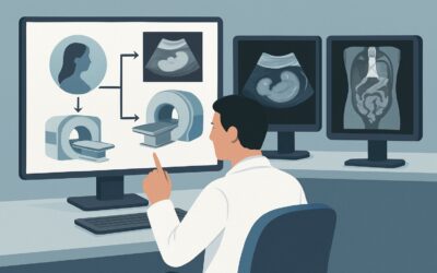
The ACR designates several forms of pelvic ultrasound as Usually Appropriate for the initial evaluation of a postmenopausal woman with suspected gynecologic pelvic pain. These include US pelvis transabdominal and US pelvis transvaginal, often performed together, as well as US duplex Doppler pelvis. This strong recommendation is based on the modality’s high diagnostic yield, safety profile, and accessibility.
The combination of transabdominal and transvaginal ultrasound provides a comprehensive evaluation. The transabdominal view offers a wide field of view, essential for assessing large masses that may extend beyond the pelvis and for orienting the overall anatomy. The transvaginal approach uses a higher-frequency transducer placed closer to the uterus and ovaries, yielding superior spatial resolution for detailed assessment of the endometrium, myometrium, and ovarian morphology. The addition of Duplex Doppler is critical for evaluating blood flow to the ovaries (to assess for torsion) and for characterizing the vascularity of any identified mass, which can help differentiate benign from suspicious lesions.
Most importantly, all ultrasound modalities are non-invasive and use no ionizing radiation (adult RRL: O 0 mSv). This avoids unnecessary radiation exposure, a key principle of diagnostic stewardship.
Why Other Studies Are Rated Lower
It is equally important to understand why other common imaging studies are not recommended for this specific initial workup:
- CT pelvis with IV contrast is rated Usually not appropriate. While excellent for evaluating bowel or urinary tract pathology, CT has inferior soft-tissue contrast compared to ultrasound for visualizing the endometrium and internal ovarian architecture. It also delivers a significant radiation dose (adult RRL: ☢☢☢ 1-10 mSv) without providing superior information for the primary gynecologic differential.
- MRI pelvis without and with IV contrast is also rated Usually not appropriate as a first-line test. MRI offers outstanding soft-tissue detail and is a superior problem-solving tool, but it is more costly, less widely available, and not necessary for the initial evaluation. Its role is preserved for when ultrasound findings are indeterminate or when further characterization of a complex mass is needed prior to surgical planning.
What’s Next After Pelvic Ultrasound? Downstream Workflow
The results of the initial pelvic ultrasound will guide the subsequent clinical pathway. The goal is to triage patients toward reassurance, further specialized imaging, or specialist consultation.
If the ultrasound is positive for a suspicious finding: A finding such as a complex adnexal mass with solid components, an ovarian mass with significant vascularity on Doppler, or an abnormally thickened and irregular endometrium requires urgent action. The next step is typically a referral to a gynecologist or gynecologic oncologist. They may order a follow-up MRI for pre-operative planning or proceed directly to endometrial biopsy or surgical evaluation.
If the ultrasound is negative: A completely normal pelvic ultrasound, showing an atrophic uterus and normal-appearing ovaries, makes a significant gynecologic cause of pain much less likely. At this point, the clinical focus should pivot to non-gynecologic etiologies. A careful re-evaluation of the patient’s history and a physical exam focused on musculoskeletal, gastrointestinal, and urologic systems is warranted. Further workup may include imaging or consultation targeted at these other organ systems.
If the ultrasound is indeterminate: Sometimes, ultrasound cannot fully characterize a finding. An adnexal structure might be obscured by bowel gas, or a mass may have features that are not definitively benign or malignant. This is the precise scenario where a problem-solving study is needed. The next step is often an MRI pelvis without IV contrast, which the ACR rates as May be appropriate in this context and is the focus of the sibling scenario on indeterminate ultrasound results.
Pitfalls to Avoid (and When to Get Help)
In this clinical scenario, several common pitfalls can delay diagnosis or lead to unnecessary testing. Be mindful to avoid ordering a CT scan as the first imaging test when the clinical suspicion is primarily gynecologic; this often leads to a non-diagnostic result for the uterus and ovaries and adds needless radiation. Another pitfall is failing to obtain both transabdominal and transvaginal views, as each provides unique and complementary information. Finally, do not dismiss an adnexal mass in a postmenopausal woman as a simple cyst without careful sonographic characterization; any complex features warrant further investigation. If the ultrasound reveals a highly suspicious mass, immediate escalation to a gynecologic subspecialist is the most appropriate next step, rather than ordering more imaging without a clear clinical question.
Related ACR Topics and Tools
For a comprehensive overview of all clinical variants related to this topic, please consult our parent guide. For other tools to assist in evidence-based imaging decisions, see the resources below.
- For breadth across all scenarios in Postmenopausal Subacute or Chronic Pelvic Pain, see our parent guide: Postmenopausal Subacute or Chronic Pelvic Pain: ACR Appropriateness Decoded.
- To look up other clinical scenarios, use the ACR Appropriateness Criteria Lookup tool.
- To review technical details for performing imaging studies, see the Imaging Protocol Library.
- To discuss cumulative radiation exposure with patients, the Radiation Dose Calculator can be a helpful aid.
Frequently Asked Questions
Is a transabdominal ultrasound alone sufficient for this workup?
No, a transabdominal ultrasound alone is generally not sufficient. While it provides a good overview of the pelvis, the transvaginal approach is critical for detailed evaluation of the endometrium and ovarian morphology. The ACR considers the combination of both transabdominal and transvaginal ultrasound to be ‘Usually Appropriate’.
What endometrial thickness is considered abnormal in a postmenopausal woman with pain?
In a postmenopausal woman not on hormone replacement therapy, an endometrial thickness greater than 4-5 mm is typically considered abnormal and warrants further investigation, such as a sonohysterogram or endometrial biopsy, especially if pain is the presenting symptom. However, this threshold is most validated for postmenopausal bleeding, and any focal thickening or irregularity should be viewed with suspicion regardless of the absolute measurement.
Why is MRI with contrast ‘Usually not appropriate’ for the initial workup?
For the initial workup, MRI with contrast is considered ‘Usually not appropriate’ because it is a higher-cost, lower-availability resource that is not needed in most cases. Ultrasound provides sufficient information to rule out or identify major pathology. MRI is reserved as a problem-solving tool for indeterminate ultrasound findings or for pre-operative staging of a known or highly suspected malignancy.
If the ultrasound is normal, can I completely rule out a gynecologic cause of pain?
A normal, high-quality pelvic ultrasound makes a significant structural gynecologic cause (like a large mass or endometrial cancer) highly unlikely. However, it cannot rule out all causes, such as microscopic endometriosis (rarely symptomatic postmenopause), pelvic adhesions, or pelvic floor muscle dysfunction. If pain persists despite a normal ultrasound, the focus should shift to non-gynecologic or functional causes.
Should I order a CA-125 blood test along with the initial ultrasound?
Ordering a CA-125 tumor marker is a clinical decision that depends on the ultrasound findings. It is not typically ordered before imaging. If the ultrasound reveals a suspicious adnexal mass, a CA-125 can be a useful adjunct for risk stratification and is often obtained by the consulting gynecologist as part of their evaluation.
Reviewed by Pouyan Golshani, MD, Interventional Radiologist — May 30, 2026