What’s the Best Initial Imaging for Retrosternal Dysphagia in Immunocompetent Patients?

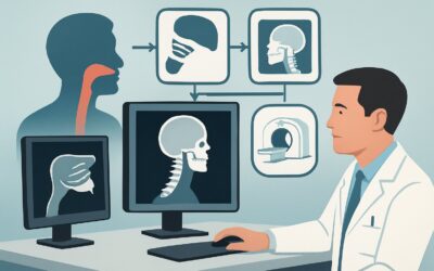
A 58-year-old male presents to your outpatient clinic with a three-month history of intermittent difficulty swallowing solids. He describes a sensation of food “getting stuck” behind his sternum, which sometimes resolves on its own and other times requires him to drink water. He denies any trouble initiating a swallow, coughing, or choking. His medical history is notable only for gastroesophageal reflux disease (GERD), and he is not immunocompromised. You suspect an esophageal cause and must decide on the most appropriate initial imaging study to evaluate his retrosternal dysphagia. This article details the American College of Radiology (ACR) evidence-based workflow for this specific clinical scenario. For this presentation, the ACR rates a Fluoroscopy biphasic esophagram as “Usually Appropriate.”
Who Fits This Clinical Scenario?
This guidance applies specifically to adult patients with a competent immune system who present with retrosternal dysphagia for initial evaluation. The key inclusion criteria are:
- Symptom Localization: The patient clearly localizes the sensation of food sticking or obstruction to the chest or lower neck, after the swallow has been initiated. This is distinct from oropharyngeal dysphagia.
- Immune Status: The patient is immunocompetent. This is a critical distinction, as the differential diagnosis and initial workup can change significantly in immunocompromised individuals.
- Timing: This is the initial imaging workup for this symptom. The patient has not had prior relevant imaging or endoscopic evaluation for this complaint.
It is crucial to distinguish this presentation from similar but distinct clinical scenarios that follow different diagnostic pathways. This workflow does not apply if the patient has:
- Oropharyngeal Dysphagia: Difficulty initiating a swallow, often accompanied by coughing, choking, or nasal regurgitation. This points to a different set of neuromuscular or structural causes evaluated in the unexplained oropharyngeal dysphagia variant.
- An Immunocompromised State: Patients with HIV/AIDS, on chronic steroids, or post-transplant have a higher risk of infectious esophagitis (e.g., Candida, CMV, HSV), which alters the pre-test probability and imaging choice.
- A Recent Postoperative History: Dysphagia occurring soon after thoracic, neck, or esophageal surgery has its own dedicated workup to assess for anastomotic leaks, strictures, or motility changes.
What Diagnoses Are You Working Up in This Scenario?
In an immunocompetent patient with retrosternal dysphagia, the differential diagnosis is primarily focused on structural and motor abnormalities of the esophagus. The initial imaging study is selected to effectively evaluate these possibilities.
Mechanical Obstruction: This is the most common category of causes. Benign peptic strictures, often a complication of chronic GERD, are a frequent finding. A Schatzki ring, a thin mucosal ring at the gastroesophageal junction, is another classic cause of intermittent solid food dysphagia. Less commonly, esophageal webs or diverticula can be responsible. Critically, imaging must also be sensitive enough to detect signs of esophageal malignancy, which can present as a stricture or mass.
Esophageal Motility Disorders: If a structural blockage is not present, the issue may be functional. Achalasia, characterized by failed relaxation of the lower esophageal sphincter and absent peristalsis, is a primary consideration. Other disorders like diffuse esophageal spasm or ineffective esophageal motility can also cause retrosternal dysphagia. These conditions are characterized by abnormal muscular contractions rather than a fixed physical blockage.
Eosinophilic Esophagitis (EoE): An increasingly recognized inflammatory condition, EoE is a common cause of dysphagia, particularly in younger patients and those with a history of atopy. It can cause esophageal narrowing, rings (sometimes described as a “feline” or “ringed” esophagus), or strictures that are readily identifiable on a fluoroscopic study.
Extrinsic Compression: While less common, structures outside the esophagus can compress it and cause dysphagia. This can include an enlarged left atrium (dysphagia megalatriensis), a thoracic aortic aneurysm, or a mediastinal mass. While fluoroscopy can show the resulting esophageal narrowing, a cross-sectional study like CT is often required for definitive characterization of the external cause.
Why Is a Fluoroscopy Biphasic Esophagram the Recommended Study for This Presentation?
The ACR designates the Fluoroscopy biphasic esophagram as “Usually Appropriate” for this scenario because it provides a comprehensive evaluation of both esophageal structure and function, directly addressing the primary differential diagnoses.
The “biphasic” nature of the study is key. The first phase uses a high-density barium suspension to coat the mucosal lining, followed by an effervescent agent to distend the esophagus with gas. This double-contrast technique provides exquisite detail of the esophageal wall, making it highly sensitive for subtle strictures, rings, webs, ulcers, and mucosal irregularities suggestive of malignancy or eosinophilic esophagitis.
The second phase involves the patient swallowing a lower-density, single-contrast barium solution. This phase is performed with dynamic fluoroscopic imaging, allowing for a real-time assessment of esophageal motility. The radiologist can observe peristaltic waves, identify abnormal contractions seen in spasm, or recognize the classic “bird’s beak” tapering and aperistalsis of achalasia. This dual-capability makes the biphasic esophagram a highly efficient first-line test.
In contrast, other studies are rated lower for this specific initial workup:
- CT of the neck and chest is rated “Usually Not Appropriate.” While excellent for evaluating extrinsic compression or advanced malignancy, it has a significantly higher radiation dose (☢☢☢☢ 10-30 mSv vs. ☢☢☢ 1-10 mSv for esophagram) and is far less sensitive for mucosal detail and motility disorders. It cannot reliably diagnose Schatzki rings, mild strictures, or achalasia.
- A modified barium swallow is rated “May Be Appropriate” but is not the primary recommendation. This study is optimized to evaluate the oropharyngeal phase of swallowing, which is not the patient’s complaint. While it includes imaging of the esophagus, it is not as detailed or comprehensive for distal pathology as a dedicated biphasic esophagram.
The biphasic esophagram offers a superb balance of diagnostic yield and safety, providing a clear roadmap for subsequent management in most patients with retrosternal dysphagia.
What’s Next After a Fluoroscopy Biphasic Esophagram? Downstream Workflow
The results of the biphasic esophagram will guide the next steps in the patient’s management. The workflow typically branches based on whether the findings suggest a structural, motor, or inflammatory cause.
If the study is positive for a structural abnormality (e.g., stricture, mass, ring, or web): The definitive next step is an upper endoscopy (esophagogastroduodenoscopy, or EGD). EGD allows for direct visualization of the lesion, biopsy to rule out malignancy or confirm eosinophilic esophagitis, and potential therapeutic intervention. For instance, a benign stricture or Schatzki ring can often be dilated during the same endoscopic procedure, providing immediate symptom relief.
If the study is positive for a motility disorder (e.g., features of achalasia or spasm): While the esophagram is highly suggestive, the gold standard for diagnosing and classifying motility disorders is high-resolution esophageal manometry. The patient should be referred to a gastroenterologist for this test, which measures the pressure and coordination of esophageal muscle contractions and sphincter function. The results of manometry will confirm the diagnosis and guide specific treatments, which can range from medications to endoscopic or surgical procedures.
If the study is negative or non-diagnostic: A normal biphasic esophagram does not entirely rule out esophageal pathology. Subtle mucosal changes, such as those in early eosinophilic esophagitis or mild reflux esophagitis, may not be visible. If the patient’s symptoms of retrosternal dysphagia persist despite a normal esophagram, the next appropriate step is typically an EGD to perform a direct visual inspection and obtain biopsies.
Pitfalls to Avoid (and When to Get Help)
When working up retrosternal dysphagia, several common pitfalls can delay diagnosis or lead to unnecessary testing. Be mindful of the following:
- Misinterpreting the patient’s history: Carefully distinguish between oropharyngeal dysphagia (trouble starting a swallow) and esophageal dysphagia (food sticking after swallowing). Ordering an esophagram for a patient with clear oropharyngeal symptoms is a low-yield choice.
- Accepting an incomplete study: Ensure the patient is appropriately NPO before the esophagram. Retained food or liquid can obscure pathology and lead to a non-diagnostic result, requiring a repeat study.
- Stopping the workup prematurely: A normal esophagram in a patient with persistent, credible symptoms is not the end of the line. These patients often require referral for endoscopy.
- Ignoring alarm features: The presence of “red flag” symptoms such as odynophagia (painful swallowing), significant unintentional weight loss, anemia, or hematemesis warrants an expedited workup. In these cases, consider a direct referral to a gastroenterologist for prompt endoscopy, which may be more appropriate than starting with an esophagram.
Related ACR Topics and Tools
This article focuses on a single, common clinical scenario. For a comprehensive overview of imaging for all types of dysphagia, from oropharyngeal to postoperative, please see our parent guide. For tools to help with ordering, protocoling, and patient communication, see the resources below.
- Parent Topic Hub: For breadth across all scenarios in Dysphagia, see our parent guide: Dysphagia: ACR Appropriateness Decoded.
- ACR Criteria Lookup: To explore adjacent scenarios or other clinical questions, use the ACR Appropriateness Criteria Lookup.
- Protocol Library: For detailed technical specifications of imaging studies, including the biphasic esophagram, consult the Imaging Protocol Library.
- Dose Calculator: To discuss radiation exposure with patients, the Radiation Dose Calculator can help contextualize the 1-10 mSv dose for this procedure.
Frequently Asked Questions
Why not just order an upper endoscopy (EGD) first for all patients with retrosternal dysphagia?
While EGD is an excellent test, the biphasic esophagram offers unique advantages as an initial study. It is less invasive and provides a comprehensive assessment of esophageal motility, which EGD cannot evaluate. For suspected motor disorders like achalasia, the esophagram is often the key to making the initial diagnosis and guiding the next step (manometry). It provides a functional and structural ‘road map’ before a more invasive procedure.
What if the patient’s main symptom is heartburn, not dysphagia?
If the primary symptom is heartburn or other signs of GERD without dysphagia, the diagnostic pathway is different. Imaging is generally not the first-line investigation for uncomplicated GERD. The workup typically starts with a trial of proton pump inhibitors (PPIs) or EGD if alarm features are present. The workflow in this article is specifically for the presenting complaint of dysphagia.
Does the recommendation change if the patient is older, for example, over 75?
The recommendation for a biphasic esophagram as the initial study does not change based on age alone, provided the patient is immunocompetent. However, the pre-test probability of certain conditions, particularly malignancy, increases with age. Therefore, if an esophagram shows a stricture in an older patient, the urgency for a subsequent EGD with biopsy is higher.
Can a CT scan of the chest ever be the right first test for this type of dysphagia?
According to the ACR, a CT is ‘Usually Not Appropriate’ as the *initial* test for uncomplicated retrosternal dysphagia. However, if the clinical suspicion for extrinsic compression is very high (e.g., the patient has a known large thoracic aortic aneurysm, a history of mediastinal cancer, or symptoms like hoarseness suggesting nerve involvement), a CT with contrast might be considered. In most cases, though, the esophagram should come first.
What specific information should I include in the order for a biphasic esophagram?
To ensure the radiologist performs the correct study, the order should clearly state the clinical indication: ‘Retrosternal dysphagia, rule out structural or motility disorder.’ It is helpful to specify ‘biphasic esophagram’ or ‘double-contrast esophagram.’ Mentioning key symptoms, like ‘intermittent solid food dysphagia,’ helps the radiologist tailor the examination and focus on the most likely pathologies.
Reviewed by Pouyan Golshani, MD, Interventional Radiologist — May 21, 2026