What’s the Best Initial Imaging for Tertiary Hyperparathyroidism? An ACR-Guided Workflow
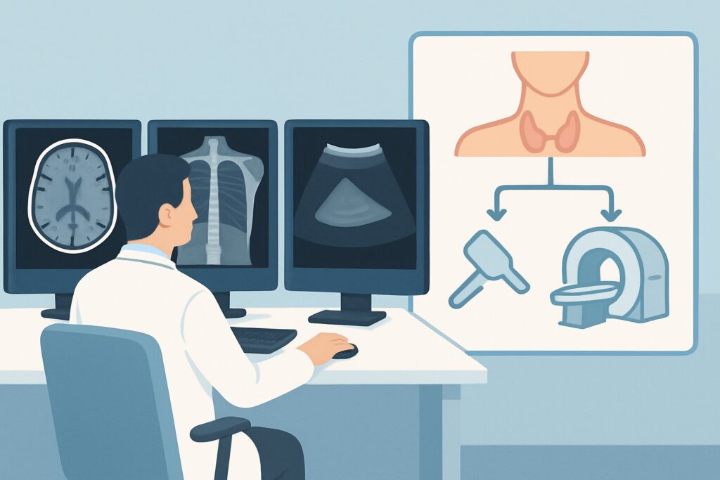
A 52-year-old patient with end-stage renal disease, on hemodialysis for the past 12 years, presents for a pre-operative evaluation. Their nephrologist has been managing secondary hyperparathyroidism for years, but recently, the patient’s serum calcium has risen to 11.8 mg/dL, and their parathyroid hormone (PTH) level remains markedly elevated despite maximal medical therapy. The diagnosis is tertiary hyperparathyroidism, and a parathyroidectomy is planned. As the consulting physician, you need to order the initial imaging study to localize the hyperfunctioning glands for the surgeon. This article details the American College of Radiology (ACR) recommended workflow for this specific clinical question. For the initial imaging of tertiary hyperparathyroidism, the ACR rates `US parathyroid` as ‘Usually Appropriate’.
Who Fits This Clinical Scenario?
This guidance applies to adult or pediatric patients with a confirmed biochemical diagnosis of tertiary hyperparathyroidism who require initial, pre-operative imaging. The key inclusion criteria are a history of chronic kidney disease (CKD) leading to long-standing secondary hyperparathyroidism, which has progressed to an autonomous state characterized by hypercalcemia and persistently elevated PTH levels.
This workflow is specifically for the initial imaging workup. It is crucial to distinguish this scenario from others that may present similarly but require a different diagnostic approach:
- Primary Hyperparathyroidism: This applies to patients without a history of CKD who present with hypercalcemia and elevated PTH. Their imaging workup follows a different ACR variant.
- Secondary Hyperparathyroidism: This applies to patients with CKD and elevated PTH but with normal or low serum calcium. Imaging is not typically performed in this stage unless surgery is being considered for severe, medically refractory disease.
- Recurrent or Persistent Hyperparathyroidism: This applies to patients who have already undergone parathyroid surgery and have persistently or recurrently high PTH levels. Their workup is more complex and often involves different imaging modalities.
Applying this workflow to the correct patient population—those with biochemically proven tertiary hyperparathyroidism undergoing their first imaging evaluation—is essential for appropriate test selection.
What Diagnoses Are You Working Up in This Scenario?
In tertiary hyperparathyroidism, the primary goal of imaging is not to make the diagnosis—which is established biochemically—but to localize the abnormal parathyroid tissue for surgical planning. The underlying pathology is different from primary hyperparathyroidism and influences the imaging findings.
Multi-gland Parathyroid Hyperplasia: This is the most common finding. Unlike primary hyperparathyroidism, which is most often caused by a single adenoma, tertiary disease results from the chronic stimulation of all parathyroid glands by hypocalcemia and hyperphosphatemia in CKD. This leads to diffuse, often asymmetric, enlargement of all four glands. Imaging aims to identify the location and size of all glands to guide the surgeon, who may need to perform a subtotal (3.5 gland) or total parathyroidectomy with autotransplantation.
Dominant Adenoma on a Background of Hyperplasia: Over time, one or more of the hyperplastic glands can undergo clonal proliferation and develop into an autonomous, hyperfunctioning adenoma. Imaging is critical to identify if there is a dominant gland that might be the primary driver of the severe hypercalcemia, which could influence the surgical strategy.
Ectopic Parathyroid Glands: In a minority of patients, parathyroid glands are located in ectopic positions, most commonly in the thymus, mediastinum, or within the thyroid gland. Failure to identify an ectopic gland pre-operatively is a common cause of persistent hyperparathyroidism after surgery. Imaging must be sensitive enough to survey these potential locations.
Why Is US Parathyroid a Recommended Initial Study?
For the initial imaging of tertiary hyperparathyroidism, the ACR designates `US parathyroid` as ‘Usually Appropriate’. This recommendation is based on its safety profile, accessibility, and diagnostic capability in this specific clinical context.
Ultrasound offers excellent spatial resolution of the neck without using ionizing radiation (adult and pediatric radiation relative level: O, 0 mSv). This is a significant advantage, particularly in younger patients or those who may require future imaging. It can readily identify enlarged parathyroid glands, distinguish them from thyroid nodules and lymph nodes, and provide precise anatomical information regarding their relationship to surrounding structures like the trachea, esophagus, and carotid artery. For a surgeon planning a minimally invasive approach, this information is invaluable.
However, the ACR also rates several other studies as ‘Usually Appropriate’, acknowledging the complexity of tertiary hyperparathyroidism. These include:
- Sestamibi Scans: Modalities like `Sestamibi dual-phase scan with SPECT or SPECT/CT neck` (RRL=☢☢☢ 1-10 mSv) are also highly valuable. These functional studies identify hypermetabolic parathyroid tissue. They are particularly useful for detecting ectopic glands in the chest that are beyond the field of view of a standard neck ultrasound. Many centers use both ultrasound and a Sestamibi scan as complementary first-line studies.
- CT Neck: A `CT neck without and with IV contrast` (Adult RRL=☢☢☢ 1-10 mSv) provides superb anatomical detail and can also help locate ectopic glands. Its use of radiation and iodinated contrast makes it a strong second-line or complementary option rather than the universal first choice.
Conversely, a study like `Venous sampling parathyroid` is rated ‘Usually not appropriate’ for initial imaging. This is an invasive procedure with risks, reserved for complex cases where non-invasive imaging has failed to localize the source of excess PTH.
What’s Next After US Parathyroid? Downstream Workflow
The results of the initial parathyroid ultrasound will guide the subsequent steps in the patient’s pre-operative workup. The goal is to provide the surgeon with a clear roadmap to the abnormal glands.
If the US is positive and localizing: When ultrasound clearly identifies one or more enlarged parathyroid glands in the neck that correlate with the clinical picture, this may be sufficient for surgical planning. However, given the high incidence of multi-gland disease and ectopic glands in tertiary hyperparathyroidism, many surgeons will proceed with a complementary functional study, such as a Sestamibi SPECT/CT, to confirm the findings and survey the mediastinum before heading to the operating room.
If the US is negative or equivocal: A negative neck ultrasound in a patient with confirmed tertiary hyperparathyroidism is a strong indication for further imaging. This result suggests the hyperfunctioning glands may be in an ectopic location (e.g., mediastinum) or are not significantly enlarged and thus are difficult to resolve with ultrasound. The logical next step is a functional imaging study, with `Sestamibi dual-phase scan with SPECT or SPECT/CT neck` being a ‘Usually Appropriate’ choice to localize metabolically active tissue anywhere from the skull base to the diaphragm.
If the US is indeterminate: Sometimes, ultrasound may identify a structure that could be a parathyroid gland, a thyroid nodule, or a lymph node. In this situation, a Sestamibi scan can be decisive. If the indeterminate nodule demonstrates tracer uptake, it is likely parathyroid tissue. If it does not, another cause should be considered. A contrast-enhanced CT or MRI of the neck may also be used to further characterize the finding.
Pitfalls to Avoid (and When to Get Help)
Navigating the workup for tertiary hyperparathyroidism requires careful attention to the nuances of the disease. Here are a few common pitfalls to avoid:
- Stopping after a negative ultrasound: Unlike in primary hyperparathyroidism, a negative US in tertiary disease does not rule out operable pathology. It should prompt further investigation with functional imaging.
- Ignoring the possibility of ectopic glands: Tertiary HPT has a significant incidence of ectopic glands. Relying solely on an imaging modality that only evaluates the neck (like US) can lead to missed glands and surgical failure.
- Misinterpreting multi-gland disease: It is crucial to remember that this condition typically involves all four glands. The goal of imaging is to locate all of them, not just the largest one.
- Failing to correlate with biochemistry: Imaging findings must always be interpreted in the context of the patient’s lab values. An enlarged gland on US in a patient with normal calcium and PTH is unlikely to be a surgical target.
If initial non-invasive imaging (e.g., US and Sestamibi) is negative despite a strong biochemical diagnosis, it is time to escalate. This may involve consulting with a radiologist specializing in endocrine imaging or a high-volume endocrine surgeon to consider advanced imaging like 4D-CT or, in rare cases, venous sampling.
Related ACR Topics and Tools
The ACR Appropriateness Criteria are a comprehensive resource for evidence-based imaging decisions. For a broader overview of imaging across all forms of parathyroid disease, please see our parent topic article. For additional tools to help with ordering and patient communication, explore the resources below.
- For breadth across all scenarios in Parathyroid Adenoma, see our parent guide: Parathyroid Adenoma: ACR Appropriateness Decoded.
- To explore other clinical scenarios and their corresponding imaging recommendations, visit the ACR Appropriateness Criteria Lookup.
- For detailed procedural techniques for the recommended studies, see the Imaging Protocol Library.
- To discuss radiation exposure with patients, use the Radiation Dose Calculator to estimate cumulative dose.
Frequently Asked Questions
Why are both ultrasound and a Sestamibi scan rated ‘Usually Appropriate’ for tertiary hyperparathyroidism?
They provide complementary information. Ultrasound offers superior anatomical detail of the neck without radiation but can miss ectopic glands in the chest. Sestamibi scans are functional studies that detect hypermetabolic tissue anywhere from the neck to the mediastinum but have lower spatial resolution. Many centers use both to maximize pre-operative localization, combining the strengths of anatomy and function.
Is 4D-CT a good first-line test for tertiary hyperparathyroidism?
While 4D-CT is a powerful tool for localizing parathyroid glands, the ACR does not rate it as a primary first-line study in this specific variant, instead listing ‘CT neck without and with IV contrast’ as ‘Usually Appropriate’. 4D-CT involves a higher radiation dose and is often reserved for complex cases, such as negative or discordant results from ultrasound and Sestamibi scans, or for re-operative surgery.
Does the imaging approach change for a pediatric patient with tertiary hyperparathyroidism?
The fundamental approach is similar, but there is a stronger emphasis on minimizing ionizing radiation. This makes ultrasound an even more attractive initial study (Pediatric RRL: O, 0 mSv). If further imaging is needed, the risks and benefits of radiation from studies like Sestamibi or CT must be carefully weighed. For CT, pediatric-specific low-dose protocols are mandatory.
If a patient has a large thyroid goiter, is ultrasound still the best first test?
A large, nodular goiter can make ultrasound of the parathyroid glands technically challenging, as thyroid nodules can obscure or be mistaken for parathyroid adenomas. While US is still a reasonable start, the ordering clinician should anticipate a higher likelihood of needing a complementary functional study like a Sestamibi scan with SPECT/CT to differentiate parathyroid from thyroid tissue.
What is the role of MRI in this scenario?
According to the ACR, ‘MRI neck without and with IV contrast’ is rated ‘May be appropriate’. It does not involve ionizing radiation, which is an advantage. However, it is generally considered less sensitive and specific than high-quality ultrasound or Sestamibi scans for initial localization. MRI is typically reserved as a problem-solving tool for cases where other non-invasive imaging is negative or conflicting.
Reviewed by Pouyan Golshani, MD, Interventional Radiologist — May 29, 2026