What’s the Next Step for a 2-Year Stable Pulmonary Nodule in a Smoker?

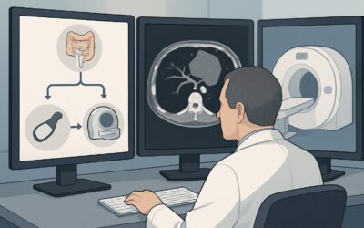
A primary care physician reviews the morning’s imaging results. The patient is a 68-year-old man with a 40-pack-year smoking history, in for a routine follow-up. The chest CT report confirms the 1.5 cm solid nodule in his right upper lobe is unchanged in size and morphology compared to a scan from two years prior. The clinical question is immediate: Is two years of stability enough to stop surveillance in a high-risk individual, or does the smoking history warrant a more aggressive approach? This article provides a detailed clinical workflow for this specific scenario, navigating the decision between continued observation and intervention. Based on the American College of Radiology (ACR) Appropriateness Criteria, the next step of ‘Follow-up imaging only’ is rated May be appropriate, reflecting a nuanced decision that balances risk factors with the high probability of benignity.
Who Fits This Clinical Scenario for a Stable Pulmonary Nodule?
This guidance applies to a specific subset of patients. Correctly identifying if your patient fits this profile is the critical first step to ensure the recommended workflow is applicable.
Inclusion Criteria for This Workflow:
- Patient: An adult.
- Nodule Characteristics: A solitary, solid (not ground-glass or subsolid) pulmonary nodule measuring between 1 and 3 cm in diameter.
- History: The nodule’s diameter has been documented as stable (unchanged) on prior imaging for a minimum of two years.
- Risk Factor: The patient has a history of smoking.
Exclusion Criteria (These Scenarios Require a Different Approach):
- Enlarging Nodule: If the nodule has shown any interval growth, even if minor, it represents an active process and falls under a different, more aggressive management pathway. This is covered in the sibling scenario: Adult. Serially, enlarging solitary solid pulmonary nodule, now 1 to 3 cm in diameter. Person who smokes. Next step.
- New or Incompletely Followed Nodule: If the nodule is newly discovered or lacks a full two-year history of stability, this guidance does not apply.
- Subsolid or Ground-Glass Nodules: These nodules have a different differential diagnosis and natural history, often requiring longer follow-up periods.
- History of Extrathoracic Malignancy: In a patient with a known primary cancer elsewhere, a stable pulmonary nodule raises the possibility of an indolent metastasis, altering the risk calculation and management strategy.
What Diagnoses Are You Working Up with a Stable Solid Nodule?
Even with two years of stability, the goal of continued evaluation is to differentiate between benign entities and the small possibility of an indolent malignancy. The differential diagnosis in this specific context is narrow but consequential.
Benign Granuloma
This is the most common and most likely diagnosis for a solid, calcified, or non-calcified pulmonary nodule that has remained stable for years. Granulomas are small, organized collections of immune cells that form in response to chronic inflammation, most often from a healed infection like histoplasmosis or tuberculosis. Their stability over a two-year period is a hallmark feature, indicating a resolved, inactive process.
Slow-Growing Adenocarcinoma
This is the primary concern that justifies continued attention. While most malignant nodules demonstrate growth within two years, some well-differentiated adenocarcinomas (particularly those with a lepidic growth pattern) can be remarkably indolent. Their growth rate may be so slow that it is not perceptible over a 24-month interval. The patient’s smoking history elevates this risk, making it the key reason why simply stopping surveillance may not be the preferred option for every patient.
Carcinoid Tumor
A less common consideration is a typical carcinoid tumor. These are low-grade neuroendocrine tumors that are often slow-growing and can remain stable in size for many years before declaring themselves. While less frequent than adenocarcinoma, they remain a possibility for a stable solid nodule, especially if centrally located or demonstrating avid enhancement on a contrast-enhanced CT.
Hamartoma
A hamartoma is a benign, disorganized growth of normal lung tissue, including cartilage, fat, and connective tissue. They are a common cause of solitary pulmonary nodules and are characteristically slow-growing or stable. While many have classic imaging features (e.g., “popcorn” calcification, intralesional fat), some can appear as nonspecific solid nodules, and their stability is a key diagnostic clue.
Why Is ‘Follow-up Imaging Only’ a Reasonable Next Step for This Nodule?
The ACR panel rates ‘Follow-up imaging only’ as May be appropriate for this scenario, a rating that underscores the central clinical dilemma: balancing the high likelihood of benignity against the small but real risk of a slow-growing cancer in a high-risk individual.
The rationale for this approach is rooted in risk stratification. Two years of size stability is a powerful predictor of a benign etiology. For many nodules, this finding is sufficient to cease further imaging. However, the patient’s smoking history acts as a significant countervailing risk factor. Therefore, extending surveillance with another follow-up CT scan in 12-24 months is a reasonable, conservative strategy. It allows for the detection of delayed growth in a very indolent malignancy while avoiding the immediate risks of an invasive procedure.
This decision should be made in conversation with the patient. Discussing the very low probability of malignancy after two years of stability, weighed against the cumulative radiation exposure and potential anxiety associated with continued surveillance, is a key component of shared decision-making.
Why Alternative Procedures Are Rated Lower:
- Percutaneous Lung Biopsy: This procedure is rated May be appropriate (Disagreement). The “Disagreement” qualifier indicates that expert opinion is divided. For some clinicians and patients, the desire for diagnostic certainty may outweigh the procedural risks (e.g., pneumothorax, hemorrhage). For others, subjecting a patient to an invasive procedure for a nodule with a >95% chance of being benign is not justifiable. This option is best reserved for patients with very high-risk features (e.g., spiculated margins, upper lobe location, severe emphysema) or significant patient anxiety that cannot be assuaged by surveillance.
- Surgical Management: Resection is rated Usually not appropriate. Performing a lobectomy or wedge resection for a nodule that has been stable for two years is considered overly aggressive. The morbidity and mortality associated with thoracic surgery are not warranted given the extremely high likelihood that the nodule is benign. Surgery is reserved for nodules that are proven to be malignant or demonstrate clear growth on follow-up imaging.
What’s Next After Follow-up Imaging? Downstream Workflow
The decision to continue surveillance initiates a new branch in the clinical workflow. The results of the next scan will determine the subsequent steps.
- If the Nodule Remains Stable: If a follow-up CT scan in another 12-24 months shows continued stability, the likelihood of malignancy becomes exceptionally low. At this point, after three to four years of documented stability, most guidelines would support discharging the patient from nodule-specific surveillance. They should, however, continue to be considered for annual low-dose CT lung cancer screening if they meet the established criteria based on age and smoking history.
- If the Nodule Shows Growth: Any unequivocal growth in a solid nodule that was previously stable is highly suspicious for malignancy. This finding immediately changes the management pathway. The patient should be referred to a pulmonologist or thoracic surgeon for further evaluation, which will almost certainly involve either a percutaneous biopsy or surgical resection to obtain a definitive tissue diagnosis. This patient’s case now aligns with the sibling scenario for an enlarging nodule.
- If the Result is Indeterminate or Equivocal: In rare cases, there may be subtle changes in morphology (e.g., development of a new lobulation) or a borderline change in size that does not meet the threshold for definite growth. In this situation, the next step is often a shorter-interval follow-up CT (e.g., in 6 months) or a discussion about proceeding to a percutaneous biopsy, especially if the patient’s risk profile is high.
Pitfalls to Avoid (and When to Get Help)
Navigating this scenario requires careful attention to detail to avoid common errors in judgment and measurement.
- Pitfall 1: Measurement Error. Relying on inconsistent measurement techniques between scans can create the illusion of stability or growth. Always compare to the same prior study and use electronic calipers to measure the long and short axes of the nodule on the same imaging plane (typically axial).
- Pitfall 2: Ignoring Morphologic Changes. Stability is not just about size. The development of new spiculation, lobulation, or an increase in density, even with a stable diameter, is a concerning feature that should prompt escalation rather than continued routine surveillance.
- Pitfall 3: Conflating Nodule Surveillance with Screening. Do not discharge a high-risk patient from annual lung cancer screening just because a specific nodule has been deemed stable. The stable nodule is resolved, but the patient’s underlying risk for developing a new cancer persists.
- Escalation Trigger: If there is any documented growth, new suspicious morphologic features, or the patient develops related symptoms (e.g., hemoptysis), escalate immediately with a referral to Pulmonology or Thoracic Surgery for consideration of tissue sampling.
Related ACR Topics and Tools
For a comprehensive overview of all clinical variants and imaging modalities covered by the ACR for this topic, please consult the parent guide. Additional GigHz tools can help you apply these criteria accurately in your practice.
- For breadth across all scenarios in Radiologic Management of Pulmonary Nodules and Masses, see our parent guide: Radiologic Management of Pulmonary Nodules and Masses: ACR Appropriateness Decoded.
- To explore other clinical presentations, use the ACR Appropriateness Criteria Lookup.
- For detailed procedural techniques on the recommended studies, visit the Imaging Protocol Library.
- To discuss cumulative radiation exposure with your patients, consult the Radiation Dose Calculator.
Frequently Asked Questions
Does the 2-year stability rule apply if the patient is a non-smoker?
Yes, and the evidence for benignity is even stronger. For a low-risk individual (non-smoker) with a solid nodule stable for two years, most guidelines, including the Fleischner Society guidelines, would recommend stopping follow-up entirely. The ACR’s ‘May be appropriate’ rating for continued follow-up in this scenario is driven specifically by the elevated baseline risk in a person who smokes.
What if the nodule has benign-type calcifications? Does that change management?
Absolutely. The presence of diffuse, central, laminated, or ‘popcorn’ calcifications is a reliable sign of a benign granuloma or hamartoma. If these features are unequivocally present, no further follow-up is necessary for the nodule, regardless of the patient’s smoking history.
Is a PET/CT scan helpful for a nodule that has been stable for two years?
A PET/CT is rated ‘Usually not appropriate’ in this specific scenario. Because the pre-test probability of malignancy is very low after two years of stability, a PET/CT has a high likelihood of producing a false-positive result from inflammatory changes in a benign granuloma. This can lead to unnecessary anxiety and invasive procedures. Its use is generally reserved for nodules with a higher suspicion of malignancy.
How should I measure the nodule to confirm stability?
Consistency is key. The nodule’s diameter should be measured on thin-section, non-contrast CT images in the axial plane. The average of the long-axis and short-axis diameters should be calculated and compared to the same measurement from the prior study. Volume-based assessment is more sensitive but less widely used; diameter-based stability is the current standard in most guidelines.
If we opt for continued surveillance, what is the recommended interval for the next CT scan?
There is no single mandated interval, but a common approach is to obtain the next non-contrast chest CT in 12 months. If that scan also shows stability, the interval for a subsequent scan can often be extended to 24 months. This decision should be guided by the nodule’s specific features and the patient’s overall risk profile and preferences.
Reviewed by Pouyan Golshani, MD, Interventional Radiologist — August 4, 2026