What Supplemental Breast Cancer Screening Is Best for Intermediate-Risk Women with Extremely Dense Breasts?

It’s 4:00 PM on a Tuesday, and you’re reviewing the day’s imaging reports. A 46-year-old patient’s screening mammogram result is in: BI-RADS category D, “extremely dense breasts,” which may obscure small masses. The finding itself is negative (BI-RADS 1), but you know her family history places her at intermediate risk for breast cancer. The density notification law in your state requires you to inform her of this finding and discuss supplemental screening. Now, you face the clinical decision: what is the most appropriate next step to offer this patient for effective cancer detection?
This article provides a focused clinical workflow for ordering supplemental breast cancer screening in an adult female at intermediate risk with extremely dense breasts. According to the American College of Radiology (ACR) Appropriateness Criteria, the primary recommended study, Digital breast tomosynthesis screening, is rated Usually appropriate.
Who Fits This Clinical Scenario for Supplemental Screening?
This guidance applies to a specific patient population: adult females undergoing supplemental breast cancer screening who meet two key criteria: intermediate risk and extremely dense breasts.
Inclusion Criteria:
- Intermediate Risk: This category includes women with a calculated lifetime risk of 15% to 19.9% using a validated model (e.g., Tyrer-Cuzick). It also includes women with a personal history of lobular carcinoma in situ (LCIS), atypical ductal hyperplasia (ADH), or atypical lobular hyperplasia (ALH).
- Extremely Dense Breasts: This corresponds to Breast Imaging Reporting and Data System (BI-RADS) breast composition category D. In these cases, more than 75% of the breast tissue is fibroglandular, which can significantly lower the sensitivity of standard 2D mammography by masking underlying lesions.
Exclusion Criteria (Who This Article Is NOT For):
- Average-Risk Women: Patients with a lifetime risk below 15% and no other major risk factors fall into a different screening paradigm. While supplemental screening may still be considered for dense breasts, the risk-benefit calculation differs.
- High-Risk Women: Patients with a lifetime risk of 20% or greater, a known pathogenic genetic mutation (e.g., BRCA1, BRCA2), or a history of chest radiation therapy between ages 10 and 30 are considered high-risk. For this group, breast MRI is more strongly recommended.
What Diagnoses Are You Working Up in This Scenario?
The primary goal of supplemental screening in women with extremely dense breasts is to detect clinically occult malignancies that are masked by the overlying fibroglandular tissue on a standard 2D mammogram. The differential diagnosis in this screening context is focused on identifying early-stage, non-palpable breast cancers.
Invasive Ductal Carcinoma (IDC): This is the most common type of invasive breast cancer. On mammography, IDC often presents as a spiculated mass or architectural distortion. In extremely dense breasts, these signs can be completely obscured, making supplemental imaging critical for early detection when the cancer is most treatable.
Invasive Lobular Carcinoma (ILC): ILC is the second most common type of invasive breast cancer and is notoriously difficult to detect on mammography. It tends to grow in a diffuse, single-file pattern rather than forming a discrete lump, leading to subtle findings like architectural distortion that are easily hidden by dense tissue. Supplemental imaging significantly improves the detection rate for ILC.
Ductal Carcinoma in Situ (DCIS): This is a non-invasive, or pre-invasive, form of breast cancer. While often detected by calcifications on mammography, some forms of DCIS, particularly non-calcified or low-grade DCIS, can present as non-specific densities or architectural distortion that are masked by breast density. Identifying DCIS early is key to preventing its progression to invasive cancer.
Supplemental screening also helps characterize benign findings, such as cysts or fibroadenomas, potentially reducing the need for unnecessary recalls and biopsies that might otherwise be prompted by ambiguous findings on a 2D mammogram.
Why Is Digital Breast Tomosynthesis the Recommended First Step?
For an intermediate-risk woman with extremely dense breasts, the ACR rates Digital breast tomosynthesis (DBT) screening as Usually appropriate. DBT, often called 3D mammography, acquires multiple low-dose X-ray images of the breast from different angles, which are then reconstructed into thin, one-millimeter slices. This technique mitigates the primary limitation of 2D mammography: the masking effect of overlapping breast tissue.
By “scrolling” through the breast tissue slice by slice, DBT improves the radiologist’s ability to identify the margins of potential lesions and detect subtle architectural distortions. This leads to a higher cancer detection rate and, importantly, a lower recall rate (fewer false positives) compared to 2D mammography alone in women with dense breasts.
Comparison to Other Modalities:
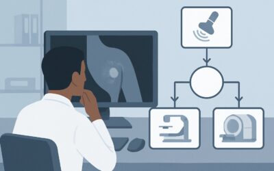
- Breast MRI with and without IV contrast is also rated Usually appropriate. It is the most sensitive imaging modality for detecting breast cancer. However, it is more costly, less widely available, and requires intravenous gadolinium-based contrast. While a valid option, its higher rate of false-positive findings and associated costs often position DBT as a more practical initial supplemental test for the intermediate-risk group.
- Breast Ultrasound (US) is rated May be appropriate. Handheld or automated whole-breast ultrasound can detect additional mammographically occult cancers. However, it is highly operator-dependent and is associated with a significant increase in false-positive findings, leading to more benign biopsies compared to other modalities.
From a safety perspective, DBT involves a low level of ionizing radiation (ACR RRL ☢☢, 0.1-1 mSv), which is comparable to a standard 2D mammogram. In contrast, Breast MRI and US involve no ionizing radiation. The decision between DBT and MRI should involve a shared decision-making conversation with the patient, weighing the enhanced cancer detection of both modalities against factors like cost, availability, and the potential for false positives. For detailed technical specifications on performing this study, see our guide on the Screening Mammography (with DBT) protocol.
What Is the Downstream Workflow After Supplemental Screening?
The results of the supplemental screening study, reported using the BI-RADS classification, will dictate the next steps in the patient’s care. The workflow is designed to ensure suspicious findings are addressed promptly while avoiding unnecessary interventions for benign results.
If the Result is Negative (BI-RADS 1 or 2):
A negative or benign finding is reassuring. The patient should be advised to continue with her annual screening schedule. The conversation about the optimal supplemental screening modality should be revisited annually, as guidelines and evidence evolve.
If the Result is Incomplete (BI-RADS 0):
This indicates that the radiologist needs more information to make a final assessment. The patient will be called back for additional imaging, which typically involves diagnostic mammographic views (e.g., spot compression, magnification) and/or a targeted breast ultrasound of the area in question.
If the Result Suggests a Probably Benign Finding (BI-RADS 3):
This category is for findings that have a very high probability ( >98%) of being benign. The standard recommendation is short-interval follow-up imaging, usually with a diagnostic mammogram or ultrasound in six months, to ensure stability. Biopsy is typically not immediately warranted.
If the Result is Suspicious or Highly Suggestive of Malignancy (BI-RADS 4 or 5):
These findings warrant tissue sampling. The patient should be referred for an image-guided biopsy. The specific biopsy method (e.g., stereotactic, ultrasound-guided, or MRI-guided) will depend on which modality best visualizes the lesion. A definitive management plan is then based on the pathology results.
Common Pitfalls in Screening Women with Extremely Dense Breasts
Navigating supplemental screening for this patient population requires careful consideration to avoid common missteps.
- Over-reassurance from a “Normal” 2D Mammogram: The most significant pitfall is accepting a negative 2D mammogram report at face value without considering the masking effect of extreme breast density. For intermediate and high-risk women, a negative mammogram is not the end of the screening conversation.
- Risk Misclassification: Failing to use a formal risk assessment model can lead to under- or over-screening. Incorrectly classifying a high-risk patient as intermediate may lead to recommending DBT when MRI would have been more appropriate.
- Neglecting Shared Decision-Making: Not discussing the pros and cons of each supplemental modality (e.g., radiation from DBT, contrast for MRI, false positives with US, out-of-pocket costs) can lead to patient anxiety and dissatisfaction. The “best” test depends on both clinical factors and patient preferences.
If a patient presents with a new palpable lump or other clinical concern (e.g., nipple discharge, skin changes), this supersedes any screening algorithm. Such findings require a full diagnostic workup, typically starting with a diagnostic mammogram and ultrasound, regardless of recent screening results. Escalation to a breast specialist or surgeon is warranted for any clinically suspicious finding.
Related ACR Topics and GigHz Tools
This article covers one specific clinical scenario. For a comprehensive overview of all patient presentations related to supplemental screening and breast density, or to explore the tools used to make these decisions, please see the following resources.
- For breadth across all scenarios in Supplemental Breast Cancer Screening Based on Breast Density, see our parent guide: Supplemental Breast Cancer Screening Based on Breast Density: ACR Appropriateness Decoded.
- To look up ACR ratings for other clinical presentations, use the ACR Appropriateness Criteria Lookup.
- For detailed procedural techniques on recommended studies, explore the Imaging Protocol Library.
- To discuss cumulative radiation exposure with patients, consult the Radiation Dose Calculator.
Frequently Asked Questions
Why is breast MRI also ‘Usually Appropriate’ but not always the first choice for this scenario?
Breast MRI is the most sensitive test for detecting breast cancer, which is why it earns a ‘Usually Appropriate’ rating. However, it has a higher false-positive rate than DBT, is more expensive, requires IV contrast, and is less accessible. For intermediate-risk women, many guidelines and clinical pathways recommend starting with DBT as a practical and effective first step, reserving MRI for higher-risk patients or specific clinical indications.
Does insurance typically cover supplemental screening for extremely dense breasts?
Coverage varies significantly by state and insurance plan. Many states have passed legislation mandating that insurers cover supplemental screening for women with dense breasts. However, the specifics of which tests are covered and what the patient’s out-of-pocket cost will be (e.g., copays, deductibles) can differ. It is essential to verify coverage with the patient’s insurer before ordering.
If a patient is intermediate-risk but has heterogeneously dense breasts (BI-RADS C), does this recommendation change?
Yes, the recommendations can differ. While supplemental screening is still a consideration for women with heterogeneously dense breasts, the degree of masking is less severe than with extremely dense breasts. The ACR panel provides separate guidance for that specific scenario, and the decision-making process may weigh the risks of false positives more heavily against the potential benefits.
What if my institution only offers 2D mammography, not DBT?
If Digital Breast Tomosynthesis (DBT) is unavailable, the next best step would be to consider the other rated modalities. In this case, a discussion about whole-breast ultrasound (‘May be appropriate’) or referring the patient to a center that offers breast MRI (‘Usually appropriate’) would be the logical next steps, guided by a shared decision-making conversation about the pros and cons of each.
How is ‘intermediate risk’ formally calculated?
Intermediate risk is typically defined as a lifetime risk of 15% to 19.9%. This is calculated using validated statistical models that incorporate factors like age, family history of breast and ovarian cancer in first- and second-degree relatives, age at first live birth, age at menarche, and personal history of breast biopsies. The Tyrer-Cuzick (IBIS) model is one of the most commonly used tools for this assessment.
Reviewed by Pouyan Golshani, MD, Interventional Radiologist — May 30, 2026