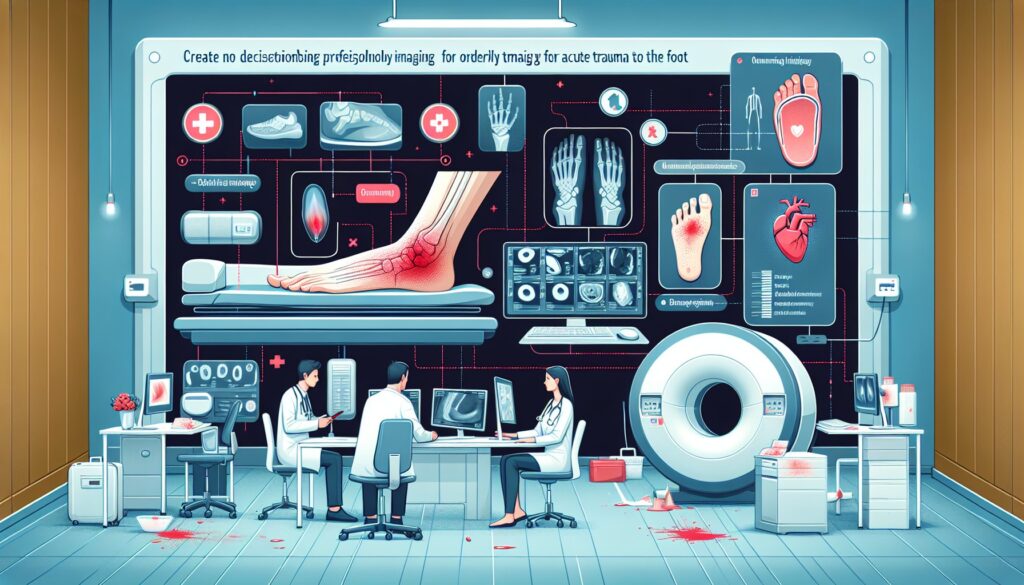
When to Order Imaging for Acute Trauma to the Foot: ACR Appropriateness Decoded
When to Order Imaging for Acute Trauma to the Foot: ACR Appropriateness Decoded
It’s a busy shift in the emergency department when a patient presents with a swollen, painful foot after a fall. They are tender over the midfoot and navicular, but your clinical exam is otherwise ambiguous. The decision to order imaging, and which type, is critical for accurate diagnosis and avoiding unnecessary radiation exposure. The American College of Radiology (ACR) provides evidence-based guidelines to navigate these common clinical questions. This article decodes the ACR Appropriateness Criteria for acute trauma to the foot, helping you choose the right study for the right patient, every time.
What Does ACR Acute Trauma to the Foot Cover?
The ACR guidelines for Acute Trauma to the Foot apply to adults and children over five years of age presenting with a recent injury. The criteria are heavily structured around the application of the Ottawa Ankle and Foot Rules, a validated clinical decision tool used to determine the need for radiography. These scenarios cover initial imaging decisions as well as next steps when initial radiographs are negative or equivocal but clinical suspicion for a significant injury remains high.
This topic specifically addresses acute traumatic injuries. It does not cover chronic foot pain, evaluation for stress fractures in the absence of acute trauma, suspected foreign bodies without a history of penetrating trauma, or complications related to underlying conditions such as diabetic foot ulcers or Charcot neuroarthropathy. For those presentations, different ACR Appropriateness Criteria may apply.
What Imaging Should I Order for Acute Trauma to the Foot? Recommendations by Clinical Scenario
The choice of imaging for acute foot trauma hinges on the clinical exam, particularly the findings of the Ottawa Foot Rules. The ACR provides clear guidance for several common scenarios.
For an adult or child over 5 with acute foot trauma where the Ottawa rules can be evaluated and are negative, and there is no suspicion for injury in areas not covered by the rules, initial imaging is Usually not appropriate. This includes radiography, CT, MRI, and ultrasound. In this case, the clinical decision rule is strong enough to confidently rule out a clinically significant fracture, and no imaging is the recommended course.
Conversely, if the Ottawa rules are positive, initial imaging with Radiography foot is Usually appropriate. Weightbearing views may also be obtained and are also rated as Usually appropriate. Radiographs are the foundational imaging modality for detecting fractures and dislocations in this setting. Similarly, if the Ottawa rules cannot be evaluated due to exclusionary criteria (e.g., intoxication, distracting injuries, diminished sensation), Radiography foot is also Usually appropriate.
In situations where the Ottawa rules are negative but there is suspected pathology in an anatomic area not addressed by the rules (such as the toes or metatarsals), Radiography foot (with or without weightbearing) is Usually appropriate. The Ottawa rules are specific to midfoot and malleolar fractures, and clinical suspicion for injury elsewhere warrants imaging.
When initial radiographs are normal or equivocal but there is a high suspicion for a Lisfranc injury, tendon injury, or an occult fracture, advanced imaging is necessary. Both MRI foot without IV contrast and CT foot without IV contrast are considered Usually appropriate as the next imaging study. CT excels at defining complex osseous anatomy and subtle fractures, while MRI is superior for evaluating ligamentous injuries, tendon pathology, and bone marrow edema. The choice between them often depends on the specific suspected injury and local availability.
Finally, for suspected penetrating trauma with a retained foreign body where radiographs are negative, US foot is Usually appropriate. Ultrasound is highly effective for identifying and localizing radiolucent foreign bodies like wood or plastic. If ultrasound is unavailable or non-diagnostic, CT or MRI without contrast may be appropriate.
ACR Imaging Recommendations Table
| Clinical Scenario | Top Procedure | ACR Rating | Adult RRL | Pediatric RRL |
|---|---|---|---|---|
| Adult or child >5 years. Ottawa rules negative. No other suspected abnormalities. Initial imaging. | Radiography foot | Usually not appropriate | ☢ <0.1 mSv | ☢ <0.03 mSv [ped] |
| Adult or child >5 years. Ottawa rules positive. Initial imaging. | Radiography foot (with or without weightbearing) | Usually appropriate | ☢ <0.1 mSv | ☢ <0.03 mSv [ped] |
| Adult or child >5 years. Ottawa rules cannot be evaluated. Initial imaging. | Radiography foot | Usually appropriate | ☢ <0.1 mSv | ☢ <0.03 mSv [ped] |
| Adult or child >5 years. Ottawa rules negative. Suspected pathology in area not addressed by Ottawa rules. Initial imaging. | Radiography foot (with or without weightbearing) | Usually appropriate | ☢ <0.1 mSv | ☢ <0.03 mSv [ped] |
| Adult or child >5 years. Suspect Lisfranc injury, tendon injury, or occult fracture. Radiographs are normal or equivocal. Next imaging study. | MRI foot without IV contrast / CT foot without IV contrast | Usually appropriate | O 0 mSv / ☢ <0.1 mSv | O 0 mSv [ped] / ☢ ☢ 0.03-0.3 mSv [ped] |
| Adult or child >5 years. Suspect penetrating trauma with a foreign body. Radiographs are negative. Next imaging study. | US foot | Usually appropriate | O 0 mSv | O 0 mSv [ped] |
Adult vs. Pediatric Acute Trauma to the Foot Imaging: Radiation Dose Tradeoffs
The imaging recommendations for acute foot trauma are generally consistent for adults and children over five years old. However, the relative radiation level (RRL) associated with ionizing radiation studies like radiography and CT warrants special consideration in younger patients. Children have a longer life expectancy, giving more time for potential long-term effects of radiation to manifest, and their developing tissues are more radiosensitive.
The ACR guidelines reflect this by providing distinct pediatric RRLs, adhering to the ALARA (As Low As Reasonably Achievable) principle. For foot radiography, the pediatric dose is minimal (☢ <0.03 mSv). For a non-contrast CT of the foot, the pediatric dose is categorized as ☢ ☢ (0.03-0.3 mSv), a higher tier than the adult dose (☢ <0.1 mSv). This highlights the importance of judicious use of CT in children. Whenever possible, non-ionizing modalities like MRI or ultrasound should be considered as alternatives for follow-up imaging after inconclusive radiographs, especially when soft tissue or ligamentous injury is the primary concern.
Imaging Protocol Details for Acute Trauma to the Foot
Once you’ve decided on the right study, the specific imaging protocol is crucial for diagnostic quality. Technical parameters, patient positioning, and contrast administration can significantly impact the utility of an exam. Our protocol guides provide detailed, scannable information for the key studies recommended in these ACR criteria.
Tools to Help You Order the Right Study
Navigating imaging guidelines can be complex, but several tools can streamline the process. GigHz offers a suite of resources designed to support evidence-based clinical practice and enhance patient safety.
For scenarios beyond acute foot trauma, the ACR Appropriateness Criteria Lookup provides a searchable interface to the full library of ACR guidelines, covering thousands of clinical variants across all specialties. It helps ensure you’re always aligning your orders with the latest expert consensus.
To ensure technical excellence in the studies you order, the Imaging Protocol Library offers detailed, step-by-step guides for hundreds of common and advanced imaging procedures. These are invaluable for trainees and for clarifying protocol questions with technologists.
When discussing radiation with patients, especially parents, the Radiation Dose Calculator is a powerful communication aid. It helps contextualize the dose from medical imaging and supports shared decision-making by providing clear, understandable comparisons.
What are the Ottawa Foot Rules?
The Ottawa Foot Rules are a clinical decision tool to help determine if foot radiographs are required after acute trauma. An x-ray is indicated if there is any pain in the midfoot zone AND any of the following: bone tenderness at the base of the fifth metatarsal, bone tenderness at the navicular bone, or an inability to bear weight both immediately and in the emergency department for four steps.
When is CT better than MRI for follow-up foot imaging?
After inconclusive radiographs, CT is generally preferred for its superior ability to delineate complex fractures, assess articular involvement, and identify subtle cortical breaks. It is faster and more widely available than MRI. MRI is superior for evaluating soft tissue structures, including ligaments (like the Lisfranc ligament), tendons, and for detecting bone marrow edema indicative of an occult fracture that may not be visible on CT.
Why is radiography the first step even if it might be negative?
Radiography is fast, inexpensive, widely available, and uses a very low dose of radiation. It is highly effective at identifying the majority of clinically significant fractures and dislocations. Even when negative, it serves as a crucial baseline and helps guide the decision for more advanced imaging like CT or MRI, which are more resource-intensive and, in the case of CT, involve a higher radiation dose.
What is the best imaging modality for a suspected foreign body?
For a suspected retained foreign body after penetrating trauma, the imaging choice depends on the material. Radiographs are excellent for radiopaque objects like metal or glass. If radiographs are negative and a radiolucent object like wood, plastic, or a thorn is suspected, ultrasound is the modality of choice. It is highly sensitive for identifying such objects and can guide removal. CT and MRI are typically reserved for cases where ultrasound is equivocal or the object is in a deep or complex location.
Is a contrast-enhanced study ever needed for acute foot trauma?
For the vast majority of acute traumatic foot injuries, intravenous contrast is not necessary. The ACR guidelines rate contrast-enhanced CT and MRI as “Usually not appropriate” for initial evaluation and for follow-up of suspected occult fractures or Lisfranc injuries. Contrast is typically reserved for specific indications such as concern for infection (abscess), vascular injury, or tumor, which fall outside the scope of standard acute trauma evaluation.
Reviewed by Pouyan Golshani, MD, Interventional Radiologist — May 12, 2026