When to Order Imaging for Congenital or Acquired Heart Disease: ACR Appropriateness Decoded
When to Order Imaging for Congenital or Acquired Heart Disease: ACR Appropriateness Decoded
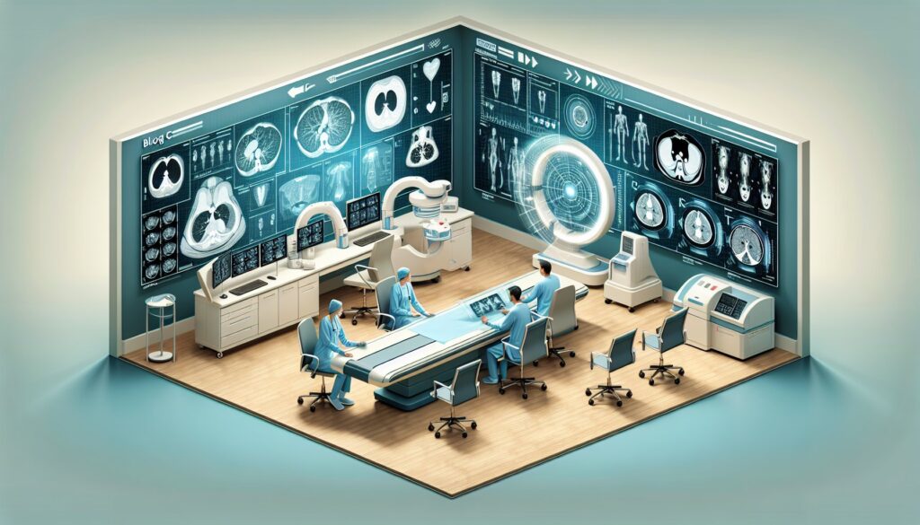
You’re evaluating a patient with a history of surgically corrected congenital heart disease. Transthoracic echocardiography (TTE) provided some answers but left key questions about right ventricular function and potential pulmonary artery stenosis unanswered. The next step is advanced imaging, but the choice between cardiac magnetic resonance imaging (MRI) and computed tomography angiography (CTA) isn’t always clear, especially when considering lifetime radiation exposure versus diagnostic yield. This guide decodes the American College of Radiology (ACR) Appropriateness Criteria for congenital or acquired heart disease, providing evidence-based recommendations to help you select the right test for your patient.
What Does ACR Congenital or Acquired Heart Disease Cover?
The ACR Appropriateness Criteria for Congenital or Acquired Heart Disease focus on selecting the next imaging study after an initial, often incomplete, evaluation with transthoracic echocardiography. These guidelines apply to both pediatric and adult patients across a range of common and complex scenarios. The criteria cover postoperative surveillance, such as after repair of Tetralogy of Fallot or transposition of the great arteries (TGA), as well as the initial workup for suspected conditions like aortic coarctation or anomalous pulmonary venous return. They also address specific preoperative planning for staged single ventricle palliation. This topic is intended for non-invasive diagnostic imaging decisions and generally does not cover routine, uncomplicated follow-up where TTE is sufficient, nor does it detail interventional procedures, though it does rate their appropriateness as a diagnostic next step.
What Imaging Should I Order for Congenital or Acquired Heart Disease? Recommendations by Clinical Scenario
After an inconclusive transthoracic echocardiogram, the choice of advanced imaging depends heavily on the specific clinical question and the patient’s underlying anatomy. The ACR provides detailed guidance for these common scenarios.
For a child or adult with repaired Tetralogy of Fallot or pulmonary valve stenosis, where there is concern for pulmonary valve dysfunction or branch pulmonary artery stenosis, multiple modalities are rated Usually appropriate. These include cardiac MRI (with or without contrast) for detailed functional and morphological assessment without radiation, and cardiac CTA or CTA chest with IV contrast for excellent anatomic detail. Chest radiography is also Usually appropriate as a baseline. Invasive procedures like pulmonary arteriography are considered May be appropriate if intervention is contemplated.
In patients who have undergone an atrial switch procedure for transposition of the great arteries (TGA), both cardiac MRI and CTA are Usually appropriate to assess the systemic right ventricle and identify potential baffle leaks or stenosis. MRI is often preferred for its ability to quantify ventricular function and flow without ionizing radiation.
For patients with TGA who have had an arterial switch procedure, the concerns shift to the pulmonary arteries and coronary arteries. Cardiac MRI and CTA remain Usually appropriate for evaluating the great vessels. However, stress imaging with MRI is also Usually appropriate to assess for myocardial ischemia, a known long-term risk due to coronary artery reimplantation. A CTA of the coronary arteries with IV contrast may be appropriate to directly visualize coronary anatomy.
When evaluating a child with a suspected or confirmed congenital or acquired coronary artery abnormality, both cardiac MRI and CTA coronary arteries are Usually appropriate. MRI can delineate the proximal course of the arteries without radiation, while CTA provides superior spatial resolution for detailed anatomy.
For children with single ventricle physiology, imaging choices depend on the surgical stage. For preoperative evaluation for stage 2 palliation, cardiac MRI, CTA, and invasive cardiac catheterization (arteriography) are all Usually appropriate to define the complex anatomy. Similarly, for preoperative planning for stage 3 palliation (Fontan), these same modalities are Usually appropriate. Postoperatively, after stage 3 palliation, non-invasive cardiac MRI and CTA chest with IV contrast are the preferred Usually appropriate methods for surveillance of the Fontan circuit.
In cases of suspected anomalous pulmonary venous return or aortic coarctation, cardiac MRI and CTA chest are again the primary Usually appropriate modalities. They provide comprehensive, three-dimensional views of the thoracic vasculature essential for diagnosis and surgical planning.
Finally, for surveillance in a child with known aortopathy or connective tissue disease (e.g., Marfan syndrome), MRA of the chest, abdomen, and neck (with or without contrast) is Usually appropriate to monitor for aortic dilation or dissection. CTA chest is also Usually appropriate, though MRA is often favored to limit cumulative radiation dose from serial imaging.
ACR Imaging Recommendations Table
| Clinical Scenario | Top Procedure | ACR Rating | Adult RRL | Pediatric RRL |
|---|---|---|---|---|
| Repaired tetralogy of Fallot or pulmonary valve stenosis with concern for pulmonary valve dysfunction. | MRI heart function and morphology without and with IV contrast | Usually appropriate | O 0 mSv | O 0 mSv [ped] |
| Transposition of the great arteries after atrial switch. | MRI heart function and morphology without and with IV contrast | Usually appropriate | O 0 mSv | O 0 mSv [ped] |
| Transposition of the great arteries after arterial switch. | MRI heart function and morphology without and with IV contrast | Usually appropriate | O 0 mSv | O 0 mSv [ped] |
| Child. Suspected or confirmed congenital or acquired coronary artery abnormality. | CTA coronary arteries with IV contrast | Usually appropriate | ☢ ☢ ☢ 1-10 mSv | ☢ ☢ ☢ ☢ 3-10 mSv [ped] |
| Child. Known single ventricle physiology. Preoperative evaluation for stage 2 palliation. | MRA chest without and with IV contrast | Usually appropriate | O 0 mSv | O 0 mSv [ped] |
| Child. Known single ventricle physiology. Preoperative evaluation for stage 3 palliation. | Arteriography pulmonary | Usually appropriate | ☢ ☢ ☢ ☢ 10-30 mSv | ☢ ☢ ☢ ☢ 3-10 mSv [ped] |
| Child or adult. Known single ventricle physiology. Postoperative evaluation after stage 3 palliation. | MRA chest without and with IV contrast | Usually appropriate | O 0 mSv | O 0 mSv [ped] |
| Known or suspected anomalous pulmonary venous return. | MRA chest without and with IV contrast | Usually appropriate | O 0 mSv | O 0 mSv [ped] |
| Suspected aortic coarctation. | MRA chest without and with IV contrast | Usually appropriate | O 0 mSv | O 0 mSv [ped] |
| Child. Known aortopathy or connective tissue disease. Surveillance of the aorta. | MRA chest without and with IV contrast | Usually appropriate | O 0 mSv | O 0 mSv [ped] |
Adult vs. Pediatric Congenital or Acquired Heart Disease Imaging: Radiation Dose Tradeoffs
Managing radiation exposure is a central theme in imaging for congenital heart disease, particularly in children. Patients with complex congenital conditions often require multiple imaging studies throughout their lives, leading to significant cumulative radiation doses. The ACR guidelines reflect this by assigning distinct pediatric Relative Radiation Levels (RRLs), which are often in a higher category than their adult counterparts for the same CT-based study. This is because children have a longer life expectancy during which radiation-induced effects can manifest, and their developing tissues are more radiosensitive.
This concern drives the frequent recommendation of non-ionizing modalities like MRI and MRA as Usually appropriate, especially for surveillance imaging in conditions like aortopathy. While CTA offers exceptional spatial resolution and speed, which can be critical in uncooperative children or for detailed coronary assessment, MRI is often the preferred first choice for functional analysis and follow-up. The principle of As Low As Reasonably Achievable (ALARA) is paramount. Each imaging decision should balance the immediate diagnostic need against the long-term risk of radiation, favoring MRI/MRA whenever it can provide the necessary clinical information.
Imaging Protocol Details for Congenital or Acquired Heart Disease
Once you’ve decided on the right study, the specific imaging protocol is critical for obtaining diagnostic-quality images. Our protocol guides provide detailed, practical information on technique, contrast administration, and interpretation principles for many of the studies recommended in these guidelines.
- CT Chest/Abdomen/Pelvis with IV Contrast
- CT Chest Without Contrast
- CTA Coronary Arteries (CCTA)
- CT Abdomen/Pelvis Without Contrast (Renal Stone)
Tools to Help You Order the Right Study
Selecting the optimal imaging study involves navigating complex guidelines and considering patient-specific factors like radiation dose. GigHz offers a suite of tools designed to support clinicians in making evidence-based decisions at the point of care.
The ACR Appropriateness Criteria Lookup tool provides rapid access to the full spectrum of ACR guidelines, allowing you to find recommendations for clinical scenarios beyond congenital heart disease. It helps ensure your imaging orders are aligned with national standards.
For detailed procedural information, the Imaging Protocol Library offers a comprehensive collection of imaging protocols. This resource is invaluable for understanding the technical aspects of the studies you order, from patient prep to sequence selection.
To help manage and communicate radiation risk, the Radiation Dose Calculator is a practical tool for estimating cumulative radiation exposure for your patients. It supports informed conversations about the benefits and risks of imaging, particularly for patients requiring lifelong surveillance.
Frequently Asked Questions about Imaging for Congenital or Acquired Heart Disease
Why is cardiac MRI often preferred over CT for congenital heart disease follow-up?
Cardiac MRI is often preferred for several reasons. First, it does not use ionizing radiation, which is a major advantage for patients, especially children, who may need many scans over their lifetime. Second, it provides excellent assessment of ventricular size and function, myocardial tissue characterization (e.g., fibrosis), and quantification of blood flow and shunts, which are often superior to CT. CT’s main advantages are faster acquisition times and higher spatial resolution for anatomical detail, particularly for coronary arteries and small vessels.
When is invasive cardiac catheterization (arteriography) still necessary?
Invasive cardiac catheterization is typically reserved for situations where direct pressure measurements are needed (e.g., pulmonary artery pressures before a Fontan procedure), when a therapeutic intervention is planned at the same time as the diagnostic procedure (e.g., stent placement in a stenotic pulmonary artery), or when non-invasive imaging has failed to provide definitive anatomical or hemodynamic information.
What is the role of transesophageal echocardiography (TEE) in these scenarios?
Transesophageal echocardiography (TEE) is rated as May be appropriate in many of these scenarios. It offers superior visualization of posterior cardiac structures, such as the atrial septum, mitral valve, and atrial baffles after a switch operation, compared to transthoracic echo. However, it is semi-invasive, often requires sedation or general anesthesia (especially in children), and provides a more limited field of view than CT or MRI for extracardiac structures like the great vessels. It is often used in the intraoperative setting or when specific structures are poorly seen on TTE and a cross-sectional study like MRI or CT is not readily available or contraindicated.
How do I choose between a cardiac MRI and an MRA of the chest?
The choice depends on the primary clinical question. A “cardiac MRI” (MRI heart function and morphology) focuses on the heart itself, using cine sequences to evaluate wall motion, ventricular volumes, and ejection fraction, as well as sequences to assess valve function and tissue characterization. An “MRA of the chest” focuses on the vasculature, using contrast-enhanced sequences to create detailed 3D maps of the aorta, pulmonary arteries, and systemic/pulmonary veins. In practice, many congenital heart disease protocols are comprehensive and include elements of both, but the naming and emphasis will shift based on whether the main question is about cardiac function or vascular anatomy.
Are there contraindications to MRI in patients with prior cardiac surgery?
Most modern sternal wires, clips, and valve prostheses are MRI-safe or MRI-conditional. However, older devices, particularly certain types of epicardial pacemaker leads or retained wires, can be a contraindication. It is critical to verify the specific type of implanted hardware before proceeding with an MRI. An institutional protocol for screening patients with prior surgery is essential for safety.
Reviewed by Pouyan Golshani, MD, Interventional Radiologist — May 12, 2026