When to Order Imaging for Headache-Child: ACR Appropriateness Decoded

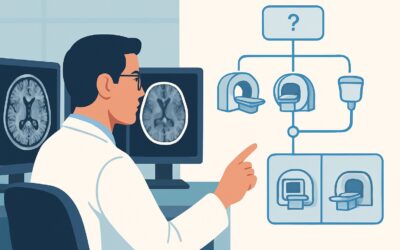
It’s 2 AM in the pediatric emergency department. A 9-year-old presents with a week of worsening headaches. The parents are anxious and asking about a brain scan. You know that most pediatric headaches are benign, but you’re weighing the small risk of missing a serious diagnosis against the risks of radiation or sedation for an MRI. What’s the right call? The American College of Radiology (ACR) Appropriateness Criteria for Headache-Child provide evidence-based guidance to navigate this exact decision, helping you choose the right test for the right patient while minimizing unnecessary imaging.
This reference article decodes the ACR guidelines for pediatric headache, providing a scannable summary of the recommendations. It outlines when imaging is warranted, which modality to choose based on the clinical scenario, and the key considerations for radiation safety in a young population. Use this guide to make confident, appropriate imaging decisions for your pediatric patients.
What Does ACR Headache-Child Cover?
The ACR Appropriateness Criteria for Headache-Child focus specifically on neuroimaging for children presenting with various headache patterns. The guidelines are designed to help clinicians differentiate between patients who require imaging and those for whom it is unlikely to be beneficial. This topic applies to the initial imaging workup for several distinct clinical scenarios, including:
- Children with a suspected primary headache (e.g., migraine, tension-type) and a normal neurological exam.
- Children with a suspected secondary headache, where red flag signs or symptoms suggest an underlying structural cause.
- Presentations of a sudden, severe “thunderclap” headache, which can be a sign of a vascular emergency.
- Headaches attributed to a suspected infection of the central nervous system.
- Headaches occurring as a sequela of remote trauma (i.e., not in the acute setting).
These guidelines do not apply to headaches in the setting of acute, significant head trauma, known or suspected hydrocephalus, or in patients with a known central nervous system malignancy. For those scenarios, different ACR criteria should be consulted.
What Imaging Should I Order for Headache-Child? Recommendations by Clinical Scenario
The appropriate imaging for a child with a headache depends entirely on the clinical context. The ACR panel emphasizes that for many children, particularly those with features of a primary headache and a normal neurological exam, no imaging is necessary. The recommendations below are for initial imaging when a workup is deemed clinically indicated.
For a child with a suspected primary headache and a normal exam, the ACR’s guidance is clear: imaging is Usually not appropriate. This includes all modalities, from skull radiography to CT and MRI. The rationale is that the diagnostic yield in this population is extremely low, and the potential risks of radiation (from CT) or sedation (for MRI) outweigh the benefits.
When a secondary headache is suspected due to red flag symptoms (e.g., abnormal neurologic exam, seizure, papilledema, focal deficits), imaging is warranted. The ACR rates MRI head without and with IV contrast and MRI head without IV contrast as Usually appropriate. MRI provides superior soft-tissue contrast for evaluating for tumors, inflammation, or structural anomalies without using ionizing radiation. In this context, a CT Brain Without Contrast is rated as May be appropriate, often reserved for emergent situations where MRI is unavailable or contraindicated.
In the urgent scenario of a sudden severe headache (thunderclap headache), the primary concern is subarachnoid hemorrhage or other vascular pathology. The ACR finds three studies Usually appropriate: CT head without IV contrast to rapidly look for hemorrhage, MRI head without IV contrast to assess for parenchymal abnormalities, and MRA Brain Without Contrast (3D TOF) to evaluate the cerebral vasculature for aneurysm or dissection.
If the headache is attributed to infection (e.g., concern for meningitis complications, abscess, or encephalitis), MRI head without and with IV contrast is Usually appropriate. Contrast enhancement is critical for identifying abscesses, meningeal inflammation, and other infectious changes. A CT Brain Without Contrast or with contrast may be appropriate in certain situations but provides less detail than MRI.
Finally, for a headache attributed to remote trauma, MRI head without IV contrast is Usually appropriate. This modality is excellent for detecting sequelae such as chronic subdural hematoma, encephalomalacia, or hemosiderin deposition without radiation exposure.
ACR Imaging Recommendations Table
| Clinical Scenario | Top Procedure | ACR Rating | Adult RRL | Pediatric RRL |
|---|---|---|---|---|
| Child. Primary headache. Initial imaging. | MRI head without IV contrast | Usually not appropriate | O 0 mSv | O 0 mSv [ped] |
| Child. Secondary headache. Initial imaging. | MRI head without and with IV contrast | Usually appropriate | O 0 mSv | O 0 mSv [ped] |
| Child. Sudden severe headache (thunderclap headache). Initial imaging. | CT head without IV contrast | Usually appropriate | ☢ ☢ ☢ 1-10 mSv | ☢ ☢ ☢ 0.3-3 mSv [ped] |
| Child. Headache attributed to infection. Initial imaging. | MRI head without and with IV contrast | Usually appropriate | O 0 mSv | O 0 mSv [ped] |
| Child. Headache attributed to remote trauma. Initial imaging. | MRI head without IV contrast | Usually appropriate | O 0 mSv | O 0 mSv [ped] |
Adult vs. Pediatric Headache-Child Imaging: Radiation Dose Tradeoffs
A central theme in the ACR guidelines for pediatric headache is the principle of ALARA (As Low As Reasonably Achievable) regarding radiation exposure. Children are more radiosensitive than adults, and their longer life expectancy provides more time for potential long-term effects of radiation to manifest. Consequently, the guidelines strongly favor non-ionizing modalities like MRI whenever clinically appropriate.
This is reflected in the pediatric-specific Relative Radiation Level (RRL) values. For instance, a CT head without contrast has an adult RRL of ☢ ☢ ☢ (1-10 mSv) but a pediatric RRL of ☢ ☢ ☢ (0.3-3 mSv), reflecting dose-reduction techniques used in pediatric protocols. Despite these reductions, the cumulative dose from repeated CT scans remains a significant concern. MRI, with an RRL of O (0 mSv), avoids this risk entirely. The tradeoff is often longer scan times, potential need for sedation in younger children, and reduced availability in some settings. The decision between CT and MRI must balance the clinical urgency, the specific question being asked, and the long-term imperative to minimize radiation dose in young patients.
Imaging Protocol Details for Headache-Child
Once you’ve decided on the right study based on the clinical scenario, the specific imaging protocol is critical for diagnostic accuracy. Our protocol guides provide detailed, practical information on technique, contrast administration, and key interpretation principles for the studies recommended in these guidelines.
Tools to Help You Order the Right Study
Navigating imaging guidelines can be complex. GigHz provides a suite of tools designed to support clinicians in making evidence-based decisions at the point of care.
For clinical scenarios beyond pediatric headache, the Imaging Appropriateness Selector tool offers a fast, searchable interface to the complete ACR guidelines, helping you find the right test for hundreds of clinical presentations.
To explore the technical details of the recommended exams, the Imaging Protocol Library contains curated, device-specific protocols for a wide range of CT, MRI, and other imaging procedures.
When discussing the risks and benefits of imaging with families, especially when a CT is being considered, the Radiation Dose Calculator can help estimate cumulative exposure and frame the conversation around radiation safety.
Frequently Asked Questions
Why is imaging usually not appropriate for primary headaches in children?
For children with a presentation consistent with a primary headache disorder (like migraine or tension headache) and a normal neurological exam, the likelihood of finding a serious underlying cause on imaging is very low. Multiple studies have shown that the diagnostic yield is minimal in this group. Therefore, the ACR concludes that the potential risks associated with imaging—such as radiation from CT, the need for sedation for MRI, and the cost—outweigh the very small chance of finding a clinically significant abnormality.
When should I use IV contrast with an MRI for a pediatric headache?
Intravenous contrast is recommended when there is a concern for a secondary cause of headache that involves a breakdown of the blood-brain barrier. This includes suspected infection (abscess, meningitis, encephalitis), inflammation, or neoplasm. In these cases, contrast helps to highlight areas of abnormal enhancement, making pathology more conspicuous. For indications like remote trauma or in some cases of suspected vascular abnormality, a non-contrast MRI may be sufficient.
Is CT or MRI better for a thunderclap headache in a child?
Both can be appropriate, but they answer different questions. A non-contrast head CT is extremely sensitive for detecting acute subarachnoid hemorrhage and is very fast, making it the first-line test in many emergency departments. However, if the CT is negative, the workup is not over. An MRI/MRA is often needed to look for other causes like reversible cerebral vasoconstriction syndrome (RCVS), dissection, or posterior reversible encephalopathy syndrome (PRES), which are better visualized on MRI. The ACR rates both CT without contrast and MRI/MRA without contrast as “Usually appropriate” in this setting.
What are the “red flags” that suggest a secondary headache and warrant imaging?
Red flags are signs or symptoms that increase the suspicion of a serious underlying cause for a headache. Key red flags in children include: a headache that awakens the child from sleep, morning vomiting, an abnormal neurological exam (e.g., focal weakness, ataxia, cranial nerve palsies), papilledema on fundoscopy, seizures, a rapid change in headache frequency or severity, or a headache that is consistently located in the occipital region. The presence of any of these should prompt strong consideration for neuroimaging.
How can I explain the radiation risk of a head CT to a parent?
It’s important to be clear and use relatable comparisons. You can explain that while a CT scan uses X-rays, the dose is kept as low as possible with pediatric-specific protocols. You can compare the dose to the amount of natural background radiation a person receives over a period of several months. Acknowledge their concern and state that the scan is only being recommended because the potential benefit of finding a serious, treatable problem outweighs the small long-term risk from the radiation. Using a tool like the Radiation Dose Calculator can help provide concrete numbers for this discussion.
Reviewed by Pouyan Golshani, MD, Interventional Radiologist — June 27, 2026