What Is the Right First Imaging Study for Adolescent Scoliosis With Risk Factors?
A 14-year-old is referred from a school screening for a noticeable spinal curve. During your evaluation, you confirm a moderate thoracic curve on forward bend test, but the patient also reports intermittent, radiating leg pain and you note a subtle asymmetry in their patellar reflexes. You suspect adolescent idiopathic scoliosis (AIS), but these additional findings—the risk factors—complicate the initial imaging decision. This is no longer a routine screening case; you are now investigating for an underlying pathology that could be driving the scoliosis.
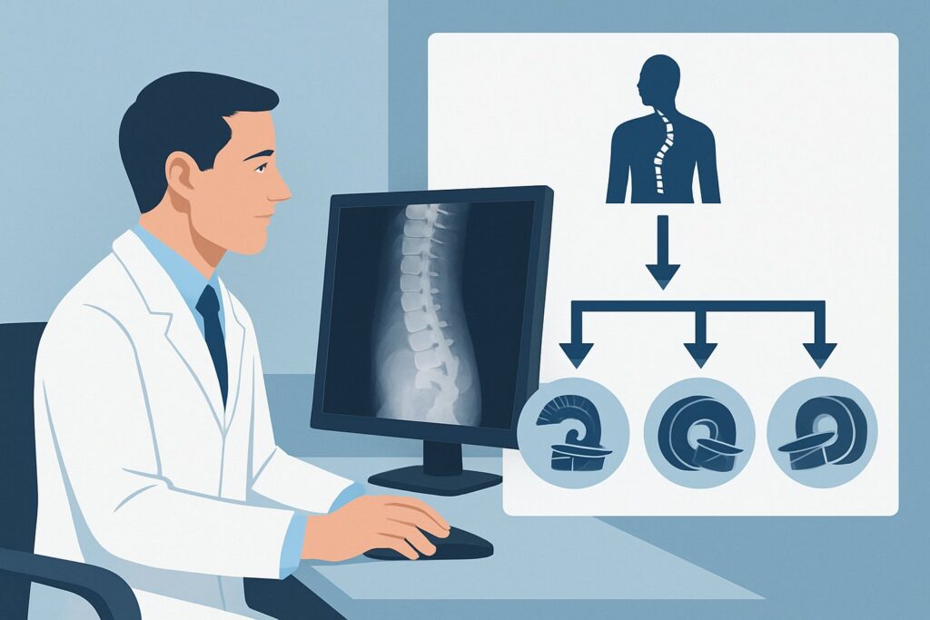
This article provides a focused clinical workflow for ordering initial imaging in an adolescent with suspected scoliosis who also presents with concerning risk factors. Based on the American College of Radiology (ACR) Appropriateness Criteria, the initial study of choice, `Radiography complete spine`, is rated Usually appropriate.
Who Fits This Clinical Scenario for Adolescent Scoliosis?
This guidance applies specifically to adolescents, defined as patients between 10 and 17 years of age, who present for an initial evaluation of scoliosis and have one or more clinical “risk factors.” These are red-flag signs or symptoms suggesting the scoliosis may not be truly idiopathic. Key risk factors include:
- Neurologic Abnormalities: Asymmetric reflexes, weakness, sensory deficits, gait disturbance, or abnormal abdominal reflexes.
- Significant Pain: Back pain that is severe, constant, occurs at night, or is disproportionate to the clinical findings.
- Atypical Curve Patterns: A left-sided thoracic curve (most idiopathic curves are right-thoracic), a short and angular curve, or a curve associated with significant kyphosis.
- Rapid Progression: A documented, unusually fast increase in the curve magnitude.
This workflow is distinct from other similar clinical situations. It does not apply to:
- Adolescents with no risk factors: A separate ACR variant covers the routine initial imaging for suspected AIS without concerning signs.
- Younger children: Patients under 10 with early-onset scoliosis or those with suspected congenital scoliosis have different underlying etiologies and require a distinct diagnostic approach.
- Patients with known conditions: This guidance is for seemingly idiopathic cases. Patients with a known diagnosis of a neuromuscular disorder (e.g., cerebral palsy, muscular dystrophy) or a connective tissue disease (e.g., Marfan syndrome) follow different evaluation pathways.
What Diagnoses Are You Working Up in This Scenario?
The presence of risk factors broadens the differential diagnosis beyond simple adolescent idiopathic scoliosis. While AIS remains a possibility, the primary goal of the workup is to exclude a secondary cause for the spinal deformity. The imaging strategy is designed to investigate these key possibilities.
Adolescent Idiopathic Scoliosis (AIS)
This remains the most common diagnosis in this age group. However, in this specific scenario, it is a diagnosis of exclusion. The clinical risk factors mandate a search for an underlying driver before settling on an “idiopathic” label.
Intraspinal Anomaly
This is the most critical category to investigate when neurologic signs or atypical curves are present. Conditions like a syrinx (syringomyelia), Chiari malformation, a tethered spinal cord, or a spinal cord tumor (e.g., astrocytoma, ependymoma) can all manifest as scoliosis. The spinal deformity is a secondary consequence of the underlying neural axis abnormality.
Benign or Malignant Bone Lesion
Pain, especially night pain relieved by nonsteroidal anti-inflammatory drugs, is a classic flag for a primary bone lesion. An osteoid osteoma or osteoblastoma located in the posterior elements of the spine can cause painful scoliosis due to muscle spasm. While less common, malignant tumors must also be considered in the differential for a painful spinal curve.
Spondylolysis or Spondylolisthesis
A defect or slippage in the pars interarticularis can cause back pain and lead to a compensatory, scoliotic posture. While not a true structural scoliosis, it is an important and treatable cause of the patient’s symptoms and clinical appearance that must be identified.
Why Is Complete Spine Radiography the Recommended First Step for Scoliosis With Risk Factors?
For an adolescent presenting with scoliosis and associated risk factors, the ACR rates `Radiography complete spine` as Usually appropriate. This is the essential first step that provides the foundational anatomic information needed for diagnosis and treatment planning, regardless of the underlying cause.
The initial radiographs—typically standing posteroanterior (PA) and lateral views of the entire spine—are critical for several reasons. They confirm the presence and direction of the curve, allow for a precise Cobb angle measurement to quantify its severity, assess vertebral rotation, and evaluate overall spinal balance. Importantly, radiographs also allow for the assessment of skeletal maturity using the Risser sign (iliac crest apophysis), which is a key factor in predicting the risk of curve progression.
While the primary concern in this scenario is an underlying non-idiopathic cause, the radiograph itself can sometimes reveal it. A bony lesion like an osteoid osteoma or evidence of spondylolysis may be directly visible. Therefore, radiographs serve a dual purpose: characterizing the scoliosis and screening for certain secondary causes.
Comparison to Other Imaging Modalities
Given the concern for intraspinal pathology, `MRI complete spine without IV contrast` is also rated as Usually appropriate. In many clinical settings, it is ordered concurrently with or immediately following radiography. However, radiography is almost always performed first to establish the orthopedic baseline. MRI is unparalleled for visualizing the spinal cord and neural elements but provides poor characterization of the osseous deformity and is not performed with the patient in a standing, weight-bearing position.
Modalities like `Bone scan complete spine` and `CT spine` are rated Usually not appropriate for this initial workup. A bone scan involves a high pediatric radiation dose (☢☢☢☢ 3-10 mSv) and lacks the specificity needed to diagnose the most likely underlying conditions. CT delivers a significant radiation dose and is typically reserved for detailed characterization of a specific bony abnormality already identified on radiographs, not as a primary screening tool.
What’s Next After Spine Radiography? Downstream Workflow
The results of the initial spine radiographs will guide the subsequent steps in the diagnostic and management pathway. The workflow hinges on whether the imaging findings can explain the clinical risk factors.
Radiographs Show Typical AIS, but Clinical Risk Factors Persist
This is a common and important outcome. If the radiographs show a curve pattern consistent with idiopathic scoliosis but the patient’s neurologic signs or atypical pain remain unexplained, the next step is to proceed with MRI. An `MRI complete spine without IV contrast` is essential to rule out the occult intraspinal anomalies (syrinx, Chiari, tumor) that can mimic AIS. This is the primary reason MRI also receives a Usually appropriate rating for this scenario.
Radiographs Reveal a Specific Bony Abnormality
If the x-rays identify a potential cause, such as a lucent nidus suggestive of an osteoid osteoma or a clear pars defect indicating spondylolysis, the downstream imaging becomes more focused. A targeted CT of the area of interest may be ordered to better characterize the bony lesion, or an MRI may be used to assess for associated soft tissue changes or nerve root compression.
Radiographs Are Negative or Indeterminate
If the radiographs do not show a significant scoliosis but the patient’s pain or neurologic symptoms are the dominant issue, the clinical focus shifts. The workup is no longer for “scoliosis” but for “back pain with neurologic signs,” which almost always proceeds directly to an MRI of the relevant spinal segment to evaluate for disc herniation, infection, or other causes.
Pitfalls to Avoid (and When to Get Help)
Navigating the workup for adolescent scoliosis with risk factors requires careful attention to avoid common missteps.
- Dismissing “Soft” Neurologic Signs: Subtle findings like asymmetric abdominal reflexes or mild clonus can be easy to overlook but are significant red flags for underlying neural axis abnormalities. A thorough neurologic exam is non-negotiable.
- Attributing All Pain to the Curve: Do not assume that back pain is simply “from the scoliosis.” In adolescents, significant pain is an atypical feature of idiopathic scoliosis and should always trigger a search for a secondary cause.
- Forgetting Radiation Dose: These patients are young and may require serial imaging over many years. Always start with low-dose techniques (e.g., PA instead of AP views) and only proceed to higher-radiation modalities like CT when absolutely necessary.
- Stopping the Workup Too Early: If initial radiographs look like typical AIS but the clinical red flags persist, do not stop. The failure to order a follow-up MRI in this situation is a major potential pitfall.
If a neurologic deficit is found or an intraspinal abnormality is identified on MRI, immediate escalation to a pediatric neurosurgeon and a pediatric orthopedic surgeon is warranted.
Related ACR Topics and Tools
For a comprehensive overview of all clinical variants related to pediatric scoliosis, including those for younger children and adolescents without risk factors, please see our parent guide. Additional GigHz resources can help refine your imaging orders and facilitate discussions with patients and families.
- For breadth across all scenarios in Scoliosis-Child, see our parent guide: Scoliosis-Child: ACR Appropriateness Decoded.
- To explore other clinical presentations, use the ACR Appropriateness Criteria Lookup.
- For details on imaging techniques, consult the Imaging Protocol Library.
- To discuss radiation exposure with families, the Radiation Dose Calculator can be a useful aid.
Frequently Asked Questions
Why is a left-sided thoracic curve considered a risk factor in adolescent scoliosis?
More than 85-90% of idiopathic thoracic scoliosis curves are convex to the right. A left-thoracic curve is an atypical pattern that significantly increases the suspicion for an underlying pathology, such as a syrinx or spinal cord tumor, which may be causing the spinal deformity. It mandates a more thorough investigation, almost always including an MRI.
If I strongly suspect an intraspinal issue, can I order an MRI at the same time as the initial radiographs?
Yes. Given that both radiography and MRI without contrast are rated ‘Usually appropriate’ in this scenario, ordering them concurrently is a reasonable and efficient approach if clinical suspicion for an underlying neurologic cause is high. The radiograph provides the necessary orthopedic measurements (like the Cobb angle), while the MRI evaluates the spinal cord and neural elements.
Does the patient need to have contrast for the follow-up MRI?
For the initial evaluation of an intraspinal anomaly causing scoliosis, an ‘MRI complete spine without IV contrast’ is typically sufficient and is rated ‘Usually appropriate’ by the ACR. Contrast is generally not needed unless a tumor is specifically suspected based on other findings or if the initial non-contrast MRI is abnormal. The ACR rates ‘MRI complete spine without and with IV contrast’ as ‘Usually not appropriate’ for the initial workup.
What is the Risser sign and why is it important on the radiograph?
The Risser sign is a measure of skeletal maturity, graded from 0 to 5, based on the ossification of the iliac crest apophysis, which is visible on the PA spine radiograph. It helps predict the remaining spinal growth. A lower Risser stage (0, 1, or 2) in a patient with a significant curve indicates a higher risk of progression, which is a critical factor in determining whether to initiate treatment like bracing.
If the MRI is normal, does that mean the scoliosis is definitely idiopathic?
If a thorough history, physical exam, radiograph, and a normal MRI of the entire spine fail to reveal an underlying cause, then a diagnosis of adolescent idiopathic scoliosis (AIS) can be made with much greater confidence. The patient can then be managed according to standard AIS protocols based on their Cobb angle and skeletal maturity.
Reviewed by Pouyan Golshani, MD, Interventional Radiologist — May 29, 2026