What Is the Best Initial Imaging for Early Onset Idiopathic Scoliosis in a Child?

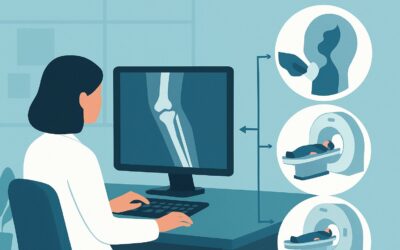
A 6-year-old girl presents for a routine check-up. During the exam, you note mild shoulder asymmetry and perform an Adams forward bend test, which reveals a right-sided rib prominence. The patient is otherwise healthy with a normal neurologic exam. You suspect early onset idiopathic scoliosis, a diagnosis that requires confirmation and characterization to guide management. The immediate clinical question is which imaging study to order first to assess the spinal curvature accurately while minimizing unnecessary radiation exposure in a young child. This article provides a detailed workflow for this specific scenario, grounded in the American College of Radiology (ACR) Appropriateness Criteria, which rate `Radiography complete spine` as `Usually appropriate` for the initial evaluation.
Who Fits This Clinical Scenario for Early Onset Scoliosis?
This guidance is specifically for the initial imaging workup of a child between 0 and 9 years of age with a clinical suspicion of early onset idiopathic scoliosis (EOIS). The term “idiopathic” is key—it implies there is no known underlying congenital, neuromuscular, or syndromic cause at the time of presentation. This workflow applies only to the first diagnostic imaging study, not to subsequent follow-up or pre-operative planning.
It is crucial to distinguish this scenario from similar but distinct clinical presentations that require different imaging pathways:
- Adolescents (10 to 17 years): Patients in this age group fall under the Adolescent Idiopathic Scoliosis (AIS) criteria, which have their own specific recommendations based on risk factors for progression.
- Known Congenital Anomalies: If the patient has a known or strongly suspected congenital vertebral malformation (e.g., hemivertebrae identified on prenatal ultrasound), the workup follows the Congenital Scoliosis pathway, which may involve different imaging modalities sooner.
- Neuromuscular or Syndromic Scoliosis: Children with established diagnoses like cerebral palsy, muscular dystrophy, or Marfan syndrome have scoliosis secondary to their condition. The imaging rationale and management goals differ significantly from the idiopathic type.
This article focuses exclusively on the otherwise healthy young child where the spinal curvature is the primary presenting problem.
What Diagnoses Are You Working Up with Initial Imaging?
The primary goal of initial imaging is to confirm the presence of scoliosis and differentiate between its potential causes. While “idiopathic” is the suspected diagnosis, imaging serves to establish this by evaluating for other etiologies.
Early Onset Idiopathic Scoliosis (EOIS) is the most common diagnosis being considered. Imaging is essential to confirm a spinal curve, measure its magnitude (Cobb angle), identify the curve pattern (e.g., right thoracic, double major), and establish a baseline to monitor for progression. Early detection and characterization are vital, as EOIS can have significant implications for thoracic development and pulmonary function if it progresses.
Congenital Scoliosis is a less common but critical differential diagnosis to exclude. This form of scoliosis results from vertebral anomalies present at birth, such as failure of formation (hemivertebrae, wedge vertebrae) or failure of segmentation (unsegmented bars, block vertebrae). These structural defects have a high rate of progression and are clearly visualized on initial radiographs, fundamentally changing the patient’s diagnosis and management plan.
Intraspinal Anomalies must also be considered, particularly in early onset cases. Conditions like a syrinx, tethered spinal cord, or Chiari malformation can cause scoliosis. While Magnetic Resonance Imaging (MRI) is the definitive modality for these diagnoses, certain radiographic findings can be red flags. Atypical features such as a left-sided thoracic curve (most idiopathic curves are right-sided), rapid curve progression, or associated vertebral anomalies (e.g., widened interpedicular distance) on the initial radiograph would prompt a downstream MRI.
Why Is Complete Spine Radiography the Recommended First Step for Early Onset Scoliosis?
The ACR Appropriateness Criteria rate Radiography complete spine as `Usually appropriate` for this clinical scenario. This recommendation is based on its ability to provide the necessary diagnostic information effectively and efficiently.
Standing posteroanterior (PA) and lateral radiographs of the entire spine are the cornerstone of the initial scoliosis evaluation. This single study achieves several critical objectives:
- Confirms the Diagnosis: It provides a clear visualization of the vertebral column to confirm the presence of a lateral curve.
- Quantifies the Curve: It allows for precise measurement of the Cobb angle, the standard method for quantifying curve magnitude and monitoring progression over time.
- Assesses Bony Anatomy: It is highly effective for identifying the congenital vertebral malformations that define congenital scoliosis, a key differential diagnosis.
- Establishes a Baseline: The initial images serve as the essential reference point against which all future imaging will be compared to determine curve progression.
The ACR panel also rates MRI complete spine without IV contrast as `Usually appropriate`. However, its role is distinct. While MRI provides exceptional detail of the spinal cord and neural elements without using ionizing radiation, it is not the standard first-line study for every child with suspected EOIS. It is typically reserved for cases with “red flags” suggesting an underlying intraspinal abnormality, such as neurologic deficits on exam, significant pain, an atypical left thoracic curve, or very early onset (infantile scoliosis). For the straightforward initial workup, radiography is the more direct and resource-appropriate starting point.
Alternative studies are rated lower for this initial diagnostic step. A Bone scan complete spine is `Usually not appropriate` because it has a significantly higher radiation dose (pediatric relative radiation level ☢☢☢☢) and is not designed to evaluate spinal curvature; its utility is for detecting metabolic, infectious, or traumatic bone processes. Similarly, CT of the spine is `Usually not appropriate` for initial diagnosis due to its much higher radiation dose compared to radiography. CT is a valuable tool for pre-operative planning or detailed characterization of complex congenital anomalies, but not as a first-line diagnostic test.
What’s Next After Radiography complete spine? Downstream Workflow
The results of the initial spine radiographs will dictate the next steps in management, which typically involve referral to a pediatric orthopedic surgeon or spine specialist.
- If the study is positive for Idiopathic Scoliosis: The primary factors guiding the next steps are the Cobb angle and the child’s age. For small curves (e.g., <20-25 degrees), the typical plan is observation with serial clinical exams and follow-up radiographs at regular intervals (e.g., every 4-6 months) to monitor for progression. For larger or progressive curves, treatment options such as casting or bracing may be initiated to guide spinal growth and prevent worsening.
- If the study is negative: If the radiographs show no significant spinal curvature, the patient’s shoulder asymmetry or rib prominence may be due to non-structural or postural causes. Reassurance and routine clinical follow-up are generally sufficient. No further imaging is typically needed unless new clinical concerns arise.
- If the study shows Congenital Anomalies: The identification of a hemivertebra, unsegmented bar, or other congenital malformation changes the diagnosis to congenital scoliosis. This finding necessitates prompt referral to a pediatric spine specialist, as the risk of progression is high. Further advanced imaging, such as MRI to screen for associated intraspinal anomalies (which occur in up to 40% of cases) and CT for pre-operative planning, is often required.
- If the study is indeterminate or shows “red flags”: If the radiographs reveal an atypical curve pattern (e.g., sharp angular curve, left thoracic curve), rapid documented progression, or vertebral changes suggestive of an underlying condition (e.g., pedicle erosion, wide spinal canal), the next step is to order an MRI of the complete spine to evaluate the spinal cord and neural elements.
Pitfalls to Avoid (and When to Get Help)
In the initial workup of early onset scoliosis, several common pitfalls can impact patient care:
- Failing to obtain a standing, full-length image: A supine or segmental image is inadequate. The patient must be standing (if able) to assess the curve under physiologic load, and the entire spine from the cervical spine to the sacrum must be included to identify all curves and assess overall balance.
- Overlooking radiation safety: Use low-dose protocols and posteroanterior (PA) projections whenever possible to minimize radiation exposure to the breast and thyroid tissue in growing children.
- Dismissing “red flags” on radiographs: Atypical curve patterns or neurologic symptoms are not features of idiopathic scoliosis and warrant an immediate escalation in the workup, typically with an MRI.
- Delaying referral: Early onset scoliosis can progress rapidly. Once a significant curve is identified, prompt referral to a pediatric orthopedic specialist is crucial for timely management.
If any neurologic signs are present or if the curve is severe or rapidly progressive, escalate care by obtaining an urgent pediatric orthopedic or neurosurgical consultation.
Related ACR Topics and Tools
For a comprehensive overview of imaging recommendations across all pediatric scoliosis scenarios, from congenital to adolescent presentations, please see our parent guide. It provides a broader context for the specific workflow detailed in this article.
- For breadth across all scenarios in Scoliosis-Child, see our parent guide: Scoliosis-Child: ACR Appropriateness Decoded.
- To explore adjacent clinical scenarios and their corresponding ACR ratings, use the ACR Appropriateness Criteria Lookup tool.
- For details on imaging techniques and patient preparation, consult the Imaging Protocol Library.
- To discuss cumulative radiation exposure with families, the Radiation Dose Calculator can be a helpful resource.
Frequently Asked Questions
Why is a PA (posteroanterior) radiograph preferred over an AP (anteroposterior) view for scoliosis?
A PA view is preferred to minimize radiation dose to the breast and thyroid glands, which are more radiosensitive, particularly in young, developing children. The X-ray beam enters from the back and exits through the front, reducing exposure to these anterior structures.
Is an MRI necessary for every child diagnosed with early onset idiopathic scoliosis?
No. While the ACR rates MRI as ‘Usually appropriate,’ it is not a routine first step for all patients. It is typically reserved for cases with specific red flags, such as neurologic symptoms, significant pain, a left-sided thoracic curve, or unusually rapid progression. Standard idiopathic cases are initially evaluated with radiography alone.
What is the minimum Cobb angle that defines scoliosis?
Scoliosis is technically defined as a lateral curvature of the spine with a Cobb angle of 10 degrees or more. Curves less than 10 degrees are often considered spinal asymmetry and typically do not require specialized follow-up unless there are other concerning features.
Should a bending radiograph be ordered as part of the initial imaging?
No, flexibility or bending radiographs are not part of the initial diagnostic workup. They are specialized views used later in the process, typically by an orthopedic surgeon, to assess the flexibility of a curve when considering surgical intervention. The initial study should consist of standing PA and lateral views only.
If the child is too young to stand, how should the radiograph be performed?
For infants or young children who cannot stand independently, the radiograph should be taken in the supine position. While a standing view is ideal for assessing the curve under gravity, a supine image is an acceptable alternative for diagnosis and baseline measurement in this age group. It is important that the ordering physician and radiologist are aware that the image was obtained supine, as curve magnitude can differ slightly compared to a standing film.
Reviewed by Pouyan Golshani, MD, Interventional Radiologist — May 29, 2026