Which Imaging Study Is Best for a Child’s First Focal Seizure?
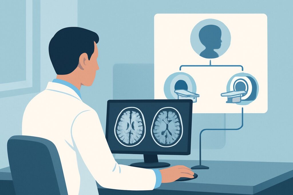
It’s 3 a.m. in the pediatric emergency department. You’re evaluating a 9-year-old boy who just had a two-minute episode of right-sided facial twitching and arm jerking while remaining aware. This is his first-ever seizure, and his post-event neurologic exam is completely normal. The immediate question is whether to image, and if so, with what modality. This scenario—a child presenting with a first-time focal seizure—requires a careful workup to identify potential underlying structural causes. This article provides a detailed workflow for this specific clinical presentation, grounded in the American College of Radiology (ACR) Appropriateness Criteria. For this patient, the ACR designates MRI head without IV contrast as Usually Appropriate, representing the most effective initial imaging step.
Who Fits This Clinical Scenario for a First-Time Focal Seizure?
This guidance applies to a specific patient population: children between 1 month and 17 years of age presenting for initial evaluation after a first-time focal seizure. A focal seizure originates in one hemisphere of the brain and can manifest with motor, sensory, or autonomic symptoms that are localized (e.g., jerking of one limb, a sensation of numbness in one area). The key inclusion criteria are the patient’s age and the focal nature of the seizure.
It is critical to distinguish this scenario from similar presentations that follow different diagnostic pathways:
- Neonatal Seizures (0 to 29 days): Seizures in the first month of life have a distinct differential diagnosis, often related to hypoxic-ischemic injury, metabolic disturbances, or congenital infections, and are evaluated under a separate ACR variant.
- Febrile Seizures: If the seizure occurred in the context of a fever in a child between 6 months and 5 years, it may be a simple or complex febrile seizure, which have their own imaging guidelines.
- Post-Traumatic Seizures: If the seizure follows a known head injury, the imaging workup is tailored to assess for traumatic brain injury.
- Generalized Seizures: Seizures that involve both hemispheres from the onset, such as primary generalized tonic-clonic or absence seizures, are considered a different clinical entity with a lower likelihood of a focal structural cause.
This article specifically excludes cases where abusive head trauma is suspected, as that scenario mandates a different and more extensive imaging protocol.
What Diagnoses Are You Working Up in a Child with a New Focal Seizure?
The primary goal of neuroimaging in a child with a new-onset focal seizure is to identify a structural lesion that could be the seizure focus. While many children will have a normal MRI, a significant minority will have an identifiable and potentially treatable cause. The differential diagnosis guides the choice of imaging modality.
Malformations of Cortical Development (MCD): These are among the most common causes of intractable focal epilepsy in children. Conditions like focal cortical dysplasia involve abnormalities in the organization of the cerebral cortex that are highly epileptogenic. These lesions can be extremely subtle and are best visualized with the high-resolution soft-tissue detail provided by MRI.
Low-Grade Neoplasms: Brain tumors are a less common but critical consideration. Slow-growing tumors, such as gangliogliomas or dysembryoplastic neuroepithelial tumors (DNETs), often present with focal seizures as their first and only symptom. MRI is the definitive modality for identifying and characterizing these lesions.
Vascular Malformations: Conditions like cavernous malformations (cavernomas) or arteriovenous malformations (AVMs) can irritate the surrounding brain parenchyma, leading to seizures. While large AVMs may be seen on CT, smaller cavernomas, especially those without recent hemorrhage, are often only visible on specific MRI sequences.
Mesial Temporal Sclerosis (MTS): This condition, characterized by hippocampal volume loss and gliosis, is a common cause of temporal lobe epilepsy. While often diagnosed in patients with a longer history of seizures, the characteristic findings can sometimes be identified on the initial MRI, particularly in adolescents.
Post-Inflammatory or Post-Ischemic Gliosis: A scar in the brain from a previous, often clinically silent, event like an infection (encephalitis), stroke, or minor trauma can become a seizure focus. MRI is superior to all other modalities for detecting these areas of gliotic brain tissue.
Why Is MRI Head without IV Contrast Usually Appropriate for This Presentation?
The ACR designates MRI head without IV contrast as “Usually Appropriate” because it offers the highest diagnostic yield for the most common underlying causes of focal seizures in children, without exposing the patient to ionizing radiation. The rationale is rooted in the modality’s superior soft-tissue contrast, which is essential for detecting the subtle structural abnormalities that cause focal seizures.
High-resolution T1-weighted, T2-weighted, and FLAIR (Fluid-Attenuated Inversion Recovery) sequences are the workhorses for this evaluation. They can clearly delineate the gray-white matter junction to identify focal cortical dysplasia, detect the abnormal signal of a low-grade tumor, and reveal the characteristic signal changes of mesial temporal sclerosis. For this initial, non-emergent workup, intravenous contrast is not typically required, as most of the key differential diagnoses (like cortical dysplasia or chronic gliosis) do not enhance. Omitting contrast avoids potential risks associated with gadolinium-based agents, particularly in children.
In contrast, other imaging modalities are rated lower for this specific scenario:
- CT head without IV contrast is rated “May be appropriate.” While readily available and fast, making it useful in emergent settings to rule out acute hemorrhage or a large mass, it has significant limitations. CT uses ionizing radiation (pediatric dose 0.3-3 mSv) and its poor soft-tissue resolution means it will miss most cases of cortical dysplasia, small tumors, and gliosis. It is considered a suboptimal first-line study for an elective workup.
- US head is rated “Usually not appropriate.” Ultrasound can be used in infants with an open anterior fontanelle to screen for major abnormalities like hemorrhage or hydrocephalus, but it provides no meaningful detail of the cerebral cortex in older infants and children and is not a suitable tool for evaluating focal seizures.
When ordering the study, it is helpful to provide the radiologist with a clear clinical history, including a description of the seizure semiology. This allows them to tailor the MRI protocol, potentially adding thin-section coronal images through the temporal lobes if mesial temporal sclerosis is suspected, to maximize the chances of finding a subtle cause.
What’s Next After MRI Head without IV Contrast? Downstream Workflow
The results of the initial MRI will guide the subsequent clinical pathway. The workflow branches based on whether the findings are positive, negative, or indeterminate.
If the MRI is positive for a structural lesion: A definitive finding, such as a tumor, vascular malformation, or clear focal cortical dysplasia, triggers a referral to pediatric neurology and potentially pediatric neurosurgery. The next step may involve a more advanced imaging study. For instance, if a tumor is suspected, an MRI head without and with IV contrast (“May be appropriate”) would be performed to better characterize the lesion’s vascularity and relationship to surrounding structures. The patient will also undergo an electroencephalogram (EEG) to correlate the electrical seizure focus with the anatomical lesion found on MRI.
If the MRI is negative: A normal MRI is a common and reassuring finding. It does not rule out epilepsy but makes a structural cause less likely. The diagnostic focus then shifts to the brain’s electrical function. The patient should be referred to a pediatric neurologist for an EEG to look for epileptiform discharges that can help classify the seizure type and guide decisions about starting anti-seizure medication. The diagnosis becomes “focal epilepsy of unknown cause,” and management is typically medical.
If the MRI is indeterminate or shows non-specific findings: Occasionally, the MRI may reveal subtle or ambiguous abnormalities. In these cases, discussion with the interpreting neuroradiologist is crucial. Further evaluation might include a follow-up MRI, a contrast-enhanced study to assess for subtle enhancement, or advanced imaging techniques like PET or SPECT, though these are “Usually not appropriate” for the initial workup.
Pitfalls to Avoid (and When to Get Help)
Navigating the workup for a child’s first focal seizure requires avoiding several common pitfalls to ensure an accurate and timely diagnosis.
- Defaulting to CT in a non-emergent setting: In a stable child with a first-time focal seizure and a normal neurologic exam, CT is a low-yield study. Opting for CT exposes the child to radiation without adequately evaluating for the most likely causes. Resist the urge for a “quick look” and arrange for an outpatient MRI.
- Forgetting the EEG: Imaging and electrophysiology are complementary. A normal MRI does not mean the child doesn’t have epilepsy. The EEG is essential for risk-stratifying recurrence and guiding treatment, regardless of the imaging results.
- Not providing adequate clinical history: The quality of a neuroradiology interpretation is enhanced by good clinical context. Describing the seizure semiology (e.g., “right-sided face and arm jerking”) can direct the radiologist’s attention to the corresponding brain region (the left frontal/parietal lobe).
If the patient presents with status epilepticus (a prolonged seizure), has a persistent focal neurologic deficit after the seizure, or shows signs of increased intracranial pressure, the situation is an emergency. In these cases, an immediate CT scan is warranted to rule out acute life-threatening processes, and escalation to a pediatric neurologist or critical care specialist is necessary.
Related ACR Topics and Tools
For a comprehensive overview of imaging recommendations across all pediatric seizure scenarios, from neonatal to post-traumatic, please consult our parent topic guide. For further exploration of the tools and criteria used in this workflow, the following resources are available.
- For breadth across all scenarios in Seizures-Child, see our parent guide: Seizures-Child: ACR Appropriateness Decoded.
- Imaging Appropriateness Selector — for adjacent scenarios
- Imaging Protocol Library — for technique on the recommended study
- Radiation Dose Calculator — for cumulative dose conversations
Frequently Asked Questions
Is an MRI necessary for every child after a single, brief focal seizure?
While practice can vary, the American Academy of Neurology and the American College of Radiology recommend neuroimaging, preferably with MRI, for any child with a focal seizure. This is because of the significant possibility of finding a treatable structural cause, such as a tumor or cortical dysplasia, which would not be apparent otherwise. A normal neurologic exam does not exclude an underlying lesion.
Why is MRI without contrast preferred over MRI with contrast for the initial study?
For the initial evaluation of a first-time focal seizure, the primary targets are non-enhancing structural abnormalities like cortical dysplasia, low-grade gliomas, or gliosis. A non-contrast MRI provides excellent visualization of these conditions. Adding gadolinium contrast is generally not necessary for the initial diagnosis and avoids the small risks associated with contrast agents. A contrast-enhanced study is typically reserved as a follow-up if the initial MRI is suspicious for a tumor or inflammatory process.
If my hospital can’t perform an MRI quickly, is it acceptable to get a CT scan instead?
In a stable, non-emergent patient, it is preferable to wait for a scheduled outpatient MRI. A non-contrast head CT has very low sensitivity for the most common causes of focal seizures in children and exposes the child to ionizing radiation. A CT is primarily useful in an emergency setting to rule out acute hemorrhage, large mass, or stroke, but it should not be considered an adequate substitute for MRI in the routine elective workup.
What if the child cannot tolerate an MRI without sedation?
Many younger children or those with anxiety or developmental delays will require sedation or general anesthesia to obtain a high-quality, motion-free MRI scan. The risks of sedation must be weighed against the diagnostic benefit of the MRI. This decision should be made in consultation with the child’s parents, the ordering clinician, and the anesthesiology/radiology departments. In most cases, the importance of identifying a potential structural lesion justifies the need for sedation.
Does a normal MRI mean the child won’t have more seizures?
No. A normal MRI is reassuring as it rules out many serious structural causes, but it does not predict the risk of seizure recurrence. The diagnosis of epilepsy is clinical and electrophysiological. The next step after a normal MRI is typically an EEG and consultation with a pediatric neurologist to determine the recurrence risk and discuss the potential need for anti-seizure medication.
Reviewed by Pouyan Golshani, MD, Interventional Radiologist — May 26, 2026