When to Order Imaging for Inflammatory Back Pain: Known or Suspected Axial Spondyloarthropathy: ACR Appropriateness Decoded

When to Order Imaging for Inflammatory Back Pain: Known or Suspected Axial Spondyloarthropathy: ACR Appropriateness Decoded
It’s late in your shift, and you’re evaluating a 28-year-old with several months of insidious-onset low back pain. The pain is worse in the morning, improves with activity, and NSAIDs help. You suspect an inflammatory process like axial spondyloarthropathy (AxSpA), but the physical exam is nonspecific. The next step is imaging, but the options are vast—radiographs, MRI, CT. Choosing the right initial study, and knowing the appropriate next step if it’s negative, is critical for timely diagnosis and management. This guide decodes the American College of Radiology (ACR) Appropriateness Criteria to help you select the most effective imaging pathway for your patient.
What Does ACR Inflammatory Back Pain: Known or Suspected Axial Spondyloarthropathy Cover?
This ACR guideline focuses specifically on imaging for patients with a clinical suspicion or established diagnosis of axial spondyloarthropathy. This includes conditions like ankylosing spondylitis and non-radiographic AxSpA. The criteria are designed to guide imaging choices across several distinct clinical scenarios: initial workup, follow-up after initial radiographs, evaluation after negative initial imaging, monitoring of known disease, and assessment for suspected fracture in an ankylosed spine.
These recommendations do not apply to the more common mechanical or degenerative back pain, which constitutes the vast majority of back pain cases in primary care and the emergency department. They also do not cover suspected spinal infection, malignancy, or acute traumatic back pain in a patient without known AxSpA. Correctly identifying the patient with inflammatory-type back pain is the crucial first step to applying these criteria effectively.
What Imaging Should I Order for Inflammatory Back Pain: Known or Suspected Axial Spondyloarthropathy? Recommendations by Clinical Scenario
The optimal imaging strategy for AxSpA depends entirely on the clinical context, from initial diagnosis to long-term follow-up. The ACR provides clear, evidence-based recommendations for each step.
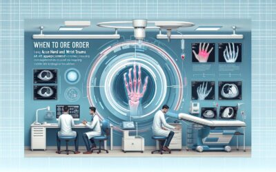
For a patient with suspected axial spondyloarthritis undergoing initial imaging, the ACR finds that Radiography of the sacroiliac joints is Usually appropriate. This is the foundational first step, as it is widely available, inexpensive, and can reveal chronic structural changes like sclerosis, erosions, and ankylosis that are part of the diagnostic criteria. Adding radiographs of the relevant area of the spine is also rated Usually appropriate. At this initial stage, more advanced modalities like MRI, CT, and bone scans are considered Usually not appropriate.
If initial radiographs are inconclusive or negative but clinical suspicion remains high, the next step is additional imaging. In this scenario, MRI of the sacroiliac joints without IV contrast is Usually appropriate. MRI is highly sensitive for detecting active inflammation (bone marrow edema/osteitis), which can precede the structural changes visible on radiographs. An MRI of both the sacroiliac joints and the spine, with or without contrast, is also Usually appropriate. In cases where MRI is contraindicated or unavailable, a CT of the sacroiliac joints without IV contrast May be appropriate to better delineate subtle structural changes. For more detail on the specific MRI sequences, see our guide on MRI Lumbar Spine Without Contrast.
In the less common scenario of a patient with negative radiographs and a negative MRI of the sacroiliac joints but persistent, compelling symptoms, the focus may shift to the spine. Here, an MRI of the spine area of interest without IV contrast is Usually appropriate to look for isolated spinal inflammation. A CT of the spine without IV contrast is also Usually appropriate to assess for subtle structural abnormalities. A dedicated protocol guide can be found here: MRI Lumbar Spine Without Contrast.
For a patient with known axial spondyloarthritis undergoing follow-up for treatment response or disease progression, repeat Radiography of the sacroiliac joints and/or spine is Usually appropriate to monitor for new or worsening structural damage over time. Routine follow-up with MRI is not typically recommended, but in cases of a significant clinical change, an MRI of the sacroiliac joints or spine May be appropriate to reassess inflammatory activity.
Finally, for a patient with established axial spondyloarthritis with spine ankylosis who has a suspected fracture (often after minor trauma), the imaging approach is different. A stiff, ankylosed spine is highly susceptible to fracture. While Radiography of the spine is Usually appropriate as a first step, these fractures can be difficult to see. Therefore, both CT of the spine without IV contrast and MRI of the spine without IV contrast are also rated Usually appropriate as initial imaging modalities. CT excels at showing bony detail and fracture lines, while MRI is superior for detecting associated ligamentous injury, epidural hematoma, or spinal cord injury. For protocol specifics, review our guide on MRI Lumbar Spine Without Contrast.
ACR Imaging Recommendations Table
| Clinical Scenario | Top Procedure | ACR Rating | Adult RRL | Pediatric RRL |
|---|---|---|---|---|
| Inflammatory back pain. Suspected axial spondyloarthritis. Initial imaging. | Radiography sacroiliac joints | Usually appropriate | ☢ ☢ 0.1-1mSv | ☢ ☢ 0.03-0.3 mSv [ped] |
| Inflammatory back pain. Suspected axial spondyloarthritis. Additional imaging following radiographs. | MRI sacroiliac joints without IV contrast | Usually appropriate | O 0 mSv | O 0 mSv [ped] |
| Inflammatory back pain. Suspected axial spondyloarthritis. Negative radiographs and negative MRI of the sacroiliac joints. | MRI spine area of interest without IV contrast | Usually appropriate | O 0 mSv | O 0 mSv [ped] |
| Known axial spondyloarthritis. Follow-up for treatment response or disease progression. | Radiography sacroiliac joints | Usually appropriate | ☢ ☢ 0.1-1mSv | ☢ ☢ 0.03-0.3 mSv [ped] |
| Axial spondyloarthritis with spine ankylosis. Suspected fracture. Initial imaging. | CT spine area of interest without IV contrast | Usually appropriate | Varies | Varies |
Adult vs. Pediatric Inflammatory Back Pain: Known or Suspected Axial Spondyloarthropathy Imaging: Radiation Dose Tradeoffs
While AxSpA typically presents in early adulthood, juvenile-onset spondyloarthropathy can occur. When evaluating pediatric patients, minimizing cumulative radiation exposure is a primary concern, guided by the As Low As Reasonably Achievable (ALARA) principle. The ACR guidelines reflect this by providing specific pediatric relative radiation levels (RRLs).
For initial radiographs of the sacroiliac joints, the pediatric dose is noted to be lower than the adult dose (RRL ☢ ☢ vs. ☢ ☢). However, the most significant consideration is the use of advanced imaging. MRI is the preferred modality for assessing active inflammation in both adults and children because it involves no ionizing radiation (RRL O). This is especially important in younger patients who may require multiple imaging studies over their lifetime. Modalities with higher radiation doses, such as bone scans with SPECT/CT (RRL ☢ ☢ ☢ ☢ [ped]) and CT, are generally avoided in the pediatric population for this indication unless absolutely necessary and MRI is not a viable option.
Imaging Protocol Details for Inflammatory Back Pain: Known or Suspected Axial Spondyloarthropathy
Once you’ve decided on the right study, the protocol matters. A non-diagnostic study due to incorrect technique can delay care. Our protocol guides cover key technical considerations, contrast administration, and interpretation principles for the studies recommended in these ACR criteria.
Tools to Help You Order the Right Study
Navigating imaging guidelines can be complex, but several tools can streamline the process. These resources are designed to bring evidence-based standards directly into your clinical workflow, ensuring you order the right test for the right reason.
For scenarios beyond inflammatory back pain, the ACR Appropriateness Criteria Lookup tool provides comprehensive access to the full library of ACR guidelines, covering thousands of clinical variants across all organ systems. It helps you find the specific recommendation you need in seconds.
To ensure the chosen study is performed correctly, the Imaging Protocol Library offers detailed, institution-vetted protocols for a wide range of CT, MRI, and ultrasound examinations. This is an essential resource for trainees and practicing physicians to understand the technical details behind the order.
When ordering studies that involve ionizing radiation, especially for younger patients or those with a history of significant exposure, the Radiation Dose Calculator is invaluable. It helps you estimate and track cumulative radiation dose, facilitating informed discussions with patients about the risks and benefits of imaging.
Why is radiography the recommended first step for suspected AxSpA instead of MRI?
Radiography is recommended first because it is cost-effective, widely accessible, and can identify the chronic, structural changes of sacroiliitis (like erosions, sclerosis, and ankylosis) that are part of the formal New York criteria for ankylosing spondylitis. While MRI is more sensitive for early inflammation, a positive radiograph can often be sufficient for diagnosis. The ACR pathway reserves the more sensitive and expensive MRI for cases where radiographs are negative or equivocal but clinical suspicion remains high.
When is IV contrast necessary for an MRI in the workup of AxSpA?
For the primary goal of detecting active inflammation (bone marrow edema), a non-contrast MRI with fluid-sensitive sequences like STIR (Short-Tau Inversion Recovery) is highly effective and often sufficient. The ACR rates MRI without contrast as “Usually appropriate.” The addition of IV gadolinium contrast is also rated “Usually appropriate” and may be helpful in specific situations, such as to increase the conspicuity of inflammation or to differentiate inflammation from other processes. However, for routine evaluation, a non-contrast study is the standard starting point.
What is the role of CT in diagnosing and managing axial spondyloarthropathy?
CT’s primary role is in evaluating structural bony changes. It is superior to radiography for detecting subtle erosions, sclerosis, and bony ankylosis of the sacroiliac joints. It is rated “May be appropriate” as a second-line test after radiographs if MRI is contraindicated. Its other key role, where it is rated “Usually appropriate,” is in the evaluation of suspected fractures in a patient with a known fused or ankylosed spine, as it provides excellent bony detail to identify complex fracture patterns.
Is ultrasound useful for evaluating the sacroiliac joints for spondyloarthropathy?
No, the ACR rates ultrasound of the sacroiliac joints as “Usually not appropriate.” While ultrasound can be used to assess for enthesitis in peripheral joints, its ability to visualize the sacroiliac joints is severely limited by the overlying bone and deep location of the joint. It is not a reliable tool for diagnosing or monitoring sacroiliitis.
How often should imaging be repeated in a patient with a confirmed diagnosis of AxSpA?
There is no fixed schedule for follow-up imaging. Routine, scheduled imaging to monitor for disease progression in an asymptomatic or clinically stable patient is generally not recommended. Repeat imaging is typically driven by clinical changes. Radiographs may be repeated every few years to assess for new structural damage, which can influence long-term management. MRI is typically reserved for situations where there is a significant change in symptoms to determine if there is a flare of active inflammation that might warrant a change in therapy.
Reviewed by Pouyan Golshani, MD, Interventional Radiologist — May 12, 2026