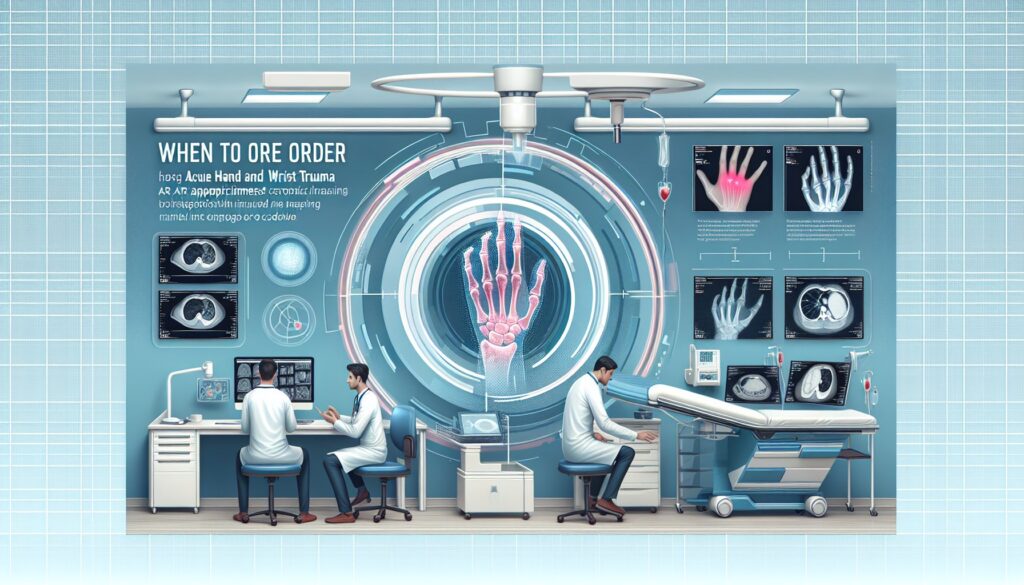
When to Order Imaging for Acute Hand and Wrist Trauma: ACR Appropriateness Decoded
When to Order Imaging for Acute Hand and Wrist Trauma: ACR Appropriateness Decoded
It’s 11 p.m. in the emergency department. A patient presents with a swollen, painful wrist after a fall onto an outstretched hand. The initial exam reveals tenderness over the scaphoid, but you’re also concerned about a potential ligamentous injury. Do you order a standard radiograph series? Should you consider a CT or MRI right away if the x-rays are negative? Making the right call quickly is critical for proper diagnosis and management, preventing long-term instability or nonunion. This guide breaks down the American College of Radiology (ACR) Appropriateness Criteria for acute hand and wrist trauma, providing clear, evidence-based pathways to help you choose the most effective initial and next-step imaging studies.
What Does ACR Acute Hand and Wrist Trauma Cover?
This ACR guideline focuses specifically on the evaluation of recent, acute trauma to the hand or wrist. The recommendations apply to a range of clinical presentations, from straightforward blunt or penetrating injuries to more nuanced scenarios involving suspected occult fractures, ligament or tendon damage, joint malalignment, or retained foreign bodies. The criteria are designed to guide imaging decisions from the initial encounter through subsequent workup if initial studies are inconclusive.
These guidelines do not cover chronic conditions such as long-standing wrist pain, suspected avascular necrosis outside of an acute fracture context, inflammatory arthropathies (e.g., rheumatoid arthritis), overuse or stress injuries, or routine post-operative evaluation. Those clinical situations are addressed in separate ACR Appropriateness Criteria documents.
What Imaging Should I Order for Acute Hand and Wrist Trauma? Recommendations by Clinical Scenario
The optimal imaging pathway for acute hand and wrist trauma depends entirely on the clinical context, including the mechanism of injury and findings from the physical exam and initial radiographs. The ACR provides guidance for several common variants.
For initial imaging of acute blunt or penetrating trauma to the hand or wrist, radiography of the area of interest is rated Usually appropriate. It is the foundational first step to assess for fractures, dislocations, and radiopaque foreign bodies. All other advanced imaging modalities, including CT, MRI, ultrasound, and bone scan, are rated Usually not appropriate for the initial evaluation.
If initial radiographs are negative or equivocal in a patient with suspected acute hand or wrist trauma, several options are available for the next step. Repeating radiographs in 10-14 days is Usually appropriate, as it may reveal a previously occult fracture line (e.g., scaphoid fracture). For a more immediate diagnosis, both MRI of the area of interest without IV contrast and CT of the area of interest without IV contrast are also rated Usually appropriate. MRI offers superior sensitivity for bone marrow edema, soft-tissue injury, and ligamentous disruption, while CT provides excellent detail of cortical bone for complex or subtle fractures.
When radiographs show an acute wrist fracture but there is clinical suspicion for tendon or ligament trauma, advanced imaging is warranted. MRI of the wrist without IV contrast and US of the wrist are both Usually appropriate to evaluate soft-tissue structures. For detailed assessment of intrinsic ligaments like the scapholunate ligament or the triangular fibrocartilage complex (TFCC), MR arthrography of the wrist and CT arthrography of the wrist are also Usually appropriate.
A similar pathway applies when initial radiographs show an acute hand fracture with suspected hand tendon or ligament trauma. In this scenario, both US of the hand and MRI of the hand without IV contrast are rated Usually appropriate to directly visualize the soft tissues. For more details on imaging these structures, see our MRI Wrist/Hand protocol guide.
For suspected ligamentous injury manifested by malalignment on radiographs in the absence of a fracture, the recommendations differ slightly based on location. For distal radioulnar joint (DRUJ) or carpal malalignment, MR arthrography of the wrist and MRI of the wrist without IV contrast are Usually appropriate. A bilateral CT of the wrist without IV contrast is also Usually appropriate to compare joint alignment with the uninjured side. For malalignment of the metacarpophalangeal (MCP), proximal interphalangeal (PIP), or distal interphalangeal (DIP) joints, US of the hand and MRI of the hand without IV contrast are Usually appropriate.
Finally, if you suspect a foreign body from penetrating trauma and initial radiographs are negative, US of the area of interest is Usually appropriate and is excellent for localizing non-radiopaque materials like wood or plastic. CT of the area of interest without IV contrast is also Usually appropriate and can detect foreign bodies with different attenuation from surrounding soft tissues. MRI without contrast May be appropriate but can be limited by artifact, especially if the foreign body is metallic.
ACR Imaging Recommendations Table
| Clinical Scenario | Top Procedure(s) | ACR Rating | Adult RRL | Pediatric RRL |
|---|---|---|---|---|
| Acute blunt or penetrating trauma to the hand or wrist. Initial imaging. | Radiography area of interest | Usually appropriate | Varies | Varies |
| Suspect acute hand or wrist trauma. Initial radiographs negative or equivocal. Next imaging study. | Radiography area of interest repeat in 10-14 days MRI area of interest without IV contrast CT area of interest without IV contrast | Usually appropriate | Varies O 0 mSv Varies | Varies O 0 mSv [ped] Varies |
| Acute wrist fracture on radiographs. Suspect wrist tendon or ligament trauma. Next imaging study. | US wrist MR arthrography wrist MRI wrist without IV contrast CT arthrography wrist | Usually appropriate | O 0 mSv O 0 mSv O 0 mSv ☢ <0.1 mSv | O 0 mSv [ped] O 0 mSv [ped] O 0 mSv [ped] |
| Initial radiographs showing distal radioulnar joint or carpal malalignment in the absence of fracture. Next imaging study. | MR arthrography wrist MRI wrist without IV contrast CT wrist without IV contrast bilateral | Usually appropriate | O 0 mSv O 0 mSv ☢ <0.1 mSv | O 0 mSv [ped] O 0 mSv [ped] |
| Acute hand fracture on radiographs. Suspect hand tendon or ligament trauma. Next imaging study. | US hand MRI hand without IV contrast | Usually appropriate | O 0 mSv O 0 mSv | O 0 mSv [ped] O 0 mSv [ped] |
| Initial radiographs showing metacarpophalangeal, proximal interphalangeal, or distal interphalangeal joint malalignment in the absence of fracture. Next imaging study. | US hand MRI hand without IV contrast | Usually appropriate | O 0 mSv O 0 mSv | O 0 mSv [ped] O 0 mSv [ped] |
| Suspect penetrating trauma with a foreign body in the soft tissues in the hand or wrist. Initial radiographs are negative. Next imaging study. | US area of interest CT area of interest without IV contrast | Usually appropriate | O 0 mSv Varies | O 0 mSv [ped] Varies |
Adult vs. Pediatric Acute Hand and Wrist Trauma Imaging: Radiation Dose Tradeoffs
The fundamental principles of imaging for acute hand and wrist trauma are similar in adults and children, but radiation safety is a paramount concern in the pediatric population. The ACR Relative Radiation Level (RRL) symbols reflect this. Modalities like ultrasound (US) and magnetic resonance imaging (MRI) are marked with “O 0 mSv,” indicating they do not use ionizing radiation, making them particularly valuable in children. The “[ped]” marker in the pediatric RRL column confirms their applicability.
While radiography and computed tomography (CT) are essential for evaluating bone, they involve ionizing radiation (indicated by “☢” symbols). The “Varies” designation for radiography reflects that the dose depends on the number of views obtained. Adhering to the As Low As Reasonably Achievable (ALARA) principle is crucial. This means using radiation-free alternatives like MRI or US when they can answer the clinical question, especially for follow-up imaging or soft-tissue evaluation after initial radiographs. For children, this often means a lower threshold for choosing MRI over CT for assessing occult fractures after negative x-rays.
Imaging Protocol Details for Acute Hand and Wrist Trauma
Once you’ve decided on the right study, the specific imaging protocol is essential for diagnostic quality. Technical parameters, such as MRI sequences or ultrasound transducer selection, can significantly impact the exam’s utility. Our protocol guides provide detailed, practical information on technique, contrast administration, and interpretation principles for the studies recommended above.
Tools to Help You Order the Right Study
Navigating imaging guidelines during a busy clinical shift can be challenging. GigHz offers several resources designed to streamline evidence-based ordering and improve communication with patients and radiologists.
The ACR Appropriateness Criteria Lookup provides a searchable interface for hundreds of clinical scenarios beyond acute hand and wrist trauma, helping you find the right study for virtually any presentation.
Our Imaging Protocol Library is a comprehensive resource for detailed, step-by-step protocols for a wide range of CT, MRI, and ultrasound examinations, ensuring you know what your order entails.
The Radiation Dose Calculator helps you estimate and track cumulative radiation exposure for your patients, facilitating informed discussions about the risks and benefits of imaging procedures that use ionizing radiation.
Why is radiography the first step for almost all acute hand and wrist trauma?
Radiography (x-ray) is the initial imaging modality of choice because it is fast, widely available, inexpensive, and highly effective at identifying most fractures and dislocations. It provides an excellent overview of bone alignment and integrity, guiding immediate management decisions like splinting or reduction. Advanced imaging is typically reserved for cases where radiographs are negative but clinical suspicion for injury remains high, or when soft-tissue structures like ligaments and tendons need to be evaluated.
My patient has a suspected scaphoid fracture but negative x-rays. What should I do next?
This is a classic and critical clinical scenario. The ACR rates three pathways as “Usually appropriate.” The traditional approach is to immobilize the wrist in a thumb spica splint and repeat radiographs in 10-14 days, which may show the fracture line after bone resorption. For a faster, more definitive diagnosis, especially in athletes or laborers, MRI without contrast is the most sensitive test for detecting an occult scaphoid fracture and any associated ligamentous injuries. CT without contrast is also an excellent alternative for visualizing the bone in fine detail.
What is the difference between an MR arthrogram and a standard MRI for the wrist?
A standard (non-arthrogram) MRI provides excellent images of bone marrow, soft tissues, and most ligaments. An MR arthrogram involves injecting a dilute gadolinium-based contrast agent directly into the radiocarpal joint under fluoroscopic or ultrasound guidance before the MRI. This distends the joint capsule and allows contrast to fill any tears in the small intrinsic ligaments, such as the scapholunate ligament or the triangular fibrocartilage complex (TFCC). This makes MR arthrography more sensitive than standard MRI for detecting and characterizing tears in these specific structures.
Why is ultrasound recommended for some hand injuries but not others?
Ultrasound is an exceptional tool for evaluating superficial soft-tissue structures. Its high resolution makes it ideal for assessing tendon integrity (e.g., extensor or flexor tendon tears), ligament sprains (e.g., ulnar collateral ligament of the thumb), and localizing non-radiopaque foreign bodies like wood splinters. However, ultrasound cannot penetrate bone, so it is not used for primary fracture diagnosis. Its utility is in clarifying suspected soft-tissue injuries, often after a fracture has already been identified on a radiograph.
Is a bone scan ever appropriate for acute hand or wrist trauma?
According to the current ACR Appropriateness Criteria for acute hand and wrist trauma, a bone scan (technetium-99m bone scintigraphy) is rated “Usually not appropriate” for all listed clinical scenarios. While historically used to detect occult fractures, it has been largely superseded by MRI and CT. A bone scan has lower spatial resolution, is less specific (it shows areas of bone turnover, which can be due to many causes), and involves a higher radiation dose than CT. MRI is more sensitive and specific for occult fractures and can simultaneously evaluate soft tissues without using ionizing radiation.
Reviewed by Pouyan Golshani, MD, Interventional Radiologist — May 12, 2026