When to Order Imaging for Lower Extremity Arterial Claudication-Imaging Assessment for Revascularization: ACR Appropriateness Decoded

When to Order Imaging for Lower Extremity Arterial Claudication-Imaging Assessment for Revascularization: ACR Appropriateness Decoded
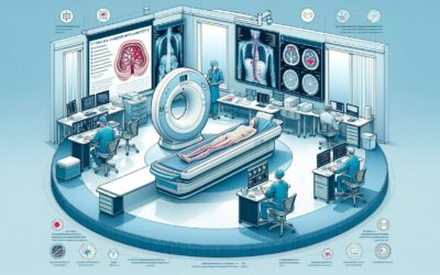
A patient with known peripheral arterial disease (PAD) presents with worsening claudication, and their symptoms are now significantly limiting their lifestyle. Conservative management has failed, and you’re considering referral for revascularization. Before the patient sees a vascular surgeon or interventional radiologist, detailed anatomical mapping is required to plan the procedure. The question is no longer *if* the patient has PAD, but *where* the hemodynamically significant lesions are located. Do you order a CTA with runoff, an MRA, or a duplex ultrasound? Each has distinct advantages in cost, radiation exposure, and detail provided. This guide breaks down the American College of Radiology (ACR) Appropriateness Criteria to help you select the optimal initial imaging study for a patient being evaluated for lower extremity revascularization.
What Does ACR Lower Extremity Arterial Claudication-Imaging Assessment for Revascularization Cover?
This specific ACR topic focuses on a well-defined clinical scenario: the imaging assessment of patients with established lower extremity arterial claudication who are candidates for revascularization. The primary goal of imaging in this context is to provide a detailed “road map” of the arterial system from the abdominal aorta down to the feet. This allows the treatment team to identify the location, severity, and length of stenoses or occlusions to determine the feasibility and optimal approach for endovascular or surgical intervention.
This guideline does *not* apply to the initial diagnosis of claudication or PAD. The initial workup typically involves a thorough history, physical examination, and non-invasive physiologic testing like the Ankle-Brachial Index (ABI). These criteria are intended for the subsequent, pre-procedural planning phase once a decision to consider intervention has been made. It also does not cover acute limb ischemia, which is a medical emergency requiring a different and more urgent diagnostic pathway. The focus here is on stable, lifestyle-limiting claudication where elective revascularization is being planned.
What Imaging Should I Order for Lower Extremity Arterial Claudication-Imaging Assessment for Revascularization? Recommendations by Clinical Scenario
For the primary clinical scenario—Lower extremity arterial claudication-imaging assessment for revascularization, initial imaging—the ACR provides several options rated as “Usually Appropriate,” reflecting that the best choice often depends on local expertise, patient factors like renal function, and the specific information required by the intervening specialist.
Non-invasive, No-Radiation Options:
Both US duplex Doppler lower extremity and MRA abdomen and pelvis with bilateral lower extremity runoff with IV contrast are rated as Usually Appropriate. Duplex ultrasound is an excellent non-invasive tool that provides both anatomic and hemodynamic information without using ionizing radiation or nephrotoxic contrast. However, it can be time-consuming and operator-dependent, and views may be limited by vessel calcification or body habitus. Contrast-enhanced MRA provides superb soft-tissue detail and a comprehensive anatomical overview from the aorta to the pedal arteries, also without radiation. It is a powerful tool, particularly for patients with contraindications to iodinated contrast. MRA without contrast may also be appropriate in certain cases, especially in patients with severe renal impairment.
Computed Tomography (CT) Options:
CTA abdomen and pelvis with bilateral lower extremity runoff with IV contrast is also Usually Appropriate. CTA is fast, widely available, and provides exceptional spatial resolution, making it excellent for visualizing calcified plaque and planning endovascular procedures. Its primary drawbacks are the use of ionizing radiation and iodinated contrast. The variant including both non-contrast and contrast phases (CTA abdomen and pelvis with bilateral lower extremity runoff without and with IV contrast) is also Usually Appropriate but carries a significantly higher radiation dose.
Invasive and Other Options:
Conventional Arteriography lower extremity is rated as Usually Appropriate. While historically the gold standard, it is an invasive procedure with associated risks (e.g., bleeding, vessel dissection) and is now often reserved for cases where a therapeutic intervention is planned for the same session (diagno-therapy) or when non-invasive imaging is inconclusive.
ACR Imaging Recommendations Table
| Clinical Scenario | Procedure | ACR Rating | Adult RRL | Pediatric RRL |
|---|---|---|---|---|
| Lower extremity arterial claudication-imaging assessment for revascularization. Initial imaging. | US duplex Doppler lower extremity | Usually appropriate | O 0 mSv | O 0 mSv [ped] |
| Lower extremity arterial claudication-imaging assessment for revascularization. Initial imaging. | Arteriography lower extremity | Usually appropriate | ☢ ☢ 0.1-1mSv | ☢ ☢ ☢ 0.3-3 mSv [ped] |
| Lower extremity arterial claudication-imaging assessment for revascularization. Initial imaging. | MRA abdomen and pelvis with bilateral lower extremity runoff with IV contrast | Usually appropriate | O 0 mSv | O 0 mSv [ped] |
| Lower extremity arterial claudication-imaging assessment for revascularization. Initial imaging. | CTA abdomen and pelvis with bilateral lower extremity runoff with IV contrast | Usually appropriate | ☢ ☢ ☢ ☢ 10-30 mSv | |
| Lower extremity arterial claudication-imaging assessment for revascularization. Initial imaging. | CTA abdomen and pelvis with bilateral lower extremity runoff without and with IV contrast | Usually appropriate | ☢ ☢ ☢ ☢ ☢ 30-100 mSv | |
| Lower extremity arterial claudication-imaging assessment for revascularization. Initial imaging. | MRA abdomen and pelvis with bilateral lower extremity runoff without IV contrast | May be appropriate | O 0 mSv | O 0 mSv [ped] |
| Lower extremity arterial claudication-imaging assessment for revascularization. Initial imaging. | MRA lower extremity without and with IV contrast | May be appropriate | O 0 mSv | O 0 mSv [ped] |
| Lower extremity arterial claudication-imaging assessment for revascularization. Initial imaging. | MRA lower extremity without IV contrast | May be appropriate | O 0 mSv | O 0 mSv [ped] |
| Lower extremity arterial claudication-imaging assessment for revascularization. Initial imaging. | CTA lower extremity with IV contrast | May be appropriate | ☢ ☢ ☢ 1-10 mSv | ☢ ☢ ☢ ☢ 3-10 mSv [ped] |
| Lower extremity arterial claudication-imaging assessment for revascularization. Initial imaging. | CTA lower extremity without and with IV contrast | May be appropriate | ☢ ☢ ☢ 1-10 mSv |
Adult vs. Pediatric Lower Extremity Arterial Claudication-Imaging Assessment for Revascularization Imaging: Radiation Dose Tradeoffs
While atherosclerotic claudication is rare in children, pediatric patients may require evaluation for revascularization due to other conditions like vasculitis (e.g., Takayasu arteritis), congenital anomalies, or post-traumatic vascular injury. For these younger patients, minimizing cumulative radiation exposure is a critical priority under the As Low As Reasonably Achievable (ALARA) principle.
The ACR guidelines reflect this by providing distinct pediatric Relative Radiation Levels (RRL). Modalities with no ionizing radiation, such as US duplex Doppler and MRA, are naturally favored and carry a pediatric RRL of ‘O 0 mSv [ped]’. When ionizing radiation is necessary, the dose considerations are paramount. For instance, conventional arteriography has a higher pediatric RRL (☢ ☢ ☢) than adult RRL (☢ ☢), reflecting the increased radiosensitivity of developing tissues. Notably, the highest-dose CTA procedures that cover the abdomen and pelvis do not have a listed pediatric RRL, underscoring that these extensive, high-dose studies should be approached with extreme caution in children and adolescents, with non-radiation alternatives exhausted first.
Imaging Protocol Details for Lower Extremity Arterial Claudication-Imaging Assessment for Revascularization
Once you’ve decided on the right study, the specific imaging protocol is crucial for obtaining diagnostic-quality images that can guide intervention. Our protocol guides cover key considerations for technique, contrast administration, and interpretation for many of the studies recommended in this guideline.
Tools to Help You Order the Right Study
Navigating imaging guidelines and radiation safety can be complex. GigHz offers a suite of free reference tools designed to support clinical decision-making at the point of care.
For scenarios beyond lower extremity arterial claudication, the ACR Appropriateness Criteria Lookup provides a searchable interface to the full ACR guidelines, helping you find evidence-based recommendations for hundreds of clinical variants.
To explore the technical details of the imaging modalities discussed here, our Imaging Protocol Library offers in-depth guides on how these studies are performed, from patient prep to imaging parameters.
When discussing radiation exposure with patients or tracking cumulative dose, the Radiation Dose Calculator is a valuable resource for estimating effective dose from various CT scans and communicating these risks in an understandable way.
What is the first-line imaging test for planning revascularization for claudication?
There is no single “first-line” test, as several are rated “Usually Appropriate.” The choice between CTA, MRA, and duplex ultrasound often depends on patient factors (renal function, contrast allergies, implanted devices), institutional preference, and the expertise of the interpreting radiologist and treating specialist. CTA is often preferred for its speed and detailed visualization of calcification, while MRA is an excellent alternative to avoid radiation and iodinated contrast. Duplex ultrasound is a valuable non-invasive option but can be more limited.
When is conventional arteriography used instead of CTA or MRA?
Digital subtraction angiography (DSA), or conventional arteriography, is an invasive procedure that is now less commonly used for purely diagnostic purposes. It is most often employed when an immediate endovascular intervention (like angioplasty or stenting) is anticipated in the same session. It may also be used when non-invasive tests like CTA or MRA are contraindicated or have provided inconclusive results.
Why are there so many MRA and CTA options with and without contrast?
The variations address specific clinical needs. For CTA, a non-contrast phase helps identify vessel wall calcification, which can be obscured by contrast. However, adding it doubles the radiation dose. For MRA, non-contrast techniques (e.g., time-of-flight) can be used in patients with severe renal dysfunction to avoid gadolinium-based contrast agents, though image quality may be reduced compared to contrast-enhanced MRA. The choice to include the abdomen and pelvis versus just the lower extremities depends on the suspected location of the disease; inflow disease from the aorta or iliac arteries is common, so comprehensive imaging is often necessary.
Is an Ankle-Brachial Index (ABI) sufficient for planning surgery?
No. The Ankle-Brachial Index (ABI) is a physiologic test used to diagnose and quantify the severity of peripheral arterial disease. It confirms the presence of hemodynamically significant disease but does not provide the anatomical information—such as the precise location, length, and morphology of stenoses or occlusions—needed to plan a revascularization procedure. Anatomic imaging with CTA, MRA, or duplex ultrasound is required after the ABI has established the diagnosis.
What is the main advantage of CTA over MRA for this indication?
The primary advantages of CTA are its rapid acquisition speed and superior spatial resolution, which provides excellent visualization of arterial calcification and metallic stents. This level of detail can be critical for planning endovascular interventions, such as choosing appropriate balloon or stent sizes. MRA, while excellent, can sometimes overestimate stenosis severity and is less effective at visualizing calcium.
Reviewed by Pouyan Golshani, MD, Interventional Radiologist — May 12, 2026