When to Order Imaging for Osteonecrosis: ACR Appropriateness Decoded
When to Order Imaging for Osteonecrosis: ACR Appropriateness Decoded
A patient presents with non-traumatic hip pain, exacerbated by weight-bearing. They have a history of long-term corticosteroid use. You suspect osteonecrosis, but the differential is broad. The initial imaging choice is critical—it sets the course for diagnosis and management, and an incorrect first step can lead to delays and unnecessary radiation exposure. Do you start with a plain film, or go directly to a more advanced modality like MRI? This guide decodes the American College of Radiology (ACR) Appropriateness Criteria for osteonecrosis, providing clear, evidence-based recommendations to help you select the right study at the right time.
What Does ACR Osteonecrosis Cover?
Osteonecrosis, also known as avascular necrosis (AVN), is the death of bone tissue due to a loss of blood supply. It most commonly affects the femoral head but can occur in other locations like the knee, shoulder, and ankle. This ACR guideline applies to patients with clinical suspicion of osteonecrosis, those with indeterminate initial radiographs, and those with confirmed disease who require further evaluation for surgical planning.
The recommendations are designed for scenarios involving non-traumatic joint pain where osteonecrosis is a primary consideration. These criteria do not apply to clinical situations where acute trauma, infection (like osteomyelitis or septic arthritis), or primary bone tumors are the leading diagnoses, as those conditions have their own distinct imaging pathways. The focus here is on the specific workup for ischemic bone injury.
What Imaging Should I Order for Osteonecrosis? Recommendations by Clinical Scenario
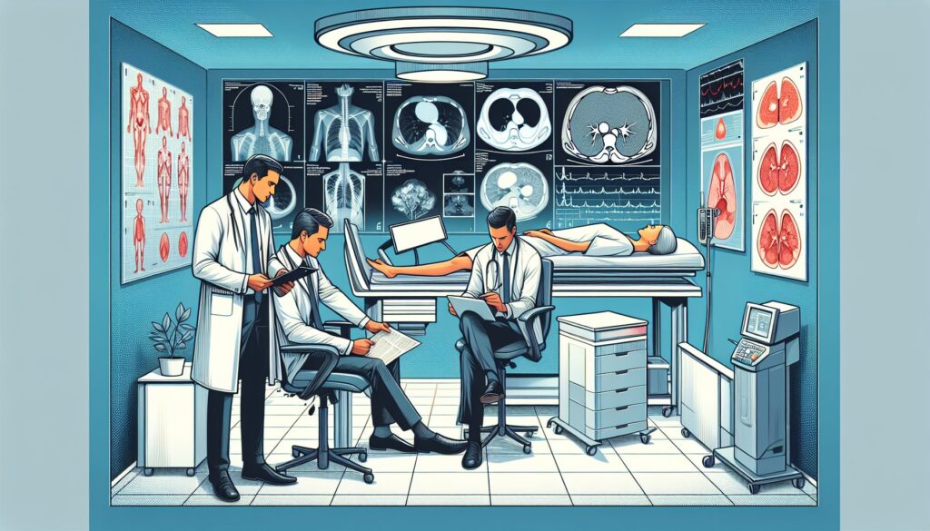
The optimal imaging strategy for osteonecrosis depends on the clinical context, from initial suspicion to pre-operative planning. The ACR provides clear guidance for each stage.
For a patient with clinically suspected osteonecrosis, the initial imaging study is straightforward. The ACR rates Radiography of the area of interest as Usually appropriate. Plain films are inexpensive, widely available, and can reveal classic findings like sclerosis, subchondral lucency (crescent sign), or femoral head collapse in later stages. At this initial stage, more advanced imaging like MRI, CT, or bone scan is considered Usually not appropriate, as radiography is the essential first-line test.
The next decision point arises when a patient has normal radiographs or radiographs that show findings suspicious for osteonecrosis. In this scenario, the ACR rates MRI of the area of interest without IV contrast as Usually appropriate. MRI is highly sensitive and specific for detecting early marrow changes of osteonecrosis long before they are visible on radiographs. An MRI without and with IV contrast may be appropriate to assess perfusion and viability but is not always necessary. A CT of the area of interest without IV contrast is rated as May be appropriate (Disagreement); it is excellent for defining the extent of subchondral fracture and articular collapse but is less sensitive than MRI for early-stage disease.
Finally, for a patient with known osteonecrosis with articular collapse by radiographs who is being considered for surgery, both MRI and CT play a key role. The ACR rates both MRI of the area of interest without IV contrast and CT of the area of interest without IV contrast as Usually appropriate. MRI provides superior detail of marrow edema, the size of the necrotic segment, and the condition of the articular cartilage. CT, meanwhile, offers a more precise evaluation of the bone structure, including the extent of subchondral collapse and any fragmentation, which is critical for surgical planning. For more on CT technique, see our guide on CT Brain Without Contrast.
ACR Imaging Recommendations Table
| Clinical Scenario | Top Procedure | ACR Rating | Adult RRL | Pediatric RRL |
|---|---|---|---|---|
| Clinically suspected osteonecrosis. Initial imaging. | Radiography area of interest | Usually appropriate | Varies | Varies |
| Clinically suspected osteonecrosis. Normal radiographs or radiographs that show findings suspicious for osteonecrosis. Next imaging study. | MRI area of interest without IV contrast | Usually appropriate | O 0 mSv | O 0 mSv [ped] |
| Known osteonecrosis with articular collapse by radiographs. Surgery planned. Next imaging study. | MRI area of interest without IV contrast | Usually appropriate | O 0 mSv | O 0 mSv [ped] |
Adult vs. Pediatric Osteonecrosis Imaging: Radiation Dose Tradeoffs
While osteonecrosis is more common in adults, it can occur in children, often related to steroid therapy for conditions like leukemia or lupus (e.g., Legg-Calvé-Perthes disease is an idiopathic osteonecrosis of the pediatric femoral head). The principles of imaging are similar, but radiation safety is paramount in younger patients due to their increased radiosensitivity and longer life expectancy, which elevates the lifetime risk of radiation-induced malignancy.
The ACR guidelines reflect this through the principle of ALARA (As Low As Reasonably Achievable). MRI is the preferred advanced imaging modality in both adults and children because it does not use ionizing radiation (0 mSv). This is particularly advantageous in pediatric cases where follow-up imaging may be required. Radiography, while using a low dose of radiation, is still the appropriate first step. CT, which involves a higher radiation dose, is generally reserved for pre-operative planning where precise bony detail is essential and is used more selectively in pediatric patients. The pediatric radiation dose for MRI is explicitly noted as “O 0 mSv [ped]” in the ACR data, reinforcing its safety and appropriateness in this population.
Imaging Protocol Details for Osteonecrosis
Once you’ve decided on the right study, the specific imaging protocol is crucial for diagnostic accuracy. Key parameters like slice thickness, field of view, and specific MRI sequences can significantly impact the quality of the images. Our protocol guides provide detailed, practical information for executing the studies recommended above.
Tools to Help You Order the Right Study
Navigating imaging guidelines during a busy clinical shift can be challenging. GigHz offers a suite of tools designed to support evidence-based decision-making at the point of care.
For clinical scenarios beyond osteonecrosis, the ACR Appropriateness Criteria Lookup provides instant access to the full library of ACR guidelines, covering thousands of clinical variants across all specialties. To ensure the selected study is performed correctly, the Imaging Protocol Library offers detailed, step-by-step protocols for a wide range of CT, MRI, and ultrasound examinations. When discussing imaging options with patients, especially those involving radiation, the Radiation Dose Calculator helps estimate and communicate cumulative radiation exposure in clear, understandable terms.
What is the first-line imaging test for suspected osteonecrosis?
The first-line imaging test is radiography (a plain X-ray) of the area of interest. According to the ACR, this is “Usually appropriate” for initial evaluation. It is readily available and can show late-stage findings like sclerosis, the crescent sign, or articular collapse.
When is an MRI indicated for osteonecrosis?
An MRI is indicated when initial radiographs are normal or inconclusive but clinical suspicion for osteonecrosis remains high. MRI is the most sensitive imaging modality for detecting early changes of osteonecrosis, such as bone marrow edema, before they are visible on an X-ray. It is also used for pre-operative planning to determine the size and location of the necrotic lesion.
Why is MRI without contrast preferred over MRI with contrast for osteonecrosis?
An MRI without intravenous contrast is rated “Usually appropriate” and is sufficient for diagnosis in most cases of osteonecrosis. The characteristic findings, such as the “double-line sign” on T2-weighted images, are well-visualized on non-contrast sequences. While contrast can help assess the perfusion of the femoral head, it is often not necessary for the initial diagnosis and adds time, cost, and a small risk of adverse reaction. Therefore, the non-contrast study is the standard follow-up after radiographs.
What is the role of CT in evaluating osteonecrosis?
CT is primarily used for pre-operative planning in patients with known osteonecrosis, especially when articular collapse has occurred. It is rated “Usually appropriate” in this setting. CT provides excellent detail of the bone structure, allowing surgeons to precisely assess the extent of subchondral fractures, femoral head deformity, and any bone fragmentation. It is less sensitive than MRI for detecting early-stage disease.
Is a bone scan useful for diagnosing osteonecrosis?
A bone scan (technetium-99m bone scintigraphy) is rated “Usually not appropriate” for the evaluation of osteonecrosis in all clinical scenarios presented by the ACR. While it can show areas of increased or decreased tracer uptake, the findings are non-specific and have been largely superseded by the superior sensitivity and specificity of MRI.
Reviewed by Pouyan Golshani, MD, Interventional Radiologist — May 12, 2026