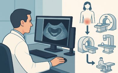
When to Order Imaging for Postmenopausal Acute Pelvic Pain: ACR Appropriateness Decoded

It’s 11 p.m. in the emergency department, and you’re evaluating a 68-year-old female with a new onset of sharp, left-sided pelvic pain. Her vitals are stable, but she has focal tenderness on exam. The differential is broad, spanning from benign gynecologic issues like ovarian torsion to more emergent non-gynecologic causes like diverticulitis or a ureteral stone. You know imaging is necessary, but the choice between ultrasound (US) and computed tomography (CT) isn’t always straightforward. Do you start with a radiation-free ultrasound, or go directly to a contrast-enhanced CT to evaluate for a wider range of pathologies? This decision involves balancing diagnostic yield, radiation exposure, and potential need for follow-up studies. This guide breaks down the American College of Radiology (ACR) Appropriateness Criteria to help you make a confident, evidence-based choice for your patient.
What Does ACR Postmenopausal Acute Pelvic Pain Cover?
The ACR Appropriateness Criteria for Postmenopausal Acute Pelvic Pain specifically address the initial imaging workup for patients who have gone through menopause and present with new, acute-onset pain in the pelvis. This guideline is designed for scenarios where the clinical suspicion is broad and could include gynecologic, gastrointestinal, or urologic etiologies. The recommendations help clinicians select the most suitable first imaging test to efficiently diagnose or exclude common causes such as ovarian torsion, tubo-ovarian abscess, diverticulitis, appendicitis, or nephrolithiasis in this specific patient population.
It is important to note what this topic does not cover. These criteria are not intended for patients with chronic pelvic pain, those with a known or highly suspected malignancy where specific staging protocols would apply, or for premenopausal patients, where the differential diagnosis and imaging considerations (especially regarding ovarian physiology) are different. The guidelines also do not apply to trauma or postoperative pain, which have their own dedicated evaluation pathways. This focus ensures the recommendations are tailored to the most likely pathologies in postmenopausal women with an acute presentation.
What Imaging Should I Order for Postmenopausal Acute Pelvic Pain? Recommendations by Clinical Scenario
For the initial imaging of a patient with postmenopausal acute pelvic pain, the ACR provides clear guidance to navigate the differential diagnosis. The primary decision point often revolves around whether the suspected etiology is gynecologic versus non-gynecologic.
For this scenario, the ACR rates both US pelvis transabdominal and US pelvis transvaginal as Usually appropriate. Ultrasound is an excellent first-line modality, particularly when a gynecologic cause like an adnexal cyst, torsion, or pyometra is suspected. It offers high-resolution imaging of the ovaries and uterus without using ionizing radiation. The combination of transabdominal views for a broad overview and transvaginal views for detailed anatomy provides a comprehensive gynecologic assessment.
Also rated as Usually appropriate is CT abdomen and pelvis with IV contrast. This study is often the preferred initial test when a non-gynecologic cause is more likely, or if the patient’s presentation is severe or non-specific. CT excels at identifying gastrointestinal and urologic emergencies such as diverticulitis with abscess, appendicitis, bowel obstruction, or pyelonephritis. The intravenous contrast is crucial for evaluating vascular structures, organ enhancement, and identifying inflammatory changes or abscess collections.
Certain studies are rated as May be appropriate in specific contexts. MRI pelvis without and with IV contrast or MRI pelvis without IV contrast can be valuable for problem-solving when ultrasound findings are indeterminate, such as for characterizing a complex adnexal mass. CT abdomen and pelvis without IV contrast may be appropriate if the primary suspicion is for renal colic, as it is the gold standard for detecting ureteral stones.
Finally, CT abdomen and pelvis without and with IV contrast is rated as Usually not appropriate. This dual-phase study imparts a significantly higher radiation dose than a single-phase scan and rarely adds diagnostic information sufficient to justify the extra radiation in the initial evaluation of acute pelvic pain.
ACR Imaging Recommendations Table
| Clinical Scenario | Top Procedure | ACR Rating | Adult RRL | Pediatric RRL |
|---|---|---|---|---|
| Postmenopausal acute pelvic pain. Initial imaging. | US pelvis transabdominal | Usually appropriate | O 0 mSv | O 0 mSv [ped] |
| Postmenopausal acute pelvic pain. Initial imaging. | US pelvis transvaginal | Usually appropriate | O 0 mSv | O 0 mSv [ped] |
| Postmenopausal acute pelvic pain. Initial imaging. | CT abdomen and pelvis with IV contrast | Usually appropriate | ☢ ☢ ☢ 1-10 mSv | ☢ ☢ ☢ ☢ 3-10 mSv [ped] |
| Postmenopausal acute pelvic pain. Initial imaging. | MRI pelvis without and with IV contrast | May be appropriate | O 0 mSv | O 0 mSv [ped] |
| Postmenopausal acute pelvic pain. Initial imaging. | MRI pelvis without IV contrast | May be appropriate | O 0 mSv | O 0 mSv [ped] |
| Postmenopausal acute pelvic pain. Initial imaging. | CT abdomen and pelvis without IV contrast | May be appropriate | O 0 mSv | O 0 mSv [ped] |
| Postmenopausal acute pelvic pain. Initial imaging. | CT abdomen and pelvis without and with IV contrast | Usually not appropriate | ☢ ☢ ☢ ☢ 10-30 mSv | ☢ ☢ ☢ ☢ ☢ 10-30 mSv [ped] |
Adult vs. Pediatric Postmenopausal Acute Pelvic Pain Imaging: Radiation Dose Tradeoffs
The clinical topic of “Postmenopausal Acute Pelvic Pain” by definition applies exclusively to an adult population. However, the ACR provides Relative Radiation Level (RRL) estimates for both adult and pediatric populations for context, highlighting a core principle of medical imaging: radiation sensitivity is age-dependent. Children and young adults are significantly more sensitive to the long-term risks of ionizing radiation than older adults. Their cells are dividing more rapidly, and they have a longer lifespan over which radiation-induced effects could potentially manifest.
This is why the principle of As Low As Reasonably Achievable (ALARA) is most stringently applied in pediatric imaging. For a study like a CT of the abdomen and pelvis, you will note the pediatric RRL is in a higher tier (☢ ☢ ☢ ☢) than the adult RRL (☢ ☢ ☢) for the same effective dose range. This reflects the greater relative biological risk attributed to the same radiation dose in a child. While not directly applicable to the postmenopausal patient, this distinction serves as a crucial reminder for clinicians to always consider cumulative radiation exposure and to preferentially choose non-ionizing modalities like ultrasound or MRI when diagnostically appropriate, especially in younger patients.
Imaging Protocol Details for Postmenopausal Acute Pelvic Pain
Once you’ve decided on the right study based on the ACR criteria, ensuring it is performed correctly is the next critical step. The specific details of an imaging protocol—such as the phases of contrast for a CT or the sequences for an MRI—can significantly impact diagnostic quality. Our protocol guides provide detailed, scannable information on technique, contrast administration, and key interpretation principles for the studies recommended above.
Tools to Help You Order the Right Study
Navigating imaging guidelines and protocols can be complex. GigHz offers a suite of free reference tools designed to support clinical decision-making at the point of care, helping you select the most appropriate study and understand its implications.
The Imaging Appropriateness Selector provides a searchable interface to the complete ACR guidelines, allowing you to quickly find evidence-based recommendations for hundreds of clinical scenarios beyond postmenopausal pelvic pain.
For detailed procedural information, the Imaging Protocol Library offers in-depth guides on how specific studies are performed, covering everything from patient prep to acquisition parameters. This is an essential resource for trainees and any clinician looking to better understand the tests they order.
To facilitate conversations with patients about radiation, the Radiation Dose Calculator helps estimate and track cumulative radiation exposure from medical imaging, contextualizing dose in understandable terms like background radiation equivalents.
What is the first-line imaging test for postmenopausal acute pelvic pain?
The choice of the first-line test depends on the suspected cause. If a gynecologic issue (e.g., ovarian torsion, adnexal mass) is most likely, transvaginal and transabdominal ultrasound are the preferred initial studies as they are rated “Usually Appropriate” and involve no radiation. If a non-gynecologic cause (e.g., diverticulitis, abscess) is more likely or the presentation is undifferentiated, CT of the abdomen and pelvis with IV contrast is also “Usually Appropriate” and provides a more comprehensive evaluation of the entire abdominopelvic region.
Why is CT with and without contrast “Usually Not Appropriate”?
A multiphase CT scan, which includes both non-contrast and post-contrast images, delivers a significantly higher dose of ionizing radiation compared to a single-phase (post-contrast only) scan. For the initial evaluation of acute pelvic pain, the additional information gained from the non-contrast phase is rarely sufficient to change management or justify the increased radiation exposure. A single post-contrast CT is typically adequate to diagnose the vast majority of relevant pathologies.
When should I consider MRI for acute pelvic pain in a postmenopausal patient?
MRI is rated “May Be Appropriate” and is typically used as a problem-solving tool rather than a first-line test in the acute setting. Its most common use is to further characterize an indeterminate adnexal mass or complex cystic structure that was first identified on ultrasound. MRI offers superior soft tissue contrast and can help differentiate benign from potentially malignant features without using ionizing radiation.
Is a non-contrast CT ever the right first choice?
Yes, a non-contrast CT of the abdomen and pelvis (“May Be Appropriate”) can be the ideal first test if there is a very high clinical suspicion for a specific condition: ureteral stones (nephrolithiasis). Non-contrast CT is the gold standard for detecting calcified stones in the urinary tract. However, if the differential diagnosis is broader, a contrast-enhanced CT is generally more informative.
Do I need to order both transabdominal and transvaginal ultrasound?
Yes, a complete pelvic ultrasound evaluation typically includes both approaches. The transabdominal portion provides a wide field of view, allowing assessment of large masses and the relationship of pelvic organs to other structures. The transvaginal portion uses a higher-frequency transducer placed closer to the uterus and ovaries, providing superior spatial resolution and more detailed anatomical characterization. They are complementary, and both are considered “Usually Appropriate.”
Frequently Asked Questions
What is the first-line imaging test for postmenopausal acute pelvic pain?
The choice of the first-line test depends on the suspected cause. If a gynecologic issue (e.g., ovarian torsion, adnexal mass) is most likely, transvaginal and transabdominal ultrasound are the preferred initial studies as they are rated “Usually Appropriate” and involve no radiation. If a non-gynecologic cause (e.g., diverticulitis, abscess) is more likely or the presentation is undifferentiated, CT of the abdomen and pelvis with IV contrast is also “Usually Appropriate” and provides a more comprehensive evaluation of the entire abdominopelvic region.
Why is CT with and without contrast “Usually Not Appropriate”?
A multiphase CT scan, which includes both non-contrast and post-contrast images, delivers a significantly higher dose of ionizing radiation compared to a single-phase (post-contrast only) scan. For the initial evaluation of acute pelvic pain, the additional information gained from the non-contrast phase is rarely sufficient to change management or justify the increased radiation exposure. A single post-contrast CT is typically adequate to diagnose the vast majority of relevant pathologies.
When should I consider MRI for acute pelvic pain in a postmenopausal patient?
MRI is rated “May Be Appropriate” and is typically used as a problem-solving tool rather than a first-line test in the acute setting. Its most common use is to further characterize an indeterminate adnexal mass or complex cystic structure that was first identified on ultrasound. MRI offers superior soft tissue contrast and can help differentiate benign from potentially malignant features without using ionizing radiation.
Is a non-contrast CT ever the right first choice?
Yes, a non-contrast CT of the abdomen and pelvis (“May Be Appropriate”) can be the ideal first test if there is a very high clinical suspicion for a specific condition: ureteral stones (nephrolithiasis). Non-contrast CT is the gold standard for detecting calcified stones in the urinary tract. However, if the differential diagnosis is broader, a contrast-enhanced CT is generally more informative.
Do I need to order both transabdominal and transvaginal ultrasound?
Yes, a complete pelvic ultrasound evaluation typically includes both approaches. The transabdominal portion provides a wide field of view, allowing assessment of large masses and the relationship of pelvic organs to other structures. The transvaginal portion uses a higher-frequency transducer placed closer to the uterus and ovaries, providing superior spatial resolution and more detailed anatomical characterization. They are complementary, and both are considered “Usually Appropriate.”
Reviewed by Pouyan Golshani, MD, Interventional Radiologist — June 27, 2026