Which Imaging Best Characterizes an Indeterminate Adnexal Mass in a Pregnant Patient?
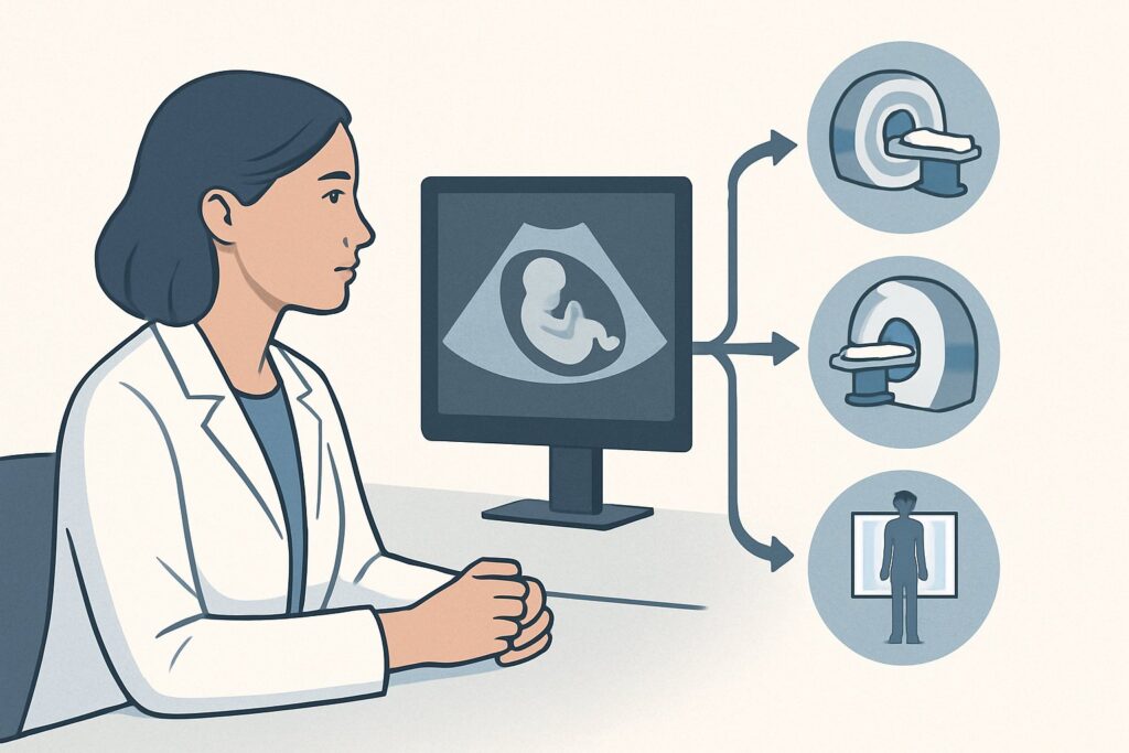
It’s a common scenario in an obstetrics practice: a routine second-trimester ultrasound reveals an incidental adnexal mass. The sonographer reports complex features—mixed cystic and solid components—that are indeterminate. The patient, a 29-year-old at 18 weeks gestation, is completely asymptomatic, but both she and her physician are concerned. The immediate clinical question is how to further characterize this mass to assess the risk of malignancy and guide management, all while ensuring the safety of the developing fetus. This article provides a step-by-step workflow for this specific situation, grounded in the American College of Radiology (ACR) Appropriateness Criteria, which designates MRI pelvis without IV contrast as Usually Appropriate.
Who Fits This Clinical Scenario?
This guidance is specifically for clinicians managing a pregnant patient with an adnexal mass that has been discovered, typically on an initial pelvic ultrasound, and could not be definitively characterized as benign. The key inclusion criteria are:
- The patient is confirmed to be pregnant.
- An adnexal mass has been identified on prior imaging (usually ultrasound).
- The features of the mass are indeterminate, meaning it is not a simple cyst and has characteristics (e.g., solid components, septations, internal vascularity) that prevent a confident benign diagnosis.
- The patient has no acute symptoms, such as severe pelvic pain, fever, or signs of hemodynamic instability, which would suggest a surgical emergency like ovarian torsion or mass rupture.
This workflow is distinct from other similar, but critically different, clinical situations. This guidance does not apply to:
- Non-pregnant patients: The risk-benefit analysis for imaging, particularly regarding contrast agents and radiation, is entirely different for premenopausal or postmenopausal patients. They are covered in a separate ACR variant.
- Patients with acute symptoms: An acute presentation necessitates an emergent evaluation, where the imaging algorithm may be altered to rapidly diagnose conditions like torsion, hemorrhage, or ectopic pregnancy.
- Masses confidently characterized as benign on ultrasound: A simple cyst or a classic-appearing dermoid on ultrasound may not require further advanced imaging and can often be managed with surveillance.
What Diagnoses Are You Working Up in This Scenario?
The primary goal of further imaging in this setting is to differentiate benign from potentially malignant masses, as this distinction dictates management during and after pregnancy. The differential diagnosis is broad, encompassing both common benign findings and rare but critical malignancies.
The most common adnexal masses in pregnancy are benign. These include functional cysts like a corpus luteum, which can appear large and complex, as well as hemorrhagic cysts. Endometriomas are also common and can undergo a process called decidualization due to hormonal changes in pregnancy, causing them to develop solid-appearing nodules and increased vascularity that can mimic malignancy on ultrasound.
Mature cystic teratomas (dermoid cysts) are one of the most frequent neoplasms found during pregnancy. While benign, they carry a risk of torsion. Less commonly, a pedunculated uterine leiomyoma (fibroid) can be mistaken for an ovarian mass on ultrasound, a distinction that MRI can readily clarify.
Pregnancy-specific conditions must also be considered. A luteoma of pregnancy is a rare, benign solid tumor that is hormonally responsive and typically regresses after delivery. It can appear as a solid, vascular mass, raising concern for malignancy.
Although the incidence of ovarian malignancy in pregnancy is low (estimated at about 1 in 1,000 to 1 in 10,000 pregnancies), it remains the most consequential diagnosis to exclude. The workup aims to identify features suspicious for epithelial ovarian cancers, germ cell tumors, or sex cord-stromal tumors, which would necessitate a multidisciplinary evaluation involving maternal-fetal medicine and gynecologic oncology.
Why Is MRI Pelvis without IV Contrast the Recommended Study for This Presentation?
When an ultrasound is inconclusive for an adnexal mass in a pregnant patient, the ACR designates MRI pelvis without IV contrast as Usually Appropriate. This recommendation is based on its superior diagnostic capability and, most importantly, its safety profile during pregnancy.
Magnetic Resonance Imaging (MRI) offers excellent soft-tissue contrast, far exceeding that of ultrasound or Computed Tomography (CT). This allows for detailed characterization of the internal architecture of the mass. MRI can reliably identify specific tissue types, such as fat within a dermoid cyst or layered blood products within an endometrioma, often providing a definitive benign diagnosis. Its multiplanar imaging capabilities are also crucial for determining the mass’s organ of origin—confirming whether it arises from the ovary, uterus, or other pelvic structures.
The key to this recommendation is the avoidance of both ionizing radiation and intravenous contrast.
- No Ionizing Radiation: Unlike CT, MRI does not use ionizing radiation. The ACR assigns it a relative radiation level of zero (O 0 mSv). This eliminates any concern for radiation-induced harm to the fetus. For this reason, all CT-based studies, such as CT pelvis without IV contrast (☢☢☢ 1-10 mSv) and CT pelvis with IV contrast (☢☢☢ 1-10 mSv), are considered Usually not appropriate.
- Avoidance of IV Contrast: Gadolinium-based contrast agents (GBCAs) are known to cross the placenta and enter the fetal circulation, with subsequent excretion into the amniotic fluid. While direct evidence of teratogenicity in humans is lacking, the long-term effects of fetal exposure are unknown. Therefore, professional societies recommend avoiding GBCAs during pregnancy unless the potential maternal benefit is critical and cannot be achieved with non-contrast imaging. In this scenario, non-contrast MRI sequences are highly effective for adnexal mass characterization, making MRI pelvis without and with IV contrast Usually not appropriate.
By providing a high-resolution, radiation-free evaluation, a non-contrast pelvic MRI offers the best balance of diagnostic yield and maternal-fetal safety for this specific clinical problem.
What’s Next After MRI Pelvis without IV Contrast? Downstream Workflow
The results of the pelvic MRI will guide the subsequent management plan, which should be developed in consultation with maternal-fetal medicine and, if necessary, gynecologic oncology specialists.
- If the MRI confirms a benign diagnosis: For masses with classic benign features on MRI (e.g., a fat-containing dermoid, a T2-dark endometrioma without suspicious features, or a simple cyst), management is typically conservative. This often involves serial ultrasound surveillance during the pregnancy to monitor for rapid growth or complications like torsion. Most of these masses do not require intervention during gestation and can be re-evaluated postpartum.
- If the MRI shows features suspicious for malignancy: If the mass demonstrates worrisome characteristics such as a large, irregular solid component, thick septations, or evidence of peritoneal spread, an urgent consultation with a gynecologic oncologist is mandatory. A multidisciplinary team will weigh the risks and benefits of surgical intervention during pregnancy versus deferring until after delivery. The timing of any potential surgery is critical and depends on the gestational age and the degree of suspicion for cancer.
- If the MRI remains indeterminate: In some cases, even MRI cannot provide a definitive diagnosis. The mass may have atypical features that do not fit a classic benign or malignant pattern. In this situation, management involves close surveillance with serial imaging (ultrasound or repeat MRI) and a high index of suspicion. The decision for surgical exploration is complex and individualized, based on the stability of the mass, patient symptoms, and the overall clinical picture.
The goal is always to avoid unnecessary surgery during pregnancy while ensuring that a potential malignancy is not left unmanaged, jeopardizing maternal health.
Pitfalls to Avoid (and When to Get Help)
Navigating an indeterminate adnexal mass in pregnancy requires careful consideration to avoid common diagnostic and management errors.
- Misinterpreting Pregnancy-Related Changes: Normal physiologic and hormone-driven changes can make benign lesions appear suspicious on ultrasound. Decidualized endometriomas and hypervascular corpus luteum cysts are classic mimics of malignancy. Relying solely on initial ultrasound findings without proceeding to MRI can lead to unnecessary anxiety and intervention.
- Inappropriate Use of CT: Reaching for CT due to familiarity or availability is a significant pitfall. The ionizing radiation exposure to the fetus is a real risk and is nearly always avoidable in this non-emergent scenario, as MRI provides superior diagnostic information without this hazard.
- Unnecessary Use of Gadolinium: Ordering an MRI “with and without contrast” by default is inappropriate in pregnancy. The non-contrast portion of the study should be performed and reviewed first. Contrast should only be considered after discussion with the radiologist if the non-contrast study is non-diagnostic and the clinical stakes are exceptionally high.
If the MRI report indicates features highly suspicious for malignancy or if the mass is growing rapidly on surveillance imaging, immediate escalation to a gynecologic oncologist experienced in managing cancer during pregnancy is essential.
Related ACR Topics and Tools
For a comprehensive overview of all clinical variants related to this topic, please consult the parent guide. For other scenarios or to explore the underlying data, the following GigHz resources provide direct access to decision-support and safety information.
- For breadth across all scenarios in Clinically Suspected Adnexal Mass, No Acute Symptoms, see our parent guide: Clinically Suspected Adnexal Mass, No Acute Symptoms: ACR Appropriateness Decoded.
- To look up other clinical scenarios, use the ACR Appropriateness Criteria Lookup.
- To review technical details for performing the recommended study, see the Imaging Protocol Library.
- To discuss cumulative radiation exposure with patients for other clinical pathways, use the Radiation Dose Calculator.
Frequently Asked Questions
Is an MRI safe for the fetus at any gestational age?
Yes, MRI is generally considered safe throughout all trimesters of pregnancy. It does not use ionizing radiation. The primary safety consideration is the avoidance of gadolinium-based contrast agents unless absolutely necessary. The American College of Obstetricians and Gynecologists (ACOG) states that MRI is not associated with known adverse fetal effects.
What if the patient is claustrophobic and cannot tolerate an MRI?
Claustrophobia is a significant challenge. Options include open MRI (which may have lower image quality), conscious sedation (which requires careful consultation with anesthesiology and MFM to ensure fetal safety), or proceeding with close ultrasound surveillance if the level of suspicion on the initial ultrasound was low to moderate. CT is not an appropriate alternative in this non-emergent setting due to radiation.
Why is contrast-enhanced MRI rated ‘Usually Not Appropriate’ if it can provide more information?
Gadolinium-based contrast agents cross the placenta and enter the fetal environment. While no definitive harm has been proven in humans, theoretical risks and a lack of long-term safety data lead experts to recommend avoiding it. Since non-contrast MRI sequences are highly effective at characterizing the vast majority of adnexal masses (by identifying fat, blood, and cystic vs. solid tissue), the added diagnostic benefit of contrast rarely outweighs the potential fetal risk in this specific scenario.
If the MRI suggests a benign dermoid cyst, does it need to be removed during pregnancy?
Generally, no. If an adnexal mass is confidently diagnosed as a benign dermoid cyst (mature cystic teratoma) on MRI, it is typically managed expectantly with ultrasound surveillance. The primary risk is ovarian torsion, which is slightly increased during pregnancy. Surgery is usually reserved for cases of torsion, extreme size causing symptoms, or rapid growth. Most are managed postpartum.
Can a transvaginal ultrasound be repeated instead of ordering an MRI?
While a follow-up ultrasound, perhaps with a more experienced sonographer or at a specialized center, can sometimes provide more clarity, it may not resolve the indeterminacy if the mass has inherently complex features. MRI is recommended when ultrasound has already proven insufficient because its superior tissue contrast can definitively answer questions that ultrasound cannot, such as confirming the presence of fat in a dermoid or clarifying the organ of origin.
Reviewed by Pouyan Golshani, MD, Interventional Radiologist — May 30, 2026