Which Imaging Studies Are Best for Staging Gestational Trophoblastic Neoplasia (GTN)?
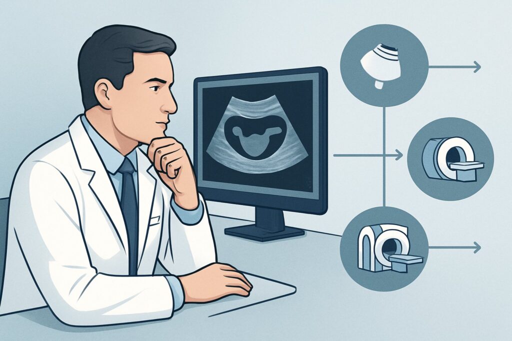
A 28-year-old patient returns to your clinic six weeks after a suction curettage for a complete hydatidiform mole. Her quantitative beta-human chorionic gonadotropin (β-hCG) levels, which were initially declining, have now plateaued for three consecutive weeks, establishing a diagnosis of postmolar gestational trophoblastic neoplasia (GTN). You need to stage her disease to determine her risk profile and select the appropriate therapy—single-agent or multi-agent chemotherapy. What is the optimal imaging pathway to assess the local uterine disease and screen for common metastatic sites? According to the American College of Radiology (ACR) Appropriateness Criteria, a US duplex Doppler pelvis is a Usually Appropriate initial study for evaluating the primary tumor site.
## Who Fits This Clinical Scenario for GTN Staging?
This guidance applies specifically to patients with a suspected or, more commonly, an established diagnosis of gestational trophoblastic neoplasia who require initial staging and risk assessment. The primary goal of imaging in this context is to determine the extent of disease, which is a critical component of the International Federation of Gynecology and Obstetrics (FIGO) staging system.
Inclusion criteria for this workflow:
- Patients with persistently elevated or rising β-hCG levels following the evacuation of a hydatidiform mole.
- Patients with a histologic diagnosis of choriocarcinoma, placental site trophoblastic tumor (PSTT), or epithelioid trophoblastic tumor (ETT).
- Patients with metastatic disease of an unknown primary where GTN is a leading consideration based on clinical context and elevated β-hCG.
This workflow does NOT apply to:
- Initial diagnosis of a molar pregnancy: A patient presenting with first-trimester bleeding and a uterine size larger than dates falls under the ACR variant for “Suspected or initial diagnosis of gestational trophoblastic disease (GTD).” The focus there is diagnosis, not staging.
- Routine surveillance after treatment: A patient who has completed chemotherapy for GTN and is being monitored with serial β-hCG levels is covered by the “Surveillance of GTN” variant.
- Acute complications: A patient with known GTN who presents with life-threatening hemorrhage, acute neurologic deficits, or other severe symptoms is managed under the “Assessment of complications” variant, which prioritizes identifying the source of the acute problem.
## What Are You Assessing in GTN Staging and Risk Assessment?
Once the diagnosis of GTN is made, imaging is not used to re-confirm the diagnosis but to answer specific questions about disease burden that directly influence FIGO staging and risk scoring. The workup is a systematic search for local invasion and distant metastases.
Local Uterine Disease: The initial and most critical step is to evaluate the uterus. You are looking for the extent of myometrial invasion by the trophoblastic tumor. Deep invasion or extension through the serosa into the parametrium significantly impacts staging. Furthermore, assessing the vascularity of the lesion is crucial, as GTN is often characterized by profound hypervascularity and the formation of uterine arteriovenous malformations.
Metastatic Disease to the Lungs: The lungs are, by a significant margin, the most common site of metastasis for GTN. All staging algorithms for GTN mandate chest imaging. The presence of lung metastases immediately classifies the disease as at least FIGO Stage III and places the patient into a higher-risk category, often necessitating multi-agent chemotherapy.
High-Risk Metastatic Disease (Brain, Liver): While less common, metastases to the brain or liver are ominous findings that automatically classify a patient as high-risk (FIGO Stage IV), regardless of other factors. Imaging of these sites is typically reserved for patients who already have evidence of other metastases (e.g., in the lungs or vagina) or who present with specific symptoms like neurologic changes or abnormal liver function tests.
## Why Is Pelvic Ultrasound the First Step for Staging Gestational Trophoblastic Neoplasia?
The initial staging of GTN is a multi-modality process, but for assessing the primary uterine site, ultrasound is the cornerstone. The ACR rates US duplex Doppler pelvis, US pelvis transabdominal, and US pelvis transvaginal as Usually Appropriate.
The transvaginal approach provides superior spatial resolution of the myometrium, allowing for detailed assessment of tumor location and depth of invasion. The addition of color and spectral Doppler is not optional—it is essential. It helps identify the characteristic chaotic, high-velocity, low-resistance flow within the tumor and can clearly delineate associated arteriovenous shunting. This information is vital for both staging and pre-procedural planning if a biopsy or surgical intervention is considered, as it highlights the risk of hemorrhage.
While ultrasound is the primary modality for the pelvis, other studies are also Usually Appropriate for a complete staging workup:
- Radiography chest: This is the standard initial screening tool for lung metastases. It is fast, low-dose (☢ <0.1 mSv), and sufficient for identifying most pulmonary nodules in this context.
- CT chest with IV contrast: This is also Usually Appropriate and is more sensitive than a plain radiograph for detecting small metastatic nodules. It is often used if the chest radiograph is negative but there is a high clinical suspicion for metastases, or to better characterize findings seen on radiography.
- MRI pelvis without and with IV contrast: This is an excellent problem-solving tool for local disease. It is considered Usually Appropriate and offers superior soft-tissue contrast compared to ultrasound for defining the precise extent of myometrial and parametrial invasion, especially in cases of large or complex tumors. It is non-ionizing (O 0 mSv), a key advantage in this young patient population.
- CT abdomen and pelvis with IV contrast: While Usually Appropriate, its primary role in GTN staging is to detect abdominal metastases (e.g., liver, kidney) rather than for primary uterine evaluation, where US and MRI are superior.
An alternative like CT abdomen and pelvis without IV contrast is rated Usually Not Appropriate because contrast is essential for characterizing vascular tumors and identifying liver metastases. Omitting it severely limits the diagnostic utility of the study.
## Downstream Workflow: What to Do After the Initial Staging Imaging?
The results of the initial imaging studies directly feed into the FIGO staging and WHO prognostic scoring systems, guiding the entire treatment plan.
- Negative Staging Workup: If a pelvic ultrasound shows disease confined to the uterus and a chest radiograph is clear, the patient has FIGO Stage I disease. Based on the WHO score (factoring in age, antecedent pregnancy, β-hCG level, and tumor size), she will likely be classified as low-risk and can proceed with single-agent chemotherapy (e.g., methotrexate or dactinomycin).
- Positive Chest Imaging: If the chest radiograph or a follow-up chest CT reveals pulmonary metastases, the patient has at least FIGO Stage III disease. This finding, along with other risk factors, will likely classify her as high-risk, requiring more intensive multi-agent chemotherapy (e.g., EMA-CO regimen). The workup must then be expanded to rule out brain and liver metastases, typically with a contrast-enhanced MRI of the head and a contrast-enhanced CT or MRI of the abdomen.
- Equivocal or Complex Pelvic Findings: If the pelvic ultrasound is indeterminate regarding the depth of invasion or suggests parametrial extension, the next step is an MRI pelvis without and with IV contrast. This will provide a definitive map of local disease extent, which can influence staging and surgical considerations.
- Suspected High-Risk Metastases: If a patient presents with neurologic symptoms or has known lung metastases, an MRI head without and with IV contrast (May be appropriate) is indicated to screen for brain involvement.
## Pitfalls to Avoid (and When to Get Help)
- Forgetting Doppler: Ordering a pelvic ultrasound without specifically requesting duplex Doppler analysis is a critical omission. The vascular signature is a key feature of GTN.
- Stopping at a Negative Chest X-Ray: In a patient with a very high β-hCG level or other high-risk features, a negative chest radiograph may not be sufficient to rule out pulmonary micrometastases. Consider proceeding to a CT chest with IV contrast.
- Incomplete Staging: Failing to complete the full staging workup (including screening for brain/liver mets) in a patient found to have lung or vaginal metastases can lead to undertreatment of high-risk disease.
- Misinterpreting Post-Evacuation Changes: Normal post-evacuation uterine changes can be complex. If the imaging findings are subtle and β-hCG levels are only mildly elevated, correlation with a gynecologic oncologist experienced in GTN is essential to distinguish expected involution from persistent neoplasia.
If you identify metastases in the lungs, liver, or brain, immediate consultation with a gynecologic oncology service is mandatory, as these patients require urgent initiation of multi-agent chemotherapy at a specialized center.
## Related ACR Topics and Tools
This article focuses on a single clinical scenario. For a comprehensive overview of all imaging variants related to this condition, from initial diagnosis to surveillance and complications, please see our parent guide.
- For breadth across all scenarios in Gestational Trophoblastic Disease, see our parent guide: Gestational Trophoblastic Disease: ACR Appropriateness Decoded.
For additional decision support and technical guidance, the following GigHz tools are available:
- ACR Appropriateness Criteria Lookup — for adjacent scenarios
- Imaging Protocol Library — for technique on the recommended study
- Radiation Dose Calculator — for cumulative dose conversations
Frequently Asked Questions
Why is a chest radiograph sufficient for initial lung screening instead of always starting with a CT?
A chest radiograph is a fast, low-radiation, and widely available initial screening tool that is sensitive enough to detect the majority of clinically significant lung metastases in GTN. According to the ACR, it is a ‘Usually Appropriate’ first step. A CT chest is reserved for cases where the radiograph is negative but clinical suspicion remains high, or to better characterize abnormalities seen on the initial film.
When should I order an MRI of the pelvis instead of an ultrasound for initial staging?
While both are rated ‘Usually Appropriate,’ pelvic ultrasound is typically the first-line modality due to its accessibility and excellent characterization of uterine vascularity with Doppler. An MRI is best used as a problem-solving tool when ultrasound findings are equivocal, when the tumor is very large and its full extent is difficult to assess with ultrasound, or when there is a strong suspicion of parametrial or adjacent organ invasion.
Is a PET/CT scan useful for staging GTN?
FDG-PET/CT is rated as ‘May be appropriate’ by the ACR. It is not a first-line staging tool. Its primary role is reserved for complex cases, such as identifying an occult primary tumor, searching for viable disease in patients with persistently elevated β-hCG after chemotherapy, or distinguishing metabolically active disease from scar tissue.
Do all patients being staged for GTN need a brain MRI?
No. A brain MRI is not required for all patients. It is indicated specifically for patients who have evidence of metastatic disease elsewhere (e.g., in the lungs or vagina) or for those who present with neurologic symptoms. In low-risk, non-metastatic (Stage I) GTN, routine brain imaging is not necessary.
What is the role of imaging for placental site trophoblastic tumor (PSTT)?
PSTT is a rare form of GTN that typically presents with lower β-hCG levels than choriocarcinoma. Pelvic ultrasound and MRI are crucial for diagnosis and staging, as PSTT often appears as a solid, less vascular uterine mass. Because PSTT is less chemosensitive and has a higher propensity for lymphatic spread, imaging to assess local invasion and rule out distant disease is critically important to guide management, which often involves hysterectomy.
Reviewed by Pouyan Golshani, MD, Interventional Radiologist — May 29, 2026