Which Imaging Best Detects Recurrence in Treated Nasopharynx Cancer?

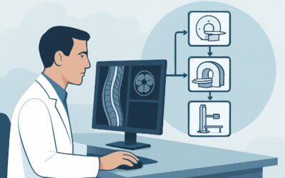
It’s late in your clinic day, and you’re seeing a 58-year-old patient for a six-month follow-up. He completed chemoradiation for nasopharyngeal carcinoma (NPC) a year ago and has been doing well, but today mentions a new, vague sense of fullness in his right ear. While his nasopharyngoscopy is unremarkable, the new symptom raises the question of local recurrence. You need to decide on the most appropriate imaging study to evaluate for recurrent disease while minimizing unnecessary radiation exposure from repeated scans. This article details the clinical workflow for surveillance and follow-up imaging in patients with treated nasopharynx cancer or an Epstein-Barr virus (EBV)-associated unknown primary of the head and neck. For this specific scenario, the American College of Radiology (ACR) finds that MRI orbits face neck without and with IV contrast is Usually appropriate.
Who Fits This Clinical Scenario?
This guidance applies specifically to patients with a history of treated nasopharyngeal carcinoma or a treated EBV-associated carcinoma of unknown primary origin. The key elements are a previously confirmed and treated malignancy and the current clinical question of surveillance or suspected recurrence. This includes both asymptomatic, routine follow-up and symptomatic evaluations where new or worsening symptoms (e.g., cranial neuropathy, neck mass, otalgia, serous otitis media) suggest a return of the disease.
This workflow should not be applied to patients in different clinical situations, as their optimal imaging strategy will differ. Key exclusions include:
- Initial Staging: Patients with a new diagnosis of nasopharynx cancer who have not yet undergone treatment require a comprehensive initial staging workup, which is a distinct clinical scenario.
- Different Tumor Locations: This guidance is specific to the nasopharynx. Patients with treated cancers of the oral cavity, oropharynx, larynx, or paranasal sinuses have different patterns of recurrence and are covered under separate ACR appropriateness criteria variants.
- Non-EBV Unknown Primary: While this scenario includes EBV-associated unknown primary tumors (where the nasopharynx is the presumed origin), patients with EBV-negative unknown primary cancers may require a different diagnostic approach.
What Diagnoses Are You Working Up in This Scenario?
When ordering follow-up imaging for treated nasopharyngeal cancer, the primary goal is to detect recurrence early while distinguishing it from expected post-treatment changes. The differential diagnosis is focused and critical.
Local Tumor Recurrence: This is the most significant concern. Recurrence can occur at the primary site in the nasopharynx, often submucosally, making it difficult to detect on physical examination alone. It can also invade adjacent structures, most critically the skull base, which has profound neurologic implications. Imaging must provide exquisite soft-tissue detail to identify subtle enhancing nodules or masses within the complex post-treatment anatomy.
Regional Nodal Recurrence: The neck is a common site for recurrence, particularly in the retropharyngeal and cervical lymph node basins (levels II, III, and V). Imaging must carefully assess for new or enlarging lymph nodes, especially those exhibiting suspicious features like central necrosis or extracapsular extension.
Post-Treatment Changes vs. Recurrence: This is the central diagnostic challenge. Radiation therapy induces changes like fibrosis, edema, and inflammation, which can mimic or obscure a tumor. Radiation necrosis is a less common but serious complication that can also appear as an enhancing mass. The ideal imaging study must help differentiate benign post-therapeutic changes from malignant recurrence.
Perineural Tumor Spread: Nasopharyngeal carcinoma has a known propensity for spreading along cranial nerves, particularly the trigeminal nerve (V) and abducens nerve (VI). Detecting perineural spread is crucial for treatment planning and is a key strength of high-resolution imaging.
Why Is MRI of the Orbits, Face, and Neck the Recommended Study?
The ACR designates MRI orbits face neck without and with IV contrast as Usually appropriate because it directly addresses the primary clinical questions in this scenario with superior diagnostic capability and no ionizing radiation.
The fundamental advantage of MRI is its exceptional soft-tissue contrast resolution. This is paramount for distinguishing recurrent tumor from post-radiation fibrosis in the intricate spaces of the nasopharynx, parapharyngeal region, and skull base. Recurrent tumors typically demonstrate T2-signal changes and avidly enhance with gadolinium contrast, whereas mature fibrosis often shows low T2 signal and less intense, more delayed enhancement. Fat-suppressed, post-contrast T1-weighted sequences are particularly valuable for identifying subtle perineural spread along cranial nerves, a task for which MRI is far superior to other modalities.
Furthermore, the lack of ionizing radiation (0 mSv) is a significant benefit for patients requiring long-term surveillance, as it avoids the cumulative radiation dose from repeated scans. This is a key consideration in a population that has already received therapeutic radiation.
Other modalities are also rated for this scenario but have specific trade-offs:
- CT neck with IV contrast is also rated Usually appropriate. It is faster to acquire and can be better for assessing cortical bone erosion at the skull base. However, its inferior soft-tissue contrast makes it significantly more difficult to differentiate tumor from post-treatment changes. It also involves a moderate radiation dose (☢☢☢ 1-10 mSv).
- FDG-PET/CT skull base to mid-thigh is also Usually appropriate and is highly sensitive for detecting metabolically active tumor. It is excellent for identifying nodal and distant metastatic disease. However, its specificity can be limited in the early post-treatment period (first 3-6 months) due to radiation-induced inflammation causing false-positive FDG uptake. It also carries the highest radiation dose (☢☢☢☢ 10-30 mSv) and is often reserved for problem-solving when MRI is equivocal or when there is high clinical suspicion for recurrence.
What’s Next After MRI? Downstream Workflow
The results of the surveillance MRI will guide the subsequent clinical pathway. A structured approach ensures appropriate and timely management.
- If the MRI is clearly positive for recurrence: A finding of a new, enhancing soft-tissue mass consistent with recurrence should prompt an urgent referral back to the multidisciplinary head and neck tumor board. The next step is typically a biopsy, often performed via endoscopy, to obtain histopathologic confirmation. The imaging findings are critical for planning the biopsy approach and determining if the patient is a candidate for salvage therapy, such as surgery or re-irradiation.
- If the MRI is negative: A definitively negative scan is reassuring. The patient can return to their standard clinical and endoscopic follow-up schedule as determined by oncology guidelines. No further immediate imaging is typically required.
- If the MRI is indeterminate or equivocal: This is a common and challenging situation where post-treatment inflammation cannot be confidently distinguished from early recurrence. The downstream workflow may involve several options, often decided in a multidisciplinary setting. One approach is a short-interval follow-up MRI (e.g., in 6-12 weeks) to assess for change. Alternatively, an FDG-PET/CT may be ordered as a problem-solving tool to see if the equivocal area is metabolically active, which would increase suspicion for malignancy. If suspicion remains high, a targeted biopsy may be pursued despite the equivocal imaging.
Pitfalls to Avoid (and When to Get Help)
Navigating post-treatment surveillance for nasopharyngeal cancer requires careful attention to detail to avoid common errors.
- Pitfall 1: Imaging too early. Performing MRI or PET/CT less than three months after completing radiation therapy can lead to false-positive results due to intense post-treatment inflammation. A baseline scan is typically recommended around 3-6 months post-treatment, with subsequent scans timed according to surveillance protocols.
- Pitfall 2: Ordering the wrong MRI protocol. Requesting a generic “MRI head” or “MRI neck” is insufficient. The order must specify “MRI orbits, face, and neck” to ensure complete coverage from the skull base through the low neck, including all relevant cranial nerves and nodal basins. Always specify “without and with IV contrast.”
- Pitfall 3: Over-reliance on imaging alone. Imaging findings must always be correlated with the clinical examination, nasopharyngoscopy, and EBV DNA titers (if applicable). A rising EBV DNA level can be an early indicator of recurrence and should lower the threshold for biopsy, even with equivocal imaging.
If there is a strong clinical suspicion of recurrence despite a negative or indeterminate MRI, escalate the case to a multidisciplinary tumor board for discussion. This collaborative review by otolaryngology, radiation oncology, medical oncology, and neuroradiology is the best way to determine the optimal next step.
Related ACR Topics and Tools
For a comprehensive overview of imaging across all head and neck cancer scenarios, consult the parent topic guide. For additional decision support, the following tools can help you select the right test, understand the technique, and discuss radiation dose with your patients.
- For breadth across all scenarios in Staging and Post-Therapy Assessment of Head and Neck Cancer, see our parent guide: Staging and Post-Therapy Assessment of Head and Neck Cancer: ACR Appropriateness Decoded.
- To explore other clinical presentations, use the ACR Appropriateness Criteria Lookup.
- To review technical details for various studies, see the Imaging Protocol Library.
- To help explain radiation exposure to patients, use the Radiation Dose Calculator.
Frequently Asked Questions
How often should surveillance imaging be performed for treated nasopharynx cancer?
Surveillance frequency varies by institutional protocols and guidelines like those from the NCCN. Generally, imaging is performed more frequently in the first 2-3 years post-treatment (e.g., every 6-12 months) and then spaced out annually for up to 5 years, after which it may be discontinued or performed only as needed for symptoms.
Why is MRI considered better than CT for nasopharynx cancer follow-up?
MRI’s superior soft-tissue contrast resolution is critical for differentiating recurrent tumor from post-radiation fibrosis and scar tissue in the complex anatomy of the nasopharynx and skull base. It is also better at detecting perineural tumor spread and involves no ionizing radiation, which is an advantage for repeated surveillance scans.
When is a PET/CT scan indicated in this surveillance scenario?
A PET/CT is rated ‘Usually appropriate’ but is often used as a second-line or problem-solving tool. It is most valuable when MRI findings are indeterminate, when there is a high clinical suspicion of recurrence despite a negative MRI, or to evaluate for distant metastatic disease if recurrence is confirmed locally.
What does ‘EBV-associated unknown primary’ mean in this context?
This refers to a situation where a patient presents with metastatic squamous cell carcinoma in the cervical lymph nodes, but no primary tumor can be found on physical exam and endoscopy. If the tumor tissue tests positive for Epstein-Barr virus (EBV), it strongly implies an occult (hidden) primary cancer in the nasopharynx, as NPC is closely linked to EBV. These patients are often treated similarly to those with known nasopharyngeal cancer.
Is IV contrast always necessary for these follow-up MRI scans?
Yes, both pre- and post-contrast sequences are essential. Comparing the two is critical for identifying and characterizing abnormal enhancement. Recurrent tumors are typically vascular and will enhance avidly, a key feature used to distinguish them from non-enhancing or slowly enhancing post-treatment scar tissue.
Reviewed by Pouyan Golshani, MD, Interventional Radiologist — May 30, 2026