Which Imaging Best Stages Distant Metastases in New Colorectal Cancer?
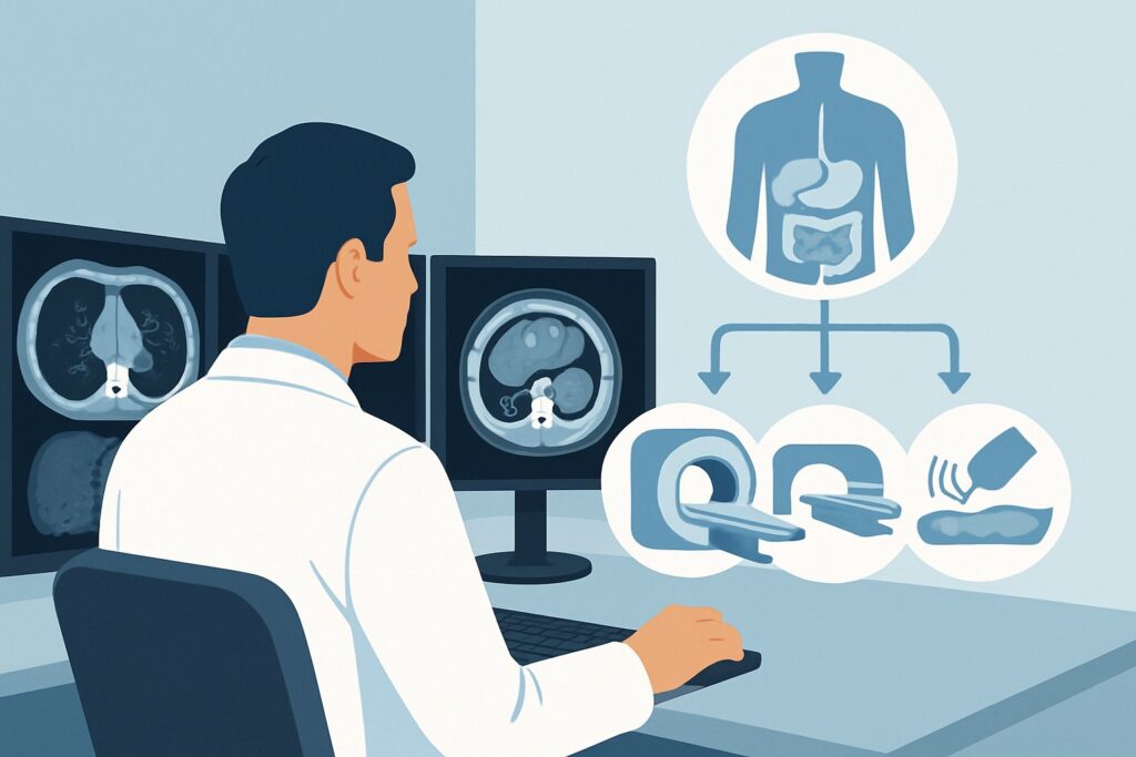
A 68-year-old male with a new diagnosis of sigmoid adenocarcinoma, confirmed on colonoscopy and biopsy, is referred to your clinic. His labs are notable only for a mild microcytic anemia. He has no specific symptoms of metastatic disease. Before you can refer him to surgical and medical oncology for a definitive treatment plan, you need to establish the extent of his disease. The critical question is whether the cancer has spread beyond the colon. This requires precise initial imaging to detect distant metastases, a decision that directly impacts his prognosis and therapeutic options. For this exact scenario—initial staging for distant metastases in colorectal cancer—the American College of Radiology (ACR) rates CT chest with IV contrast and MRI abdomen with IV contrast as Usually Appropriate.
Who Fits This Clinical Scenario?
This guidance applies specifically to patients with a new, biopsy-proven diagnosis of colorectal cancer who are undergoing their initial staging workup. The primary goal of imaging in this context is to identify distant metastatic disease, most commonly in the liver and lungs, prior to the initiation of therapy. This workflow is designed for the first comprehensive look at the patient’s disease burden outside of the primary tumor’s immediate location (locoregional staging is a separate consideration).
This article does not apply to:
- Locoregional staging of rectal cancer: Evaluating the depth of tumor invasion into the rectal wall (T-stage) and involvement of nearby lymph nodes (N-stage) requires a different imaging approach, typically with pelvic MRI. This is a distinct clinical question covered in a separate ACR variant.
- Post-treatment surveillance: Imaging to monitor for recurrence after surgery or chemotherapy follows different protocols and intervals.
- Symptomatic patients with suspected recurrence: A patient with a history of colorectal cancer who now presents with new symptoms (e.g., right upper quadrant pain, cough) may require a more tailored or urgent imaging workup, potentially including FDG-PET/CT.
Correctly identifying your patient’s situation is crucial. Applying this guidance to the initial, treatment-naïve patient ensures the most accurate baseline assessment of metastatic disease.
What Diagnoses Are You Working Up in This Scenario?
When ordering initial staging imaging for colorectal cancer, the primary goal is to confirm or exclude the presence of metastatic deposits. The imaging strategy is tailored to detect disease in the most common sites of spread.
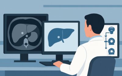
Hepatic (Liver) Metastases: The liver is the most common site for colorectal cancer metastasis due to portal venous drainage from the colon and rectum. Identifying the number, size, and location of liver lesions is paramount, as it determines whether a patient might be a candidate for curative-intent liver resection, ablation, or systemic therapy alone. MRI is highly sensitive and specific for characterizing liver lesions, distinguishing metastases from benign entities like cysts or hemangiomas.
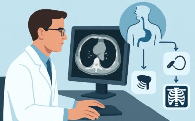
Pulmonary (Lung) Metastases: The lungs are the second most common site of distant spread. CT of the chest is the gold standard for detecting small pulmonary nodules that could represent metastases. Early detection can influence treatment, as some patients with limited lung disease may be candidates for metastasectomy or stereotactic body radiation therapy (SBRT).
Peritoneal Carcinomatosis: This refers to the spread of cancer to the peritoneum, the lining of the abdominal cavity. While less common at initial presentation, its presence significantly alters prognosis and treatment, often precluding curative-intent surgery. Both CT and MRI can detect signs of peritoneal disease, such as ascites, omental caking, or peritoneal nodules, though subtle disease can be difficult to identify.
Distant Nodal or Adrenal Metastases: Spread to lymph nodes outside the primary tumor’s regional drainage basin (e.g., retroperitoneal or supraclavicular nodes) or to the adrenal glands constitutes metastatic disease. These findings are less frequent but are readily assessed on comprehensive chest, abdomen, and pelvis imaging.
Why Is CT Chest with IV Contrast and MRI Abdomen with IV Contrast the Recommended Study?
The ACR designates the combination of CT chest with IV contrast and MRI abdomen with IV contrast as a Usually Appropriate study for the initial staging of distant metastases in colorectal cancer. This recommendation is based on a “best-of-both-worlds” approach, leveraging the strengths of each modality for the most critical anatomical regions.
CT of the chest with IV contrast is exceptionally sensitive for detecting small pulmonary nodules, the typical appearance of lung metastases. It provides a rapid and robust evaluation of the entire thorax, including the mediastinal and hilar lymph nodes. For the abdomen and pelvis, MRI with IV contrast is superior to CT for the detection and characterization of liver lesions. It can more reliably differentiate small metastases from benign liver lesions like cysts and hemangiomas, a common diagnostic dilemma on CT that can lead to unnecessary follow-up imaging or biopsies. This accuracy is critical, as the presence and extent of liver disease is a primary driver of the overall treatment strategy.
Let’s consider the alternatives:
- CT chest abdomen pelvis with IV contrast: This is also rated Usually Appropriate and is a very common and practical initial staging study. It offers the advantage of a single, faster examination. However, its sensitivity for small or indeterminate liver lesions is lower than that of MRI. It also carries a higher radiation dose (ACR Relative Radiation Level ☢☢☢☢, 10-30 mSv) compared to the recommended combination of CT chest and MRI abdomen (ACR RRL ☢☢☢, 1-10 mSv for the CT component; MRI has no ionizing radiation).
- FDG-PET/CT skull base to mid-thigh: This is rated May be appropriate. While PET/CT is excellent for detecting metabolically active disease throughout the body, it is generally not recommended as the initial imaging test for routine staging. It is often reserved for situations where conventional imaging is equivocal, in patients with a high pre-test probability of metastatic disease not seen on CT/MRI, or to clarify potential sites of oligometastatic disease being considered for local therapy.
The choice between the two Usually Appropriate options often depends on institutional preference, MRI availability, and patient-specific factors. However, the combination of CT chest and MRI abdomen provides the most detailed and accurate assessment of the two most common and prognostically significant sites of metastasis.
What’s Next After Imaging? Downstream Workflow
The results of your initial staging scans will direct the patient into one of several distinct clinical pathways. The report is not an endpoint but a critical branch point in the patient’s care journey.
If the studies are positive for widespread metastatic disease: If imaging reveals multiple, unequivocal metastases in the liver, lungs, or other sites (e.g., peritoneum), the patient is classified as having Stage IV disease. The primary treatment will be systemic therapy (chemotherapy, targeted therapy, immunotherapy). The patient should be referred to a medical oncologist to discuss treatment options. A biopsy of a metastatic site may be performed to confirm the diagnosis and obtain tissue for molecular testing, which guides systemic therapy choices.
If the studies are negative for distant metastases: If the CT chest and MRI abdomen show no evidence of distant spread, the patient has localized disease and is a potential candidate for curative-intent surgery. The next step is a referral to a colorectal surgeon. For rectal cancers, this would be followed by dedicated locoregional staging with a pelvic MRI to guide surgical planning and determine the need for neoadjuvant chemoradiation. For colon cancers, the patient typically proceeds directly to surgical resection.
If the studies are indeterminate: This is a frequent and challenging scenario. For example, the scan may show a single, small (<1 cm) liver lesion or a few tiny, non-specific lung nodules. In this case, the next step is often a multidisciplinary tumor board discussion involving radiologists, surgeons, and oncologists. Options for clarifying indeterminate findings include a short-term follow-up MRI of the liver, a dedicated FDG-PET/CT to assess for metabolic activity, or, in some cases, a percutaneous biopsy.
Pitfalls to Avoid (and When to Get Help)
Navigating the initial staging of colorectal cancer requires careful attention to detail to avoid common errors that can delay care or lead to incorrect staging.
- Omitting IV Contrast: Ordering non-contrast CT or MRI significantly limits the evaluation of the liver, vasculature, and lymph nodes. Unless there is a severe contraindication, IV contrast is essential for accurate staging.
- Misinterpreting Benign Lesions: Small, benign liver cysts or hemangiomas are common and can be mistaken for metastases on CT. This is a key reason why MRI is preferred for the abdominal evaluation.
- Ignoring Renal Function: Always check a patient’s estimated glomerular filtration rate (eGFR) before ordering studies with iodinated or gadolinium-based contrast agents to avoid contrast-induced nephropathy or other complications.
- Choosing the Wrong Modality for the Question: Using CT abdomen to “rule out” liver metastases when MRI is available can lead to indeterminate results and further imaging delays. Match the modality to the clinical question.
If the imaging results are complex, contradictory, or do not fit the clinical picture, the best next step is to escalate. Present the case at a multidisciplinary tumor board or have a direct consultation with the reporting radiologist to clarify findings and plan the next steps collaboratively.
Related ACR Topics and Tools
For a comprehensive overview of all clinical scenarios related to colorectal cancer imaging, from local staging to post-treatment follow-up, please refer to our parent guide. For tools to help with ordering, protocoling, and patient communication, see the resources below.
- For breadth across all scenarios in Staging of Colorectal Cancer, see our parent guide: Staging of Colorectal Cancer: ACR Appropriateness Decoded.
- For adjacent scenarios not covered here, use the Imaging Appropriateness Selector.
- For detailed technical parameters of the recommended studies, explore the Imaging Protocol Library.
- To discuss radiation exposure with your patients, consult the Radiation Dose Calculator.
Frequently Asked Questions
Why not just order a whole-body FDG-PET/CT for every new colorectal cancer diagnosis?
While FDG-PET/CT is very sensitive for metabolically active cancer, the ACR rates it as ‘May be appropriate’ for initial staging. High-quality, contrast-enhanced CT and MRI provide superior anatomical detail, especially for small liver and lung lesions. PET/CT is often reserved for problem-solving when initial CT/MRI results are equivocal, for high-risk patients, or for evaluating for oligometastatic disease.
Is a contrast-enhanced CT of the abdomen and pelvis good enough if MRI is not readily available?
Yes, a contrast-enhanced CT of the chest, abdomen, and pelvis is also rated as ‘Usually Appropriate’ by the ACR. It is a widely used and acceptable alternative, especially when MRI access is limited or delayed. The main trade-off is slightly lower sensitivity and specificity for characterizing small liver lesions compared to MRI, which may lead to more indeterminate findings requiring follow-up.
What should I order if my patient has a severe contraindication to both iodinated and gadolinium-based contrast?
This is a challenging situation that requires a multidisciplinary discussion. Options might include a non-contrast CT of the chest, a non-contrast MRI of the abdomen (which can still provide valuable information using diffusion-weighted imaging), or proceeding with FDG-PET/CT, which does not require IV contrast for the PET component, though the fused CT is typically non-contrast in this setting.
Does this guidance apply to surveillance imaging after a patient has completed treatment?
No, this guidance is specifically for the initial staging of a newly diagnosed, treatment-naïve patient. Surveillance imaging protocols after curative-intent treatment are different and are typically guided by society guidelines (e.g., NCCN, ASCO), which usually involve periodic contrast-enhanced CT scans of the chest, abdomen, and pelvis.
Why are the chest and abdomen evaluated with two different imaging modalities (CT and MRI)?
This approach leverages the strengths of each technology for the organ system it visualizes best. CT is the fastest and most sensitive method for detecting small lung nodules. MRI, with its superior soft-tissue contrast, is the most accurate non-invasive method for both detecting and, crucially, characterizing liver lesions, which is the most common site of metastasis.
Reviewed by Pouyan Golshani, MD, Interventional Radiologist — May 26, 2026