Which Imaging Is Best for Elevated-Risk Colorectal Cancer Screening in Patients 45-75?

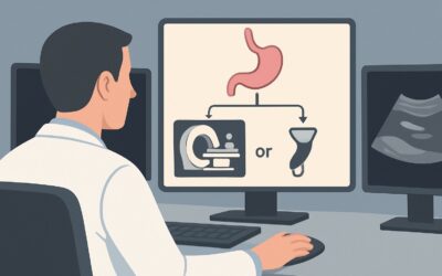
A 52-year-old patient is in your primary care office for a wellness visit. They have a family history of a single first-degree relative diagnosed with colorectal cancer at age 65, placing them in an elevated-risk category. They are due for their initial screening but are hesitant to undergo an invasive optical colonoscopy and ask about non-invasive alternatives. You need to decide on the most appropriate imaging-based screening test that is both effective and aligns with established guidelines. This article provides a detailed clinical workflow for this specific scenario: initial or subsequent screening for individuals aged 45 to 75 with elevated, but not high, risk for colorectal cancer. For this patient, the American College of Radiology (ACR) finds that CT colonography without IV contrast screening is Usually appropriate.
Who Qualifies for This Elevated-Risk Screening Protocol?
This guidance is specifically for individuals between 45 and 75 years of age who are considered to have an “elevated risk” for colorectal cancer (CRC). This category sits between average-risk and high-risk populations. While definitions can vary slightly between societal guidelines, elevated risk typically includes individuals with a personal history of polyps or a family history that does not meet the criteria for high-risk hereditary syndromes. For example, a patient with one first-degree relative diagnosed with CRC at age 60 or older, or two second-degree relatives with CRC, would often fall into this category.
It is critical to distinguish this group from others who require different screening strategies:
- Average-Risk Individuals: This protocol does not apply to patients with no personal or family history of CRC or adenomatous polyps and no history of inflammatory bowel disease or known genetic syndromes. Their screening options and intervals are different.
- High-Risk Individuals: This guidance is not for patients with a personal history of CRC, a known hereditary syndrome like Lynch syndrome or Familial Adenomatous Polyposis (FAP), a strong family history suggestive of these syndromes (e.g., first-degree relative with CRC before age 60), or long-standing inflammatory bowel disease. These patients require more intensive surveillance, typically with optical colonoscopy at shorter intervals.
- Symptomatic Patients: This workflow is for asymptomatic screening. Patients presenting with symptoms like rectal bleeding, a change in bowel habits, or unexplained weight loss require a diagnostic workup, not a screening study.
What Diagnoses Are You Working Up in This Scenario?
The primary goal of screening in this elevated-risk population is the early detection of colorectal neoplasia before it becomes symptomatic. The imaging study is designed to identify specific precursor lesions and early-stage cancers, allowing for timely intervention.
Adenomatous Polyps: These are the most common precursor lesions for colorectal adenocarcinoma. The primary objective of any screening modality is to detect and facilitate the removal of these polyps, particularly advanced adenomas (≥10 mm in size, or with villous histology or high-grade dysplasia), which carry a higher risk of progressing to cancer. CT colonography is highly sensitive for detecting polyps of this size.
Colorectal Carcinoma (CRC): The second major target is early-stage, asymptomatic colorectal cancer. Detecting CRC before it has invaded deeply into the bowel wall or metastasized significantly improves prognosis and treatment outcomes. Screening aims to find these cancers when they are most treatable, often curable with local resection.
Sessile Serrated Lesions/Polyps (SSL/Ps): While historically less emphasized, these flat or minimally raised lesions are now recognized as an important alternative pathway to CRC. They can be more challenging to detect both optically and radiologically than traditional adenomas, but modern CT colonography techniques have improved their visualization.
It is important to note that while CT colonography can identify extracolonic findings, its primary purpose in this screening context is the evaluation of the colon and rectum for neoplasia.
Why Is CT Colonography Without IV Contrast the Recommended Study for This Presentation?
The ACR designates CT colonography (CTC) without intravenous contrast as Usually appropriate for screening this elevated-risk population, reflecting its high diagnostic performance and non-invasive nature. The rationale is based on a careful balance of sensitivity, safety, and patient tolerance compared to other options.
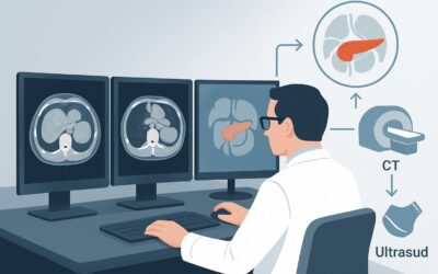
CTC, often called virtual colonoscopy, uses a low-dose CT scan to generate high-resolution 2D and 3D images of the prepared colon. Its sensitivity for detecting adenomas 10 mm or larger is comparable to that of optical colonoscopy, which is a critical benchmark for an effective screening test. While less sensitive for smaller polyps, the clinical significance of detecting and removing diminutive polyps remains a subject of debate, and the primary goal is to prevent cancer by removing advanced precursor lesions.
Let’s examine why other modalities are rated lower for this specific screening scenario:
- Fluoroscopy barium enema (double-contrast): This study is rated Usually not appropriate. While once a mainstay of colonic imaging, its sensitivity for detecting polyps and early-stage cancers is substantially lower than that of modern CTC or optical colonoscopy. Given the availability of superior alternatives, its role in screening has become obsolete.
- CT abdomen and pelvis with IV contrast: This is also rated Usually not appropriate for screening. A standard diagnostic CT is not optimized for visualizing the colonic mucosa. It requires a different patient preparation (oral contrast, not bowel cleansing and insufflation) and uses a higher radiation dose. Furthermore, the addition of IV contrast is unnecessary for polyp detection and adds risks of allergic reaction and contrast-induced nephropathy without providing benefit in a screening context.
From a safety perspective, CTC avoids the risks associated with sedation and bowel perforation inherent to optical colonoscopy, though a small risk of perforation from gas insufflation exists. The radiation dose for a screening CTC (ACR RRL ☢☢☢☢, 10-30 mSv) is a key consideration. However, this level of exposure, when performed at appropriate 5-year intervals in this age group, is generally considered to have a favorable risk-benefit profile for cancer prevention.
What’s Next After CT Colonography? Downstream Workflow
The results of a screening CT colonography (CTC) directly guide the next steps in patient management. The workflow is designed to ensure that any significant findings are addressed with a diagnostic and potentially therapeutic procedure.
Positive for a Significant Polyp (e.g., ≥10 mm): If the CTC identifies one or more polyps meeting a size threshold for clinical significance (typically 10 mm or larger, though some guidelines use 6 mm), the definitive next step is a referral for optical colonoscopy. This allows for direct visualization, confirmation, and polypectomy (removal) of the lesion(s) in a single session. The patient should be counseled that the CTC was a successful screening test that has now identified a finding requiring treatment.
Negative or Only Diminutive Polyps (≤5 mm): If the examination is negative for significant polyps or cancer, the patient can return to routine screening. For this elevated-risk group, the recommended interval after a negative CTC is 5 years. This aligns with the follow-up interval for a negative optical colonoscopy in this same risk category.
Indeterminate or Equivocal Finding: Occasionally, a finding may be indeterminate due to factors like residual stool or suboptimal colonic distention. The radiologist’s report will typically recommend a course of action. This may include a short-interval follow-up CTC or, more commonly, a direct recommendation for optical colonoscopy to resolve the ambiguity and inspect the area of concern directly.
Incomplete Study: If the CTC is technically incomplete (e.g., the patient could not tolerate the full insufflation), the next step is typically optical colonoscopy, as the colon was not fully evaluated. This situation is similar to the workflow for an incomplete optical colonoscopy, where an alternative method is needed to complete the screening.
Pitfalls to Avoid (and When to Get Help)
Ordering and interpreting imaging for elevated-risk colorectal cancer screening requires attention to detail to maximize diagnostic yield and ensure patient safety. Here are several common pitfalls to avoid:
- Ordering the Wrong CT Protocol: Do not order a standard “CT Abdomen/Pelvis” for this indication. You must specifically order a “CT Colonography for screening” to ensure the correct patient preparation (bowel cleanse, fecal tagging), low-dose radiation technique, and colonic insufflation are used.
- Inadequate Patient Preparation: The quality of a CTC is highly dependent on how well the patient has cleansed their colon. Emphasize the importance of following the preparation instructions meticulously. A poorly prepared colon can obscure polyps or mimic pathology, leading to false-negative or false-positive results.
- Misclassifying Patient Risk: Ensure the patient truly fits the “elevated-risk” category. Applying this 5-year CTC screening interval to a high-risk patient (e.g., with Lynch syndrome) would be a significant error, as they require more intensive surveillance with optical colonoscopy.
- Ignoring Extracolonic Findings: While the primary goal is colon screening, CTC visualizes all abdominal and pelvic organs. The radiologist may report incidental findings (e.g., an adrenal nodule, renal cyst, or aortic aneurysm). Have a clear plan for how you will manage and communicate these potentially significant, unexpected findings to the patient.
If there is any ambiguity about the patient’s risk category or the appropriateness of CTC versus optical colonoscopy, a consultation with a gastroenterologist is the appropriate next step before ordering the screening test.
Related ACR Topics and Tools
For a comprehensive overview of all clinical variants related to colorectal cancer screening, including workflows for average-risk and high-risk individuals, please see our parent topic hub article. For further exploration of imaging guidelines, protocols, and radiation safety, the following resources are available.
- For breadth across all scenarios in Colorectal Cancer Screening, see our parent guide: Colorectal Cancer Screening: ACR Appropriateness Decoded.
- To explore adjacent clinical scenarios and their corresponding ACR ratings, use the ACR Appropriateness Criteria Lookup.
- For details on imaging techniques and parameters for various studies, consult the Imaging Protocol Library.
- To help discuss cumulative radiation exposure with your patients, the Radiation Dose Calculator can be a useful tool.
Frequently Asked Questions
Why is CT colonography recommended every 5 years for this elevated-risk group, not every 10 years like for average-risk individuals?
The 5-year interval for elevated-risk individuals reflects their increased baseline risk of developing polyps and cancer compared to the average-risk population. This shorter interval is consistent with recommendations for optical colonoscopy in the same risk group and provides a greater safety margin for detecting new or missed lesions.
If a patient has a negative CT colonography, do they ever need an optical colonoscopy?
After a high-quality negative CT colonography (CTC), the patient can typically continue with CTC screening every 5 years. An optical colonoscopy would only be necessary if they develop symptoms (e.g., bleeding), if their risk status changes to high-risk, or if a future CTC identifies a significant polyp that requires removal.
Is intravenous (IV) contrast ever used for a screening CT colonography?
No, for routine screening in an asymptomatic patient, IV contrast is not necessary and is not recommended. It adds potential risks (allergic reaction, kidney injury) and increases radiation dose without improving the detection of mucosal polyps. IV contrast is reserved for diagnostic CT scans when evaluating a specific symptom or known abnormality.
What defines the ‘elevated-risk’ category for colorectal cancer screening?
The ‘elevated-risk’ category generally includes individuals with a personal history of certain types of polyps or a family history that increases their risk above average, but does not meet criteria for high-risk hereditary syndromes. A common example is having a single first-degree relative (parent, sibling, child) diagnosed with colorectal cancer at age 60 or older.
Can a patient with a contraindication to MRI (e.g., a pacemaker) safely undergo a CT colonography?
Yes. A CT colonography does not use magnetic fields and is safe for patients with pacemakers, defibrillators, cochlear implants, and other metallic or electronic implants that are contraindications for MRI. The primary considerations for CTC are the ability to tolerate the bowel preparation and the acceptance of the associated radiation exposure.
Reviewed by Pouyan Golshani, MD, Interventional Radiologist — May 30, 2026