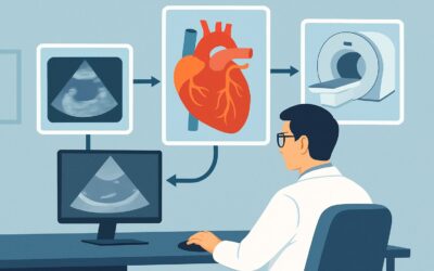
Which Imaging Is Best for Follow-Up of a Known Cardiac Mass in an Adult?

A 68-year-old patient with a history of a resected left atrial myxoma two years prior is in your clinic for annual follow-up. He is asymptomatic and feels well. The post-operative course was uneventful, and pathology confirmed a benign myxoma with clear margins. Now, the clinical question is one of surveillance: what is the most appropriate, safe, and effective imaging study to order to assess for potential recurrence? This scenario requires balancing diagnostic yield with the risks of repeated testing over a patient’s lifetime. For this specific clinical question—follow-up of a known cardiac mass with an established etiology—the American College of Radiology (ACR) Appropriateness Criteria rate US echocardiography transthoracic resting as Usually Appropriate.
Who Fits This Clinical Scenario?
This guidance applies to a specific subset of adult patients: those with a previously identified cardiac mass where the diagnosis is already established. The primary goal of imaging in this context is not initial diagnosis but surveillance.
Inclusion criteria for this workflow:
- The patient is an adult.
- A cardiac mass has been previously diagnosed.
- The etiology of the mass is known (e.g., confirmed benign tumor like a myxoma or fibroma, organized thrombus, or post-resection status).
- The purpose of the imaging is follow-up, such as monitoring for size changes, recurrence after resection, or response to therapy (e.g., anticoagulation for a thrombus).
Exclusion criteria (patients who fit a different workflow):
- Initial Evaluation of a Suspected Mass: If a patient presents with new symptoms like an embolic event, heart failure, or arrhythmia, and you suspect a cardiac mass for the first time, that constitutes an initial diagnostic workup. This workflow is covered in the ACR variant for Suspected cardiac mass. No known cardiac mass. Initial imaging.
- Characterization of an Undetermined Mass: If a mass was incidentally found on another study (like a chest CT) and its etiology is unknown, the imaging goal is tissue characterization, not surveillance. This follows the ACR variant for Known cardiac mass in echocardiography. Unknown etiology. Next imaging study.
Correctly identifying your patient’s scenario is crucial for selecting the most appropriate and resource-effective imaging pathway.
What Are You Assessing in This Follow-Up Scenario?
In surveillance imaging for a known cardiac mass, the “differential diagnosis” shifts from identifying the mass to assessing its behavior and impact over time. The key clinical questions you are trying to answer with follow-up imaging include:
Recurrence of a Resected Mass: For patients who have undergone surgical resection of a primary cardiac tumor, such as an atrial myxoma, surveillance is critical. Myxomas, while typically benign, have a known recurrence rate. Follow-up imaging is aimed at detecting any new growth at or near the resection site, ideally before it becomes large enough to cause hemodynamic compromise or embolic complications.
Change in a Conservatively Managed Mass: Not all benign cardiac masses are resected. Small, asymptomatic, and incidentally discovered masses like lipomas or fibromas may be managed with a strategy of watchful waiting. In these cases, serial imaging serves to confirm stability. Any interval growth could prompt a re-evaluation of the management plan and consideration for surgical intervention.
Response to Medical Therapy: For intracardiac thrombi, which can mimic a cardiac mass, the primary treatment is anticoagulation. Follow-up imaging is essential to monitor for resolution or reduction in the size of the thrombus, confirming the efficacy of the treatment and guiding decisions about the duration of therapy.
Development of Secondary Complications: A stable, known mass can still cause problems over time. Imaging assesses for new or worsening hemodynamic consequences, such as obstruction of a ventricular outflow tract, interference with atrioventricular valve function leading to stenosis or regurgitation, or changes in the mass’s morphology that might increase its embolic potential.
Why Is Transthoracic Echocardiography the Recommended First Step?
For routine follow-up of a known cardiac mass, the ACR designates US echocardiography transthoracic resting (TTE) as Usually Appropriate. This recommendation is based on a careful balance of diagnostic capability, safety, and practicality for serial imaging.
The primary strengths of TTE in this scenario are its safety and accessibility. It is a non-invasive procedure that uses no ionizing radiation (0 mSv), a critical consideration for any test that may be repeated annually or biannually for many years. It is widely available, relatively inexpensive, and provides excellent real-time information on cardiac structure and function. TTE can reliably measure the size of a mass, assess its mobility and point of attachment, and use Doppler to evaluate any associated hemodynamic effects on blood flow and valve function. For the core surveillance questions—is it there, has it grown, and is it causing a problem?—TTE is often sufficient.
Several other advanced imaging modalities are also rated Usually Appropriate but are typically reserved for specific situations rather than routine first-line follow-up:
- MRI heart function and morphology (with or without IV contrast): This modality offers superior soft tissue contrast and detailed tissue characterization, which is invaluable for initial diagnosis. However, for routine surveillance where the main question is a change in size, its higher cost, longer acquisition time, and limited availability make it less practical than TTE. It becomes the study of choice if TTE provides equivocal results or if there is a specific concern that requires more detailed anatomical or tissue-level assessment. Like TTE, it involves no ionizing radiation.
- CT heart function and morphology with IV contrast: While also Usually Appropriate, cardiac CT delivers a significant radiation dose (☢☢☢☢ 10-30 mSv). This cumulative exposure is a major disadvantage for a surveillance strategy that involves repeated scans. Its main advantages are excellent spatial resolution and assessment of calcification and adjacent structures, but these are not typically the primary focus of routine follow-up for a known soft-tissue mass.
In summary, TTE is the preferred initial study for surveillance due to its zero-radiation profile, cost-effectiveness, and ability to answer the most common clinical questions in this setting.
What’s Next After Echocardiography? Downstream Workflow
The results of the follow-up transthoracic echocardiogram will guide the subsequent clinical management and imaging plan. The decision tree typically branches into three main pathways.
If the study is negative for recurrence or shows a stable mass: For a patient post-resection, a negative TTE is reassuring. For a patient with a known, unresected benign mass, a study showing no interval change confirms stability. In both cases, the typical next step is to continue routine clinical and imaging surveillance. The frequency of follow-up is determined by the specific pathology of the original mass, surgical guidelines, and patient-specific factors, but may range from annually to every few years.
If the study is positive for growth or recurrence: The detection of a new or enlarging mass is a significant finding that requires further action. The next step is typically to proceed to a more advanced imaging modality for definitive characterization. Cardiac MRI without and with IV contrast is Usually Appropriate and often the best choice to delineate the mass’s tissue characteristics, precise anatomical relationships, and potential for malignancy before planning a biopsy or surgical intervention. This finding would also prompt a multidisciplinary discussion involving cardiology, cardiac surgery, and oncology.
If the study is indeterminate or technically limited: Sometimes, a TTE may be equivocal. Poor acoustic windows due to patient body habitus, lung disease, or complex post-surgical anatomy can limit visualization. In these cases, or if a suspicious but ill-defined finding is seen, the next step is to clarify the anatomy. A US echocardiography transesophageal (TEE) study, rated May be appropriate, can provide superior resolution, particularly for posterior cardiac structures like the left atrium. Alternatively, a cardiac MRI may be used to resolve the ambiguity.
Pitfalls to Avoid (and When to Get Help)
When managing follow-up for a known cardiac mass, several common pitfalls can compromise patient care.
- Over-reliance on a single modality: While TTE is the recommended first step, be aware of its limitations. If clinical suspicion for recurrence is high despite a negative or equivocal TTE, do not hesitate to proceed to a problem-solving study like TEE or cardiac MRI.
- Inadequate clinical history: The interpreting cardiologist or radiologist needs the full clinical context. Always specify the known pathology (e.g., “follow-up for resected atrial myxoma”) on the imaging requisition to focus their search and interpretation.
- Ignoring cumulative radiation dose: Avoid ordering serial cardiac CT scans for routine surveillance when a non-radiation alternative like TTE or MRI can answer the clinical question. This is especially important in younger patients with a long life expectancy.
- Losing the patient to follow-up: Establish a clear and scheduled surveillance plan with the patient. Emphasize the importance of adherence to monitoring, even when they are asymptomatic, to detect potential issues at an early and more treatable stage.
If a follow-up study shows evidence of recurrence or significant growth, immediate escalation to a cardiac surgeon and potentially a cardio-oncologist is warranted for multidisciplinary management planning.
Related ACR Topics and Tools
For a comprehensive overview of all clinical scenarios related to this topic, from initial suspicion to post-treatment evaluation, please see the parent article. Additionally, several GigHz tools can assist in applying these guidelines and communicating with patients.
- For breadth across all scenarios in Evaluation of Cardiac Masses, see our parent guide: Evaluation of Cardiac Masses: ACR Appropriateness Decoded.
- ACR Appropriateness Criteria Lookup: To explore imaging recommendations for adjacent or alternative clinical presentations.
- Imaging Protocol Library: For detailed technical specifications on how imaging studies are performed.
- Radiation Dose Calculator: To help in discussions with patients about the cumulative radiation exposure from different imaging strategies.
Frequently Asked Questions
Why is TTE preferred over TEE for routine follow-up of a known cardiac mass?
Transthoracic echocardiography (TTE) is preferred for routine surveillance because it is non-invasive, carries no sedation risk, is less expensive, and is more comfortable for the patient. Transesophageal echocardiography (TEE) is an invasive procedure that provides higher-resolution images, especially of posterior structures, but it is typically reserved for cases where TTE is technically inadequate or its findings are equivocal.
If both TTE and Cardiac MRI are ‘Usually Appropriate’, when should I choose MRI?
Choose Cardiac MRI when the results of the TTE are unclear, or when you need superior tissue characterization to assess a change in a known mass. For example, if TTE shows an increase in size, MRI can help determine if this represents simple growth, hemorrhage within the mass, or another change. For simple, routine size monitoring where TTE provides clear images, it remains the more practical first choice.
How often should follow-up imaging be performed for a resected myxoma?
The optimal frequency is not universally standardized and depends on institutional protocols and expert guidelines. However, a common approach is annual transthoracic echocardiography. The risk of recurrence is highest in the first few years post-operation, so surveillance is concentrated in that period. The schedule should be determined in consultation with the patient’s cardiologist and cardiac surgeon.
Does this follow-up guidance apply to malignant cardiac masses?
While the principles are similar, surveillance for known primary or metastatic malignant cardiac masses often involves a more aggressive and multimodal imaging strategy, frequently guided by oncology protocols. Modalities like FDG-PET/CT or FDG-PET/MRI, which assess metabolic activity, play a larger role. This article’s guidance is primarily aimed at benign masses or post-resection surveillance where recurrence is the main concern.
What if the patient has a contraindication to MRI, like an incompatible pacemaker?
If a patient requires advanced imaging beyond TTE but has a contraindication to MRI, Cardiac CT with IV contrast becomes the next best option, despite the radiation exposure. The diagnostic need to clarify an ambiguous finding on TTE would outweigh the risk of a single, indicated CT scan. The protocol should be optimized to minimize the radiation dose.
Reviewed by Pouyan Golshani, MD, Interventional Radiologist — May 30, 2026