Which Imaging Is Best for Follow-up of an Untreated Pancreatic Neuroendocrine Tumor?
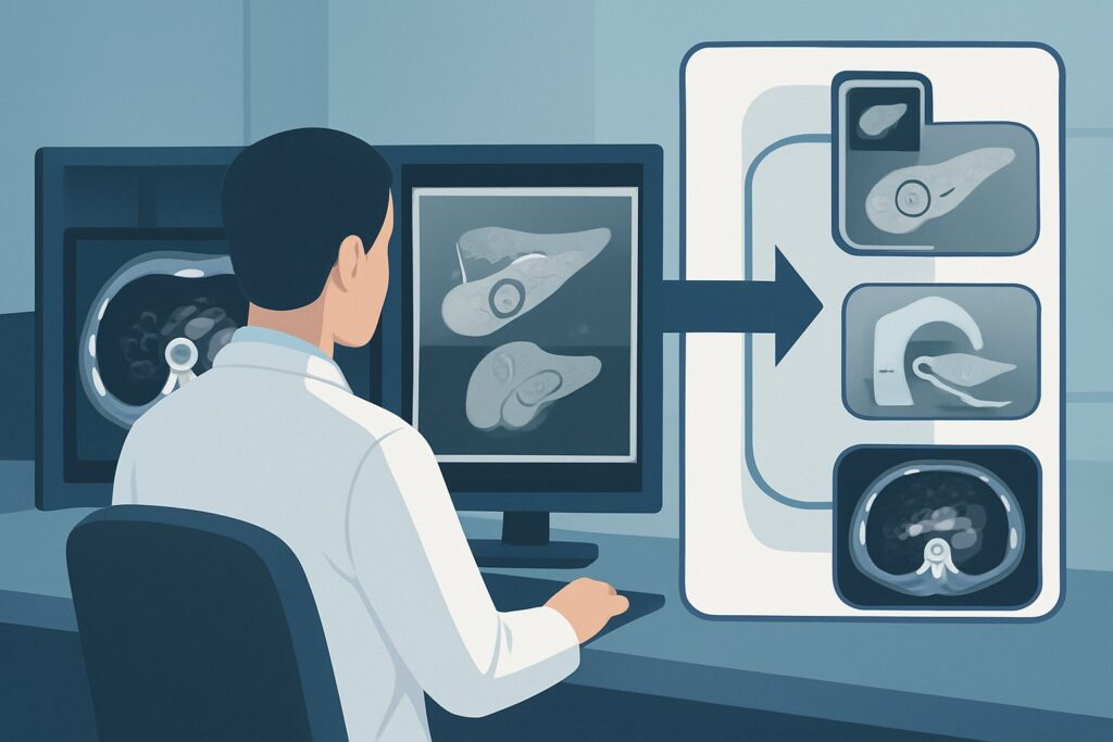
A 62-year-old patient with a known 1.5 cm, non-functional pancreatic neuroendocrine tumor (pNET) in the pancreatic tail returns for their annual follow-up. The tumor was discovered incidentally and, given its small size and low grade, has been managed with active surveillance. The patient remains asymptomatic. As the ordering physician, you must now decide on the most appropriate imaging study to assess for tumor stability, growth, or the development of metastatic disease. This decision requires balancing diagnostic accuracy with the long-term considerations of a surveillance strategy. For this specific scenario, the American College of Radiology (ACR) Appropriateness Criteria rate MRI abdomen and pelvis without and with IV contrast as Usually Appropriate.
Who Fits This Clinical Scenario for Pancreatic Neuroendocrine Tumor Follow-up?
This guidance applies specifically to adult patients with a previously diagnosed pancreatic neuroendocrine tumor who are undergoing follow-up of untreated disease. This typically involves a strategy of active surveillance or “watch-and-wait,” which is common for small (<2 cm), asymptomatic, well-differentiated, low-grade (G1/G2), and non-functional tumors. The goal of imaging in this population is to monitor the natural history of the disease over time, identifying any changes that would warrant a shift to active treatment. This workflow is distinct from several related clinical situations. This article does not apply to:
- Initial Staging: Patients who have a newly discovered lesion suspicious for a pNET require a comprehensive staging workup to evaluate for local invasion and distant metastatic disease. This is a separate diagnostic question.
- Post-Surgical Surveillance: Patients who have undergone surgical resection of their pNET are monitored for recurrence, not progression of known disease. Their imaging protocol and interpretation goals differ.
- Assessing Treatment Response: Patients actively receiving systemic therapy (like somatostatin analogs, targeted therapy, or chemotherapy) or liver-directed therapy require imaging to evaluate the effectiveness of the treatment, which follows a different set of criteria.
Correctly identifying the patient’s clinical context—surveillance of known, untreated disease—is critical to selecting the most appropriate and safest imaging modality.
What Diagnoses Are You Working Up in This Scenario?
In the context of following an untreated pNET, the imaging study is not meant to establish a new diagnosis but rather to assess the status of a known one. The key clinical questions you are trying to answer with surveillance imaging fall into a few critical categories.
The most fundamental question is tumor stability versus interval growth. Well-differentiated pNETs are often indolent, but they can grow. The rate of growth is a key factor in deciding when, or if, to intervene. Imaging provides objective, serial measurements of the primary tumor’s dimensions. A stable tumor supports continuing surveillance, whereas significant growth may prompt a discussion about treatment.
A second, equally important goal is the detection of new metastatic disease. The liver is the most common site for pNET metastases, followed by regional lymph nodes. The development of new metastases is a major prognostic indicator and a clear trigger to transition from surveillance to active therapy. The chosen imaging modality must have high sensitivity for detecting small hepatic lesions.
Finally, imaging assesses for the development of local complications. Even a slow-growing tumor can begin to impinge on or invade adjacent structures. The study must evaluate the relationship of the pNET to the main pancreatic duct, the common bile duct, and major peripancreatic vessels like the splenic vein or superior mesenteric vessels. New involvement of these structures can cause symptoms (e.g., pancreatitis, biliary obstruction) and impact the feasibility and complexity of future surgical options.
Why Is MRI of the Abdomen and Pelvis the Recommended Study for This Presentation?
The ACR designates MRI abdomen and pelvis without and with IV contrast as a Usually Appropriate study for this scenario, making it the preferred first-line choice. The rationale is rooted in its superior diagnostic capabilities and excellent safety profile for long-term surveillance.
MRI offers unparalleled soft-tissue contrast, which is ideal for characterizing the primary pancreatic lesion and, crucially, for detecting and characterizing liver metastases. Small pNET liver metastases can be difficult to distinguish from benign lesions like cysts or hemangiomas on other modalities, but the multiphasic contrast-enhanced sequences of MRI often provide definitive characterization. The hypervascular nature of most pNETs results in avid enhancement on arterial phase imaging, a feature well-depicted by MRI.
The most significant advantage of MRI in a surveillance setting is its lack of ionizing radiation (0 mSv). Patients with indolent pNETs may require imaging for many years, even decades. Opting for a radiation-free modality like MRI minimizes the cumulative radiation dose and the associated long-term risks, which is a primary consideration in any surveillance program.
Why are other studies rated lower for this specific scenario?
- CT abdomen and pelvis with IV contrast: While also rated Usually Appropriate, CT exposes the patient to ionizing radiation (ACR RRL® ☢☢☢ 1-10 mSv). Given that MRI provides equivalent or superior diagnostic information for this indication, MRI is preferred to avoid unnecessary radiation exposure, especially in younger patients or those anticipated to need many years of follow-up.
- DOTATATE PET/CT: This study is rated May be appropriate (Disagreement). While Gallium-68 DOTATATE PET/CT is extremely sensitive for well-differentiated, somatostatin receptor (SSTR)-positive neuroendocrine tumors, its role in routine surveillance of stable, untreated disease is not well established and lacks consensus. It is more commonly used for initial staging, restaging in the setting of suspected progression, or to confirm SSTR expression before considering peptide receptor radionuclide therapy (PRRT). Its routine use for simple surveillance is often considered overkill, given the cost, radiation exposure, and the high performance of MRI for this task.
When ordering the MRI, it is crucial to request a multiphasic liver and pancreas protocol that includes pre-contrast, late arterial, portal venous, and delayed imaging phases to fully characterize the enhancement patterns of the primary tumor and any potential liver lesions.
What’s Next After MRI abdomen and pelvis without and with IV contrast? Downstream Workflow
The results of the surveillance MRI will directly guide the next steps in the patient’s management plan. The workflow typically branches based on three potential outcomes.
If the study demonstrates stability:
If the MRI shows no significant change in the size of the primary tumor and no new sites of disease, the patient continues on the active surveillance pathway. This is the most common and desired outcome. The next imaging study is scheduled based on established guidelines (e.g., NCCN, ENETS), with the interval typically ranging from 6 to 24 months, depending on the tumor’s grade, size, and location.
If the study shows significant progression:
Progression is defined as a notable increase in the size of the primary tumor or, more critically, the development of new metastatic disease (e.g., new liver or nodal metastases). This finding is a trigger to reconsider the management strategy. The patient’s case should be presented at a multidisciplinary neuroendocrine tumor board to discuss a transition from surveillance to active treatment. Options may include surgery, somatostatin analogs, PRRT, or other systemic therapies, depending on the extent and location of the disease.
If the study is indeterminate or equivocal:
Occasionally, MRI may reveal a new, indeterminate finding, such as a tiny liver lesion that is too small to characterize. In this situation, a short-interval follow-up MRI in 3-6 months may be appropriate to assess for stability or growth. If there is high clinical suspicion for progression despite equivocal MRI findings, one of the alternative studies, such as a DOTATATE PET/CT, may be considered to provide functional information and increase diagnostic certainty.
Pitfalls to Avoid (and When to Get Help)
When managing surveillance for an untreated pNET, several common pitfalls can compromise patient care. First, avoid inconsistency in the imaging modality. Alternating between CT and MRI makes it difficult to compare subtle changes in tumor size and characteristics accurately. Stick with MRI unless there is a new contraindication. Second, ensure the correct MRI protocol is ordered; a standard “MRI Abdomen” may not include the specific multiphasic, thin-slice sequences needed for a pancreas protocol. Be explicit in your order. Finally, do not underestimate the importance of cumulative radiation. Reaching for CT out of convenience can lead to significant long-term radiation exposure for a patient who will be monitored for life.
If the imaging results show clear evidence of progression, such as new liver metastases or vascular encasement, it is time to escalate. This patient’s care should be transitioned to or discussed with a center specializing in neuroendocrine tumors and presented at a multidisciplinary tumor board.
Related ACR Topics and Tools
This article focuses on a single clinical variant. For a comprehensive overview of imaging for all related presentations, from initial staging to post-treatment follow-up, please consult the parent topic article. Additionally, several GigHz tools can support your clinical decision-making.
- For breadth across all scenarios in Staging and Follow-up of Pancreatic Neuroendocrine Tumors, see our parent guide: Staging and Follow-up of Pancreatic Neuroendocrine Tumors: ACR Appropriateness Decoded.
- To explore other clinical scenarios, use the ACR Appropriateness Criteria Lookup.
- For detailed imaging techniques and parameters, visit the Imaging Protocol Library.
- To discuss cumulative radiation exposure with your patients, the Radiation Dose Calculator can be a helpful aid.
Frequently Asked Questions
How often should follow-up imaging be performed for an untreated pancreatic neuroendocrine tumor?
The optimal frequency depends on tumor characteristics like size, grade (G1, G2), and location, as well as prior growth rate. Guidelines from organizations like NCCN and ENETS generally recommend intervals ranging from every 6 to 12 months for the first few years, potentially extending to every 1-2 years if the tumor remains stable over a long period.
Is an MRI with MRCP also appropriate for this scenario?
Yes, the ACR rates ‘MRI abdomen without and with IV contrast with MRCP’ as ‘Usually Appropriate.’ Adding MRCP (Magnetic Resonance Cholangiopancreatography) sequences can be particularly useful if the tumor is located in the pancreatic head and there is concern for biliary or pancreatic ductal obstruction, even if subclinical. For a tumor in the body or tail with no ductal involvement, standard multiphasic MRI is typically sufficient.
What if my patient has a contraindication to MRI, like an incompatible pacemaker?
In cases where MRI is contraindicated, ‘CT abdomen and pelvis with IV contrast’ is the best alternative and is also rated ‘Usually Appropriate’ by the ACR. It is critical to use a multiphasic pancreas protocol CT to maximize diagnostic yield, though you must accept the trade-off of ionizing radiation exposure.
My patient has an allergy to gadolinium-based contrast agents. What should I order?
For patients with a severe allergy to gadolinium, an ‘MRI abdomen and pelvis without IV contrast’ is rated ‘May be appropriate.’ While non-contrast MRI can assess tumor size, it is significantly less sensitive for detecting new liver metastases and characterizing the primary tumor’s vascularity. A contrast-enhanced CT (if there is no iodinated contrast allergy) would likely be a better diagnostic choice in this situation, despite the radiation.
When should I consider a DOTATATE PET/CT in the surveillance of an untreated pNET?
While not recommended for routine surveillance of stable disease, a DOTATATE PET/CT becomes valuable in specific situations. Consider ordering it if conventional imaging (MRI or CT) is equivocal, if you suspect disease progression but can’t confirm it anatomically, or if the patient develops symptoms or rising tumor markers that suggest new disease not visible on MRI. It is also essential for determining SSTR-avidity if peptide receptor radionuclide therapy (PRRT) is being considered as a future treatment option.
Reviewed by Pouyan Golshani, MD, Interventional Radiologist — May 30, 2026