Which Imaging Is Best for Post-TKA Pain When Infection Is Excluded?
A 68-year-old patient is in your clinic, three years out from his total knee arthroplasty (TKA), reporting a persistent, deep, activity-related pain. His initial radiographs show stable components with no obvious fracture or subsidence. You’ve completed the initial workup: his C-reactive protein (CRP) and erythrocyte sedimentation rate (ESR) are normal, and a joint aspiration was negative, effectively ruling out periprosthetic joint infection. Your leading differential includes the common mechanical causes of late TKA failure: aseptic loosening, osteolysis from polyethylene wear, or subtle instability. The radiographs are inconclusive. What is the most appropriate next imaging step to confirm your suspicion and guide management? This article provides a detailed workflow for this specific clinical scenario. According to the ACR Appropriateness Criteria, an MRI of the knee without IV contrast is considered ‘Usually Appropriate’ to investigate these concerns.
Who Fits This Clinical Scenario for Post-TKA Knee Pain?
This guidance is for a well-defined patient population. The workflow applies specifically to patients who have a total knee arthroplasty and are presenting with pain, but only after two critical prerequisites have been met:
1. Initial Radiographs Are Unrevealing: Standard weight-bearing anteroposterior (AP), lateral, and skyline knee radiographs have already been obtained and do not show an obvious cause for the pain, such as a periprosthetic fracture, gross component malposition, or significant progressive radiolucent lines.
2. Infection Has Been Excluded: A thorough clinical and laboratory evaluation has been performed to rule out periprosthetic joint infection (PJI). This typically includes normal serum inflammatory markers (ESR/CRP) and may involve a joint fluid aspiration for cell count, differential, and culture.
This pathway is not appropriate for several similar-sounding, but distinct, clinical situations:
- If you suspect infection: If the patient has systemic signs like fever, or local signs like erythema and warmth, coupled with elevated inflammatory markers, the imaging workup is different. That scenario requires modalities better suited for identifying abscesses or diffuse inflammation.
- If you suspect an acute fracture: If the pain began after a specific traumatic event, the primary concern is a periprosthetic or hardware fracture, which follows a separate diagnostic algorithm.
- If you need to measure component rotation: For concerns of tibiofemoral or patellar maltracking due to component malrotation, a specialized CT protocol is the standard of care.
What Diagnoses Are You Working Up for Non-Infectious Post-TKA Pain?
With infection and acute fracture ruled out, the differential diagnosis for chronic pain after TKA narrows to a few key mechanical and reactive processes. The advanced imaging you order is intended to differentiate among these possibilities.
Aseptic Loosening
This is the most common cause of late TKA failure. It refers to the failure of fixation at the implant-bone or cement-bone interface without an infectious cause. Over time, micromotion can lead to the development of a fibrous membrane at this interface, causing pain and progressive instability of the components. While large radiolucent lines on serial radiographs are the classic sign, cross-sectional imaging can detect earlier, more subtle signs like periprosthetic fluid and bone marrow edema that precede obvious radiographic changes.
Osteolysis (Particle Disease)
This is a biological process of bone resorption triggered by an inflammatory reaction to microscopic wear debris, most commonly from the polyethylene insert. The body’s macrophages attempt to clear these particles, releasing cytokines that activate osteoclasts and lead to bone loss. This process creates lytic lesions, often seen as “scalloping” around the implant, which can weaken the supporting bone and ultimately lead to component loosening and periprosthetic fracture.
Instability
Knee instability after TKA can manifest as flexion instability, extension instability, or mid-flexion instability. It can result from component malpositioning, ligamentous imbalance that was not corrected during the primary surgery, or progressive wear of the polyethylene bearing surface. Patients may report a sensation of the knee “giving way.” Imaging can help identify signs of component liftoff during stress, excessive fluid, or significant polyethylene wear that contributes to the mechanical imbalance.
Why Is MRI Without Contrast the Recommended Study for Suspected Aseptic Loosening?
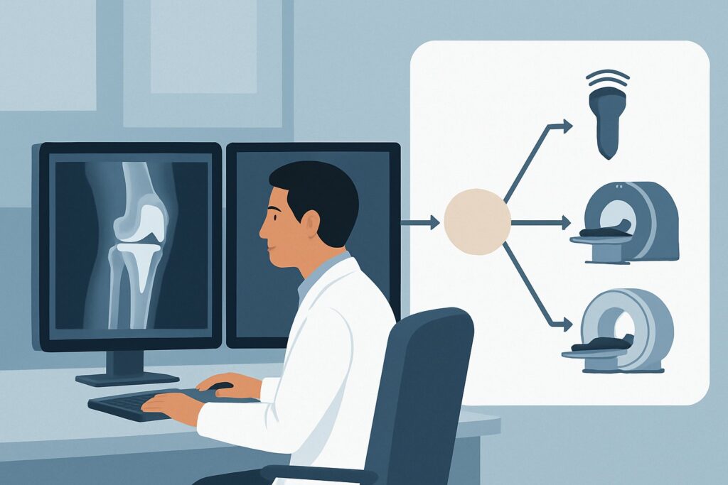
When radiographs are inconclusive but suspicion for mechanical failure remains high, the ACR recommends moving to advanced cross-sectional imaging. Both MRI and CT are highly rated, but MRI offers distinct advantages for evaluating the specific differential in this scenario.
MRI knee without IV contrast is rated ‘Usually Appropriate’ by the ACR. Its primary strength is the superior evaluation of soft tissues and the bone-implant interface. Modern MRI scanners equipped with metal artifact reduction sequences (MARS) can significantly mitigate the signal distortion caused by metallic implants. This allows for clear visualization of:
- The Bone-Implant Interface: MRI can detect subtle fluid and granulation tissue in this region, which are early indicators of aseptic loosening.
- Periprosthetic Tissues: It can identify and characterize synovitis, fluid collections, and the inflammatory response associated with particle disease (osteolysis).
- Polyethylene Components: MRI can directly visualize the polyethylene liner to assess for wear, fracture, or delamination.
- Bone Marrow: It is highly sensitive for detecting bone marrow edema patterns that may indicate stress reactions or early osteolysis.
CT knee without IV contrast is also rated ‘Usually Appropriate’. CT provides excellent spatial resolution and is superior for delineating the precise size and location of osteolytic lesions and assessing bone stock. It is less affected by metal artifact than older MRI sequences. However, it provides limited information about the soft tissues, synovium, and polyethylene components. It is an excellent alternative if MRI is contraindicated or unavailable.
Why are other studies rated lower for this specific scenario?
- 3-phase bone scan knee (‘May be appropriate’): While sensitive for detecting increased metabolic activity around an implant, its findings are nonspecific. Increased uptake can be seen with loosening, infection, or even normal postoperative remodeling, making it difficult to distinguish between the key differentials.
- MRI knee without and with IV contrast (‘Usually not appropriate’): In the absence of suspected infection or tumor, gadolinium-based contrast adds little diagnostic information for evaluating aseptic loosening or osteolysis and introduces unnecessary risk and cost. The key findings are related to fluid and tissue at the interfaces, which are well-visualized on non-contrast sequences.
When ordering the study, specifying the use of MARS protocols is crucial for obtaining diagnostic-quality images. Once you’ve decided on MRI knee without IV contrast, our protocol guide covers the technique, contrast, and reading principles: MRI Knee Without Contrast.
What’s Next After MRI knee without IV contrast? Downstream Workflow
The results of the MRI will guide the subsequent clinical and surgical decision-making. The workflow typically branches based on whether a clear mechanical cause is identified.
- If the MRI is positive for aseptic loosening or significant osteolysis: This finding often confirms the need for revision arthroplasty. The imaging helps the surgeon plan the procedure by defining the extent of bone loss, assessing the integrity of the remaining bone stock, and determining the type of revision components that may be required.
- If the MRI is negative for mechanical failure: When the MRI shows well-fixed components and no evidence of significant osteolysis or synovitis, the focus shifts to other potential pain generators. This may include evaluating for patellofemoral tracking issues, tendinopathies, bursitis, or referred pain from the hip or spine. The workup may proceed to a different clinical pathway, potentially involving diagnostic injections or physical therapy.
- If the MRI is indeterminate or shows unexpected findings: In some cases, the MRI may reveal nonspecific findings like mild synovitis or small fluid collections. If clinical suspicion for a low-grade infection resurfaces despite negative initial tests, a labeled white blood cell scan might be reconsidered. If a soft-tissue mass or other unexpected abnormality is found, further characterization, potentially with a contrast-enhanced MRI or biopsy, may be warranted.
Pitfalls to Avoid (and When to Get Help)
Navigating the workup of a painful TKA requires careful attention to detail to avoid common missteps.
- Inadequate Radiographs: Failing to obtain high-quality, weight-bearing radiographs with proper penetration can obscure subtle signs of loosening or subsidence, leading to premature ordering of advanced imaging.
- Forgetting MARS: Ordering a standard knee MRI without specifying metal artifact reduction sequences will likely result in a nondiagnostic study due to overwhelming artifact from the implant. Always communicate the presence of hardware to the radiology department.
- Over-reliance on Imaging: Imaging findings must be correlated with the patient’s history and physical exam. Mild periprosthetic fluid or tiny, stable radiolucent lines can be incidental and may not be the source of a patient’s pain.
- Ignoring the Possibility of Low-Grade Infection: While initial tests may be negative, a low-grade, indolent infection can mimic aseptic loosening. If the clinical picture is at all ambiguous, maintaining a low threshold for repeat aspiration or further infectious workup is critical.
If the diagnosis remains elusive after a comprehensive non-invasive workup, consultation with a musculoskeletal radiologist or an orthopedic surgeon specializing in revision arthroplasty is the appropriate next step.
Related ACR Topics and Tools
This article focuses on a single, common scenario. For a broader view of all clinical variants or to explore the tools used in this workflow, the following resources are available. For breadth across all scenarios in Imaging After Total Knee Arthroplasty, see our parent guide: Imaging After Total Knee Arthroplasty: ACR Appropriateness Decoded.
- ACR Appropriateness Criteria Lookup — For official guidance on thousands of clinical scenarios.
- Imaging Protocol Library — For detailed technical specifications on how imaging studies are performed.
- Radiation Dose Calculator — For discussing cumulative radiation exposure with patients.
Frequently Asked Questions
Why is MRI without contrast preferred over CT if both are ‘Usually Appropriate’?
While both are excellent, MRI without contrast provides superior soft-tissue detail. This allows for better evaluation of the synovium, polyethylene wear, and the bone-implant interface for subtle fluid or granulation tissue indicative of loosening. CT is better for quantifying bone loss (osteolysis) but provides less information on the soft-tissue reaction.
What if my patient has a contraindication to MRI, like a non-compatible implanted device?
In cases where MRI is contraindicated, CT of the knee without IV contrast is the best alternative. It is also rated ‘Usually Appropriate’ by the ACR for this scenario and provides excellent detail of the bony structures and osteolytic lesions, though with less soft tissue information.
My initial lab workup for infection was negative, but the MRI shows a large joint effusion. Should I reconsider infection?
Yes. While a sterile effusion can be seen with aseptic loosening or particle disease, a large or complex effusion on MRI should lower your threshold to reconsider a low-grade periprosthetic joint infection. Correlating with clinical signs and potentially repeating a joint aspiration for culture and cell count is a prudent next step.
Do I need to order a special MRI protocol for a patient with a TKA?
Yes, it is critical to specify that the patient has metallic hardware and to request metal artifact reduction sequences (MARS). Standard MRI protocols will be severely degraded by artifact from the implant, rendering the study nondiagnostic. Most modern imaging centers have established MARS protocols for joint arthroplasty.
The radiologist reported ‘mild synovitis’ but no clear signs of loosening. What does that mean?
Mild, nonspecific synovitis can be a reactive change to wear debris (particle disease) or simply a finding in a symptomatic joint. If there are no other signs of loosening or significant osteolysis, and the synovitis is mild, it may not be the primary pain driver. This result requires careful clinical correlation and may shift the workup toward other non-implant-related causes of knee pain.
Reviewed by Pouyan Golshani, MD, Interventional Radiologist — May 30, 2026