Which Imaging Is Best for Renal Cell Carcinoma Follow-up After Ablation?
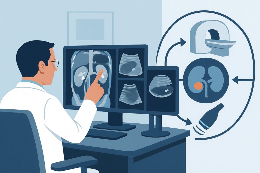
A 68-year-old male is in your urology clinic for his first follow-up visit, three months after a successful percutaneous cryoablation of a 2.5 cm renal cell carcinoma (RCC) in his right kidney. He is asymptomatic and his renal function is at baseline. You need to order the initial post-treatment imaging to confirm technical success and establish a new baseline for surveillance. The key clinical question is which imaging modality best assesses the ablation zone for any residual or recurrent enhancing tumor while minimizing long-term risks for a patient who will require years of follow-up. According to the American College of Radiology (ACR) Appropriateness Criteria, for this specific scenario, an MRI abdomen without and with IV contrast is rated Usually appropriate.
Who Fits This Clinical Scenario for Post-Ablation RCC Follow-up?
This guidance applies specifically to patients who have undergone percutaneous thermal ablation for clinically localized renal cell carcinoma. This includes common ablative techniques such as radiofrequency ablation (RFA), cryoablation, and microwave ablation (MWA). The primary goal of imaging in this population is to assess the treatment zone for completeness of ablation and to monitor for local tumor recurrence over time.
This workflow is distinct from other RCC management pathways. It is crucial to differentiate this scenario from:
- Patients post-surgical resection: Individuals who have had a partial or radical nephrectomy have different patterns of potential recurrence (e.g., at the surgical margin, in the nephrectomy bed) and follow a separate imaging surveillance protocol.
- Patients on active surveillance: Untreated patients being monitored for small renal masses require imaging to assess tumor growth rate and changes in morphology, not to evaluate a post-treatment zone.
- Patients with metastatic disease: Individuals with known metastatic RCC at diagnosis or follow-up require systemic imaging to assess treatment response in all sites of disease, a scope beyond localized post-ablation surveillance.
Applying this guidance is most appropriate for asymptomatic patients undergoing routine, scheduled surveillance after a curative-intent ablative procedure for localized disease.
What Are You Looking For in Post-Ablation RCC Imaging?
The primary purpose of follow-up imaging is to distinguish successful treatment from treatment failure or recurrence. The differential considerations for imaging findings within the ablation zone are narrow but critical.
Complete Ablation (Treatment Success): This is the desired outcome. On imaging, a successful ablation zone should appear as a well-defined, non-enhancing area at the site of the original tumor. Immediately after the procedure, there may be some surrounding inflammation or hemorrhage, but the treated tumor itself should not show enhancement. Over months and years, this zone is expected to involute and scar down, decreasing in size.
Local Tumor Recurrence or Residual Disease: This is the most important finding to detect. It is defined as the appearance of nodular or mass-like enhancement within or at the margin of the ablation zone on post-contrast imaging. This enhancement signifies viable, vascularized tumor tissue that was either incompletely treated (residual disease) or has grown back after initial treatment (recurrence). Differentiating this from benign post-procedural changes is the central challenge.
Benign Post-Ablation Changes: In the early post-procedure period (first few weeks to months), the ablation zone can be surrounded by a thin, smooth, uniform rim of enhancement representing benign inflammatory granulation tissue. This is a normal healing response and typically subsides over time. It is crucial to distinguish this benign rim enhancement from the thick, nodular, or irregular enhancement characteristic of recurrence.
Why Is MRI of the Abdomen the Recommended Study for Post-Ablation Follow-up?
The ACR rates MRI abdomen without and with IV contrast as Usually appropriate because it provides the optimal balance of diagnostic accuracy and patient safety for this specific surveillance task.
The primary advantage of MRI is its superior soft-tissue contrast resolution. This allows for a more confident assessment of enhancement patterns within and around the ablation zone. MRI is particularly adept at distinguishing non-enhancing post-treatment scar tissue from subtly enhancing foci of recurrent tumor, which can be challenging on other modalities. Furthermore, MRI achieves this without using ionizing radiation (0 mSv). This is a significant benefit for RCC patients who often require many scans over a decade or more of follow-up, minimizing cumulative radiation exposure.
While MRI is the top-rated study, other modalities are also considered:
- CT abdomen without and with IV contrast: This study is also rated Usually appropriate and is an excellent alternative, especially if MRI is contraindicated, unavailable, or if the patient has claustrophobia. CT is fast and widely accessible. However, it involves a significant dose of ionizing radiation (☢☢☢☢ 10-30 mSv) and may be slightly less sensitive than MRI for detecting very subtle enhancement in small recurrences.
- US kidneys retroperitoneal: Rated as May be appropriate, ultrasound is not typically used as the primary modality for assessing post-ablation enhancement. While contrast-enhanced ultrasound (CEUS) can be effective, standard grayscale and Doppler ultrasound are often limited by operator dependence, patient body habitus, and bowel gas. It is not considered a reliable tool for ruling out recurrence.
When ordering the recommended MRI, it is essential to specify “without and with IV contrast” and to ensure the patient’s renal function (eGFR) is adequate for the administration of a gadolinium-based contrast agent.
What Are the Next Steps After the Post-Ablation MRI?
The imaging report will guide the subsequent clinical workflow, which generally follows one of three paths.
1. Findings Consistent with Successful Ablation: If the MRI shows a non-enhancing ablation zone with no evidence of nodular enhancement, the patient can continue with their scheduled surveillance protocol. The frequency of imaging is typically highest in the first two years post-procedure (e.g., every 6 months) and then decreases over time (e.g., annually) according to institutional or societal guidelines (such as those from the AUA or NCCN).
2. Findings Suspicious for Recurrence: If the MRI demonstrates new or persistent nodular enhancement within or at the margin of the ablation zone, this is highly concerning for local tumor recurrence. The next step is typically a discussion at a multidisciplinary urologic oncology conference. Management options include a biopsy of the enhancing area to confirm recurrence, followed by consideration of repeat ablation, surgical resection (salvage partial or radical nephrectomy), or, in some cases, active surveillance of the small recurrence.
3. Indeterminate or Equivocal Findings: Sometimes, imaging findings are not definitive. For example, there may be subtle or questionable enhancement that is difficult to distinguish from benign post-procedural changes. In this situation, the most common next step is a short-interval follow-up scan, often with the same modality (MRI), in 3 to 6 months to assess for stability or progression. If the finding is still equivocal, a biopsy may be considered.
Pitfalls to Avoid (and When to Get Help)
Navigating post-ablation surveillance requires attention to detail to avoid common errors.
- Misinterpreting Benign Enhancement: Be aware that a thin, smooth, peripheral rim of enhancement can be a normal finding in the first 6-12 months post-ablation. Do not mistake this for recurrence, which is typically nodular and irregular.
- Ordering Non-Contrast Studies: Surveillance for recurrence is entirely dependent on assessing enhancement. Ordering a CT or MRI without IV contrast is insufficient and will miss the key diagnostic finding.
- Inconsistent Imaging Modality: Whenever possible, use the same imaging modality (preferably MRI) and protocol for serial follow-up exams. This allows for the most accurate comparison and assessment of change over time.
- Ignoring the Baseline Scan: The first post-ablation scan (typically at 3-6 months) is critical as it establishes the new baseline. All subsequent scans should be compared directly to this initial post-treatment study.
If imaging findings are complex or the clinical picture is unclear, escalate by presenting the case at a multidisciplinary tumor board with urologists, interventional radiologists, and medical oncologists.
Related ACR Topics and Tools
For a comprehensive overview of all clinical scenarios related to this topic, including follow-up after surgical resection and active surveillance, please refer to the parent topic hub article. Additional tools can assist in ordering the appropriate imaging and discussing it with your patients.
- For breadth across all scenarios in Post Treatment Follow-up and Active Surveillance of Renal Cell Carcinoma, see our parent guide: Post Treatment Follow-up and Active Surveillance of Renal Cell Carcinoma: ACR Appropriateness Decoded.
- To explore adjacent clinical presentations, use the ACR Appropriateness Criteria Lookup.
- For details on imaging techniques, consult the Imaging Protocol Library.
- To facilitate conversations about cumulative radiation exposure from imaging, use the Radiation Dose Calculator.
Frequently Asked Questions
How soon after renal ablation should the first follow-up scan be performed?
The first post-ablation imaging study is typically performed between 3 and 6 months after the procedure. This timing allows acute post-procedural inflammation to subside, providing a clearer baseline to assess for residual tumor and for comparison on future surveillance scans.
Is a CT scan an acceptable alternative if a patient cannot get an MRI?
Yes. A multiphasic CT of the abdomen without and with IV contrast is rated ‘Usually appropriate’ by the ACR and is a strong alternative to MRI. It is often used for patients with contraindications to MRI (e.g., certain implants, severe claustrophobia) or in centers where high-quality renal MRI is less available. The main drawback is the use of ionizing radiation.
What if the patient has chronic kidney disease (CKD) and cannot receive IV contrast?
This presents a significant clinical challenge, as assessing enhancement is key. For patients with severe CKD (e.g., eGFR < 30), the risks of both gadolinium-based and iodinated contrast must be weighed against the benefits. Options may include a non-contrast MRI or CT, or potentially a contrast-enhanced ultrasound (CEUS) if available and technically feasible. This decision should be made in consultation with a radiologist and nephrologist.
Does the type of ablation (e.g., cryoablation vs. RFA) change the imaging recommendation?
No, the primary imaging recommendation for MRI or CT with contrast remains the same regardless of the thermal ablation modality used. While the appearance of the ablation zone may differ slightly between techniques (e.g., size, initial inflammatory response), the fundamental principle of looking for nodular enhancement to detect recurrence is unchanged.
Is chest imaging necessary for routine post-ablation surveillance of localized RCC?
For small, low-risk tumors (e.g., T1a) treated with ablation, routine chest imaging is often not required in the absence of symptoms. The ACR rates chest radiography and CT as ‘May be appropriate’. The decision to include chest imaging in the surveillance protocol often depends on the initial tumor stage, grade, and other risk factors for metastatic disease.
Reviewed by Pouyan Golshani, MD, Interventional Radiologist — May 29, 2026