Which Imaging Is Best for Suspected Deep Pelvic Hernias Like Obturator or Sciatic?

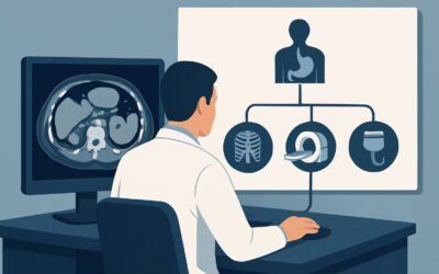
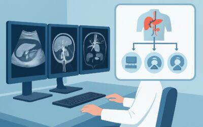
An elderly, multiparous woman presents to the emergency department with intermittent, colicky abdominal pain, nausea, and vague pain radiating down her inner thigh. A physical exam is unrevealing, with no palpable groin or abdominal wall masses. You suspect a small bowel obstruction, but the atypical thigh pain raises the possibility of a rare but consequential diagnosis: a deep pelvic hernia. This scenario, where common presentations mask an uncommon cause, requires a precise imaging strategy to avoid diagnostic delay and potential complications like bowel strangulation. This article details the American College of Radiology (ACR) workflow for this specific clinical question. For the initial imaging of a suspected obturator, sciatic, or perineal hernia, the ACR rates `MRI pelvis without and with IV contrast` as Usually appropriate.
Who Fits the Clinical Scenario for a Suspected Deep Pelvic Hernia?
This guidance applies to patients with a clinical presentation suggestive of a deep pelvic hernia, which are rare and notoriously difficult to diagnose on physical examination alone. The classic patient is an elderly, thin, multiparous female presenting with signs and symptoms of a small bowel obstruction (e.g., nausea, vomiting, abdominal distention) coupled with an atypical pain pattern. This may include pain in the medial thigh (suggesting an obturator hernia and nerve compression), the gluteal region (sciatic hernia), or the perineum.
It is critical to distinguish this scenario from more common hernia presentations that follow different diagnostic pathways:
- Exclusion 1: Palpable Groin Mass. If the patient has a reducible or non-reducible bulge in the inguinal or femoral region, the workup should follow the ACR guidance for a suspected groin hernia.
- Exclusion 2: Palpable Abdominal Wall Defect. A bulge at the umbilicus, a prior surgical incision, or elsewhere on the abdominal wall points toward the workflow for a suspected abdominal wall hernia.
- Exclusion 3: Recent Major Trauma. In the context of significant thoracic or abdominal trauma, especially with respiratory symptoms or bowel sounds in the chest, the primary concern would be a suspected diaphragmatic hernia.
This article is focused exclusively on the non-palpable, occult hernias of the pelvic floor.
What Diagnoses Are You Working Up in This Scenario?
The imaging workup for a suspected deep pelvic hernia aims to identify the specific anatomic defect and assess for complications. The differential diagnosis is narrow but includes high-morbidity conditions that require prompt identification.
Obturator Hernia
Often called the “little old lady’s hernia,” this is the most common type of deep pelvic hernia. It occurs when abdominal contents, typically a loop of small bowel, protrude through the obturator foramen. This can cause compression of the obturator nerve, leading to the classic (though not always present) Howship-Romberg sign: pain along the medial aspect of the thigh, sometimes radiating to the knee. Due to the rigid confines of the obturator canal, these hernias have a very high risk of incarceration and strangulation.
Sciatic Hernia
A much rarer entity, a sciatic hernia involves protrusion of viscera through the greater or lesser sciatic foramen. Patients may present with a tender gluteal mass, symptoms of sciatica from sciatic nerve compression, or signs of bowel obstruction. It can be challenging to differentiate from other causes of gluteal masses or radiculopathy without cross-sectional imaging.
Perineal Hernia
This type of hernia involves protrusion through a defect in the pelvic floor musculature, presenting as a bulge in the perineum, labia majora, or scrotum. Perineal hernias can be primary (congenital) or, more commonly, secondary to extensive pelvic surgery such as an abdominoperineal resection.
Beyond these specific hernias, the imaging study also serves to evaluate for other causes of pelvic pain and bowel obstruction, including adhesive disease, gynecologic or other pelvic masses, or inflammatory conditions like diverticulitis.
Why Is MRI of the Pelvis Without and With IV Contrast Usually Appropriate?
For the initial evaluation of a suspected deep pelvic hernia, the ACR designates `MRI pelvis without and with IV contrast` as a Usually appropriate study. The rationale is grounded in MRI’s superior soft-tissue contrast resolution, which is unmatched for delineating the complex anatomy of the pelvic floor muscles, fascial planes, and neurovascular structures.
MRI can precisely identify the hernia sac, its contents (bowel, omentum, or fluid), and the specific anatomic foramen through which it passes. This level of detail is crucial for both diagnosis and preoperative planning. Furthermore, the administration of intravenous contrast is key. The enhancement pattern of the bowel wall within the hernia is a critical indicator of its viability. Lack of enhancement is a specific sign of ischemia or strangulation, a finding that mandates emergent surgical intervention.
While MRI is the top-rated study, several alternatives exist with different trade-offs:
- CT abdomen and pelvis with IV contrast is also rated Usually appropriate. CT is significantly faster and more widely available than MRI, making it an excellent choice in an unstable patient or when MRI is contraindicated or unavailable. It readily diagnoses bowel obstruction and can identify the hernia. However, its soft-tissue detail of the pelvic floor is inferior to MRI, and it involves ionizing radiation (ACR Relative Radiation Level ☢☢☢ 1-10 mSv).
- US pelvis is rated Usually not appropriate. The deep location of these hernias, combined with overlying pelvic bones and bowel gas, makes them inaccessible to reliable ultrasound evaluation. Attempting to use ultrasound in this scenario is likely to produce a non-diagnostic result and delay definitive imaging.
The choice of MRI leverages its ability to provide the most detailed anatomical map of the pelvic floor without exposing the patient to ionizing radiation (ACR RRL O 0 mSv). When ordering, clearly state the clinical suspicion for an obturator, sciatic, or perineal hernia to ensure the protocol is optimized for visualizing these structures.
What Is the Downstream Workflow After a Pelvic MRI for Hernia?
The results of the pelvic MRI will directly guide the subsequent clinical management. The decision tree is typically straightforward.
If the study is positive for a deep pelvic hernia:
An urgent general surgery consultation is the immediate next step. If the MRI shows signs of bowel obstruction, and particularly if there is evidence of strangulation (e.g., lack of bowel wall enhancement, free fluid), this constitutes a surgical emergency. The detailed anatomical information from the MRI is invaluable for the surgeon in planning the operative approach, which may be transabdominal, laparoscopic, or through a direct local approach depending on the hernia type and clinical circumstances.
If the study is negative for a hernia:
The focus shifts to the other potential causes of the patient’s symptoms. If the MRI identifies an alternative diagnosis, such as an ovarian mass, abscess, or advanced diverticulitis, the workflow proceeds down the appropriate pathway for that condition (e.g., gynecologic oncology or colorectal surgery consultation). If the MRI is entirely negative and a high clinical suspicion for intermittent obstruction remains, further investigation for causes like small bowel adhesions may be warranted.
If the study is indeterminate:
In the rare case that an MRI is equivocal, perhaps due to motion artifact, a `CT abdomen and pelvis with IV contrast` can be a valuable problem-solving tool. Given that it is also a Usually appropriate study, it can provide a rapid and comprehensive alternative assessment of the abdomen and pelvis.
Pitfalls to Avoid (and When to Get Help)
Diagnosing deep pelvic hernias requires a high index of suspicion and avoidance of common cognitive errors and logistical missteps.
- Pitfall 1: Prematurely anchoring on a musculoskeletal diagnosis. Attributing medial thigh pain to osteoarthritis or gluteal pain to lumbar radiculopathy without considering a hernia, especially when any obstructive symptoms are present, can lead to significant diagnostic delay.
- Pitfall 2: Ordering an inadequate initial imaging study. Abdominal radiographs or pelvic ultrasound are rated Usually not appropriate for this indication. Ordering them first will fail to make the diagnosis and waste critical time.
- Pitfall 3: Underestimating the urgency. Given the high rate of strangulation associated with obturator hernias, the presence of obstructive symptoms in a high-risk patient profile warrants prompt cross-sectional imaging.
If a patient presents with signs of peritonitis, hemodynamic instability, or a rigid abdomen, escalate immediately for a surgical evaluation. This should occur in parallel with, not sequentially after, ordering emergent imaging.
Related ACR Topics and Tools
This article focuses on a single, specific clinical scenario. For a comprehensive overview of imaging for all hernia types and for tools to help in ordering the correct study, the following resources are available. For breadth across all scenarios in Hernia, see our parent guide: Hernia: ACR Appropriateness Decoded.
- ACR Appropriateness Criteria Lookup: For exploring related scenarios or different patient presentations.
- Imaging Protocol Library: For detailed technical specifications of recommended imaging studies.
- Radiation Dose Calculator: For discussing cumulative radiation exposure with patients when considering CT.
Frequently Asked Questions
Why is MRI preferred over CT when CT is also rated ‘Usually Appropriate’ for a suspected deep pelvic hernia?
While both are excellent and rated ‘Usually Appropriate,’ MRI is often preferred for its superior soft-tissue contrast, which provides unparalleled detail of the pelvic floor muscles, nerves, and fascial planes without using ionizing radiation. CT is a strong alternative, particularly in emergent settings where speed is paramount or if MRI is contraindicated.
Is a non-contrast MRI of the pelvis sufficient for this diagnosis?
According to the ACR, an ‘MRI pelvis without IV contrast’ is rated ‘May be appropriate.’ However, the study ‘without and with IV contrast’ is rated ‘Usually appropriate’ because the administration of a gadolinium-based contrast agent is crucial for assessing bowel wall enhancement. This helps determine if the herniated bowel is viable or if it has become ischemic or strangulated, which is a critical factor for surgical planning.
What is the Howship-Romberg sign and why is it relevant?
The Howship-Romberg sign is pain located along the inner (medial) aspect of the thigh, sometimes extending to the knee. It is caused by the compression of the obturator nerve as it passes through the obturator canal alongside an obturator hernia. While it is a classic physical exam sign for this condition, it is only present in a minority of cases. Its presence, however, should significantly increase your clinical suspicion for an obturator hernia.
Can I use ultrasound to screen for these types of hernias?
No. The ACR rates ‘US pelvis’ as ‘Usually not appropriate’ for this clinical scenario. Unlike more superficial hernias (like inguinal or umbilical), deep pelvic hernias are located behind the pelvic bones and are often obscured by bowel gas, making them inaccessible to reliable evaluation with ultrasound. Cross-sectional imaging with either MRI or CT is required for diagnosis.
What should I order if my patient has a contraindication to MRI, such as an incompatible pacemaker?
In cases where MRI is contraindicated, ‘CT abdomen and pelvis with IV contrast’ is the best alternative imaging study. It is also rated ‘Usually appropriate’ by the ACR and provides excellent diagnostic information to identify the hernia, its contents, and any signs of bowel obstruction or compromise, albeit with the use of ionizing radiation.
Reviewed by Pouyan Golshani, MD, Interventional Radiologist — May 30, 2026