Which Imaging Is Best to Assess a Known Stress Fracture for Complications or Surgical Planning?
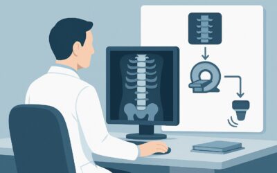
A 45-year-old competitive runner presents to your clinic for follow-up of persistent tibial pain. Initial radiographs confirmed a stress fracture of the mid-tibial diaphysis, showing a faint cortical lucency and early periosteal reaction. After a period of modified activity, she is anxious to return to training but also wants to ensure she is not at risk for a complete fracture. You need to determine the full extent of the injury to guide her return-to-play timeline and decide if surgical consultation is warranted. This article details the clinical workflow for this exact scenario: an adult with a radiographically-proven stress fracture who needs advanced imaging to assess for extent, complications, or surgical planning. For this presentation, the American College of Radiology (ACR) rates MRI of the area of interest without IV contrast as Usually Appropriate.
Who Fits This Clinical Scenario for Stress Fracture Follow-up?
This workflow is designed for a specific patient population: adults with a stress fracture that has already been identified on initial radiographs. The primary clinical question is not one of initial diagnosis, but of detailed characterization to guide subsequent management.
This guidance applies when:
- The patient is an adult.
- A stress fracture (fatigue or insufficiency) has already been diagnosed on radiographs.
- The clinical need is to determine the fracture’s extent, assess for complications like delayed healing or osteonecrosis, or acquire detailed anatomy for pre-operative planning.
- The fracture is located outside of the vertebrae (e.g., tibia, femur, metatarsals, navicular).
This guidance does NOT apply if:
- You only suspect a stress fracture, and initial radiographs are negative or indeterminate. This presentation routes to a different diagnostic algorithm.
- The patient is pregnant and has a suspected pelvic, hip, or sacral stress fracture. This is a distinct scenario with unique considerations for radiation and imaging choice.
- The primary concern is a high risk of fracture completion in a patient with a known metabolic bone disease like severe osteoporosis. While related, this has its own specific ACR variant.
- The suspected injury is a subchondral stress fracture at an extremity joint. This also follows a separate ACR workflow.
Correctly identifying your patient’s scenario is critical for selecting the most appropriate and resource-effective imaging study.
What Diagnoses Are You Working Up in This Scenario?
When a stress fracture is already known, advanced imaging is not used to find a new diagnosis but to grade the existing one and search for specific, consequential complications. The differential here is a list of potential features and associated pathologies that will directly alter the treatment plan.
Fracture Extent and Healing Status The most common goal is to precisely grade the injury. Radiographs can be subtle, but advanced imaging can differentiate a low-grade stress reaction (marrow edema only) from a high-grade injury with a distinct fracture line traversing the cortex. This information, often graded using systems like the Fredericson classification for MRI, is essential for determining weight-bearing status and predicting time to recovery. Imaging can also reveal signs of delayed union or nonunion, such as persistent edema, fracture line widening, or sclerotic fracture margins.
Osteonecrosis (Avascular Necrosis) In anatomically vulnerable locations—such as the femoral neck, talus, and tarsal navicular—a stress fracture can disrupt the tenuous blood supply, leading to osteonecrosis (ON or AVN). This is a serious complication that can lead to articular collapse and long-term disability. MRI is exceptionally sensitive for detecting the early marrow changes of ON, long before they become apparent on radiographs. Identifying this complication early is critical, as it often necessitates a shift from conservative management to surgical intervention.
Associated Soft Tissue Injury Significant stress injuries are often accompanied by damage to surrounding tissues. Advanced imaging can delineate the extent of adjacent muscle edema, hematoma, or tendinopathy. Identifying these associated injuries is important for tailoring a comprehensive rehabilitation program that addresses the entire kinetic chain, not just the bone injury.
Underlying Pathologic Lesion While less common, what appears to be a typical stress fracture on radiographs can occasionally be a pathologic fracture through a pre-existing, otherwise occult bone lesion (e.g., a non-ossifying fibroma, fibrous dysplasia, or even a malignancy). If the clinical picture or radiographic appearance is atypical, advanced imaging can provide definitive characterization of the underlying bone and exclude a more sinister cause.
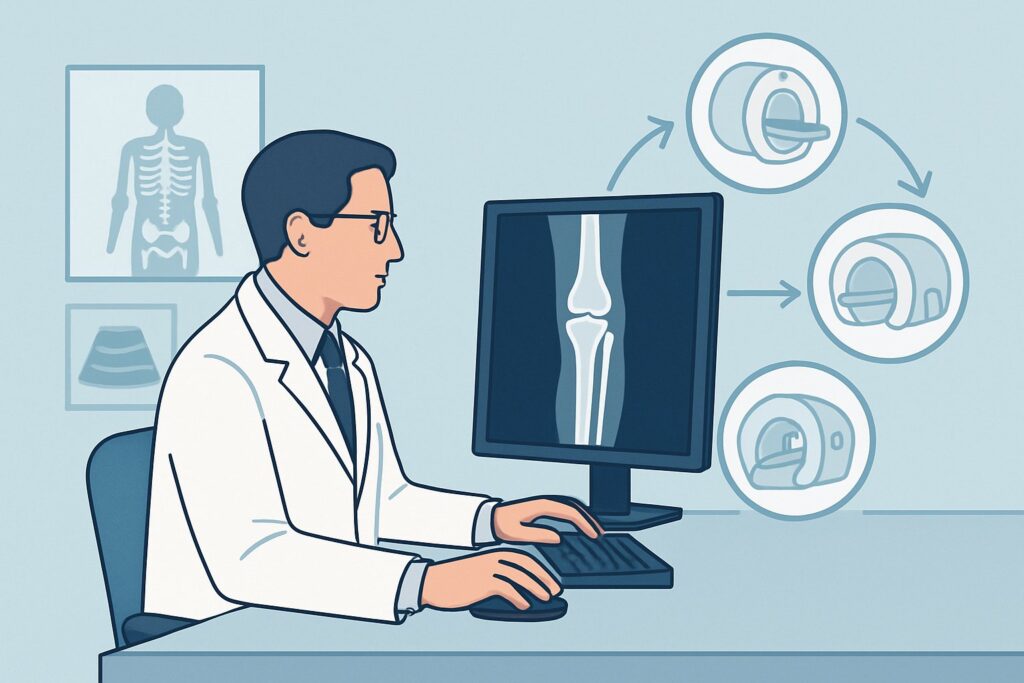
Why Is MRI Without IV Contrast the Recommended Study for This Presentation?
The ACR Appropriateness Criteria rate MRI of the area of interest without IV contrast as Usually Appropriate for this scenario, making it the primary recommendation. It provides the most comprehensive assessment of bone, marrow, and soft tissue without the use of ionizing radiation.
The rationale for this choice is rooted in MRI’s superior contrast resolution. It is highly sensitive for detecting bone marrow edema, the earliest sign of a stress response, and can clearly delineate the extent of a fracture line through both cortical and medullary bone. This capability is crucial for grading the injury and assessing for complications. For suspected osteonecrosis, MRI is the gold standard, capable of identifying early ischemic changes that are invisible on all other modalities.
Why are other studies rated lower for this specific need?
- CT area of interest without IV contrast is also rated Usually Appropriate. CT provides exquisite detail of cortical bone and is excellent for defining complex fracture patterns, especially in the foot and ankle (e.g., tarsal navicular), making it invaluable for surgical planning. However, it is less sensitive than MRI for detecting early marrow edema, soft tissue pathology, and the initial stages of osteonecrosis. It also involves ionizing radiation (RRL = Varies), a key disadvantage compared to MRI.
- Radiography area of interest repeat in 10-14 days is rated May be appropriate. While a follow-up radiograph can show signs of healing (callus) or progression (a more conspicuous fracture line), it lacks the sensitivity to answer the core questions of this scenario. It cannot reliably assess for osteonecrosis, quantify marrow edema, or evaluate surrounding soft tissues. It is a reasonable choice for low-risk fractures where the only question is interval healing, but it is insufficient for pre-operative planning or ruling out complications.
- MRI or CT with IV contrast is rated Usually not appropriate. For evaluating stress fracture extent and its primary complications, intravenous contrast adds little to no diagnostic information. The key findings—marrow edema, fracture lines, and signal changes of osteonecrosis—are readily apparent on standard non-contrast sequences. Avoiding contrast saves time, reduces cost, and eliminates the risks associated with gadolinium or iodinated contrast agents.
In summary, non-contrast MRI offers the most complete diagnostic picture with no radiation exposure (RRL = O 0 mSv), making it the preferred study for characterizing a known stress fracture.
What’s Next After MRI? Downstream Workflow
The results of the advanced imaging study will directly guide the next steps in management, creating a clear decision tree for the referring clinician.
- If the MRI shows a high-risk fracture: Findings such as a tension-side femoral neck fracture, an anterior tibial cortex fracture (“the dreaded black line”), or significant displacement of a fifth metatarsal base fracture warrant immediate orthopedic consultation. These injuries have a high rate of nonunion or completion and often require surgical fixation. The imaging will be essential for the surgeon’s pre-operative planning.
- If the MRI confirms early osteonecrosis: This finding significantly alters the prognosis and treatment plan. Management often requires strict non-weight-bearing and orthopedic consultation to discuss options ranging from core decompression to grafting or, in advanced cases, arthrodesis or arthroplasty.
- If the MRI shows a low-risk fracture with signs of healing: For a low-grade injury (e.g., significant marrow edema but no discrete cortical break) or a healing low-risk fracture, the results provide reassurance to continue with conservative management. This typically involves a period of protected weight-bearing and a gradual, criteria-based return to activity, with the MRI findings helping to establish a more accurate timeline.
- If the MRI is negative or shows an alternative diagnosis: In the rare case that the initial radiograph was misinterpreted, the MRI will reveal the true pathology (e.g., tendinopathy, tumor, infection), redirecting the entire clinical workflow toward the appropriate treatment for that condition.
Pitfalls to Avoid (and When to Get Help)
Navigating this scenario requires avoiding several common pitfalls to ensure optimal patient outcomes.
- Pitfall 1: Delaying advanced imaging for high-risk locations. For stress fractures in areas like the femoral neck, talus, or navicular, do not wait. The risk of displacement or osteonecrosis is high, and early, definitive characterization with MRI is critical.
- Pitfall 2: Ordering the wrong study for surgical planning. While MRI is the best overall study, if the primary question is surgical planning for a complex fracture pattern, a non-contrast CT may be more valuable to the surgeon for visualizing cortical detail. Often, both may be required.
- Pitfall 3: Misinterpreting “normal” follow-up radiographs. A healing stress fracture can look more prominent on follow-up radiographs due to callus formation. This should not be mistaken for worsening of the injury. Conversely, a lack of change does not rule out a problem like delayed union.
- Pitfall 4: Not providing sufficient clinical history. The radiologist’s interpretation is significantly enhanced by knowing the exact location of pain, the patient’s activity level, and the specific clinical question (e.g., “rule out AVN” or “evaluate for surgical planning”).
If MRI or CT reveals a high-risk fracture pattern, impending complication, or an unexpected underlying lesion, escalate care immediately with a referral to an orthopedic surgeon.
Related ACR Topics and Tools
For a comprehensive overview of all clinical variants related to this topic, please consult the parent guide. Additional GigHz tools can help you navigate adjacent scenarios and understand imaging techniques.
- For breadth across all scenarios in Stress (Fatigue/Insufficiency) Fracture, Including Sacrum, Excluding Other Vertebrae, see our parent guide: Stress (Fatigue/Insufficiency) Fracture, Including Sacrum, Excluding Other Vertebrae: ACR Appropriateness Decoded.
- ACR Appropriateness Criteria Lookup — for adjacent scenarios
- Imaging Protocol Library — for technique on the recommended study
- Radiation Dose Calculator — for cumulative dose conversations
Frequently Asked Questions
If both MRI and CT are ‘Usually Appropriate’, how do I choose between them?
Choose based on your primary clinical question. If you are most concerned about early complications like osteonecrosis, bone marrow edema, or associated soft tissue injury, MRI is superior. If the primary goal is to define the cortical bone and fracture lines for surgical planning, especially in complex areas like the foot, a non-contrast CT is an excellent choice. In some high-stakes cases, both may be complementary.
Why is IV contrast ‘Usually Not Appropriate’ for this scenario?
Intravenous contrast (either gadolinium for MRI or iodine for CT) typically does not add diagnostic value for assessing a stress fracture’s extent or its most common complications like osteonecrosis. The key findings are visible on non-contrast sequences, and adding contrast increases cost, scan time, and introduces small but real risks to the patient.
My patient has a known tibial stress fracture on x-ray. Can I just follow them with repeat x-rays instead of getting an MRI?
According to the ACR, repeat radiography is ‘May be appropriate’. This is a reasonable option for a low-risk stress fracture in a patient who is improving as expected. However, if you need to make a high-stakes decision about return to sport, are concerned about a complication like nonunion, or are considering surgery, a repeat x-ray is insufficient. MRI or CT is needed to get the detailed information required.
Does this guidance apply to stress fractures of the pars interarticularis (spondylolysis)?
No. This ACR topic, ‘Stress (Fatigue/Insufficiency) Fracture, Including Sacrum, Excluding Other Vertebrae,’ explicitly excludes vertebral fractures. Spondylolysis and other vertebral stress injuries are covered under different ACR Appropriateness Criteria topics related to back pain.
What if the MRI shows extensive marrow edema but no clear fracture line?
This finding represents a lower-grade stress injury (e.g., Fredericson grade 1-3). While not a complete fracture, it is still a significant bone stress response that requires treatment, typically with activity modification and protected weight-bearing. The MRI has successfully characterized the injury’s severity, allowing you to tailor a more precise and effective management plan than you could with radiographs alone.
Reviewed by Pouyan Golshani, MD, Interventional Radiologist — May 30, 2026