Which Imaging Study Best Detects HCC Recurrence After Treatment? An ACR-Guided Workflow

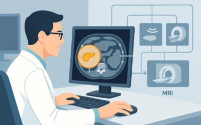
A 64-year-old man with a history of cirrhosis from nonalcoholic steatohepatitis (NASH) is in your clinic for follow-up. Six months ago, he underwent successful transarterial chemoembolization (TACE) for a 4 cm hepatocellular carcinoma (HCC) in segment 7. His alpha-fetoprotein (AFP) level has normalized, and he feels well. Now, it’s time for his first post-treatment surveillance scan to assess for residual disease and detect any new lesions. You face a critical decision: which imaging study offers the highest diagnostic confidence in this complex clinical setting? This article details the American College of Radiology (ACR) Appropriateness Criteria for post-treatment HCC monitoring, explaining the optimal imaging workflow. For this specific scenario, MRI abdomen without and with hepatobiliary contrast is rated Usually Appropriate.
Who Fits This Clinical Scenario for HCC Post-Treatment Monitoring?
This guidance applies to a specific patient population: individuals with a known history of chronic liver disease who have already received treatment for a confirmed Hepatocellular Carcinoma (HCC). The primary goal of imaging in this context is routine monitoring and surveillance for tumor recurrence or the development of new primary lesions.
Inclusion Criteria:
- Established diagnosis of chronic liver disease (e.g., cirrhosis from viral hepatitis, alcohol, or NASH).
- Confirmed prior diagnosis of HCC.
- History of completed locoregional therapy (such as ablation, TACE, or stereotactic body radiation therapy) or systemic therapy.
- The patient is undergoing scheduled surveillance, not being evaluated for an acute clinical change.
Exclusion Criteria (These Route to Different Workflows):
- Initial HCC Screening: Patients with chronic liver disease but no prior diagnosis of HCC who require screening or surveillance. This initial detection phase has a different set of imaging recommendations, often starting with ultrasound.
- Staging of Liver Fibrosis: Patients needing an initial diagnosis or staging of their underlying liver fibrosis. This workup focuses on elastography techniques, which are not the primary tools for post-treatment monitoring.
- Acute Clinical Decompensation: Patients presenting with new, acute symptoms like jaundice, worsening ascites, or hepatic encephalopathy. While imaging is required, the protocol and urgency may differ from routine surveillance.
What Are You Looking for With Post-Treatment HCC Imaging?
Post-treatment imaging is not simply a search for a new mass; it is a nuanced assessment of the treated area and the entire liver. The radiologist is evaluating for several key findings that will directly influence the patient’s prognosis and next therapeutic steps.
Viable Residual or Recurrent Tumor The most critical objective is to determine if the recent treatment was successful. This involves meticulously examining the treatment bed for any signs of viable tumor, which typically appears as areas of arterial phase hyperenhancement (APHE) followed by washout in later phases. Differentiating this from expected post-procedural inflammation or granulation tissue is the central challenge and a primary reason high-resolution, multiphasic imaging is essential.
New Hepatocellular Carcinoma Lesions The underlying cirrhotic liver remains a fertile ground for carcinogenesis. Patients who have had one HCC are at a very high risk of developing new, separate tumors. Surveillance imaging must therefore scrutinize the entire liver parenchyma, not just the previously treated site, to detect these de novo lesions when they are small and amenable to curative-intent therapy.
Post-Treatment Changes vs. True Recurrence Therapies like thermal ablation and TACE create complex imaging appearances, including coagulation necrosis, inflammation, and perfusion alterations. These benign changes can sometimes mimic or obscure small areas of recurrent tumor. The chosen imaging modality must have sufficient contrast and spatial resolution to distinguish between these possibilities, a task for which certain techniques are far superior.
Macrovascular Invasion A finding that dramatically worsens prognosis is the development of tumor thrombus within the portal or hepatic veins. Post-treatment surveillance includes a careful assessment of the major hepatic vasculature for any evidence of new or progressive tumor invasion, as this finding often shifts management toward systemic therapies.
Why Is MRI with Hepatobiliary Contrast the Top Choice for HCC Monitoring?
The ACR designates MRI abdomen without and with hepatobiliary contrast as Usually Appropriate for post-treatment HCC monitoring. This recommendation is based on the modality’s superior diagnostic capabilities in this specific and challenging clinical context.
The primary advantage of MRI is its exceptional soft-tissue contrast resolution, which allows for detailed characterization of the liver parenchyma and the complex environment of a post-treatment bed. This is crucial for distinguishing subtle residual tumor from benign post-ablation changes.
The use of a hepatobiliary contrast agent (e.g., gadoxetate disodium) provides a significant diagnostic benefit. These agents are actively taken up by functional hepatocytes. During the delayed hepatobiliary phase (typically 10-20 minutes post-injection), the normal liver parenchyma enhances and becomes bright. Most HCCs lack the necessary organic anion transporting polypeptides (OATPs) to take up this contrast, so they appear as dark (hypointense) defects against a bright background. This unique contrast mechanism substantially increases the conspicuity of small or subtle tumors that might be missed on conventional dynamic imaging alone.
A critical consideration for this patient population is the need for repeated imaging over many years. MRI carries a radiation level of O 0 mSv, completely avoiding the cumulative risk of ionizing radiation. This stands in stark contrast to CT.
Comparing Alternatives:
- CT abdomen with IV contrast multiphase: This study is also rated Usually Appropriate and is an excellent alternative when MRI is contraindicated (e.g., incompatible metallic implants, severe claustrophobia) or unavailable. It provides robust assessment of arterial enhancement and washout. However, its primary drawback is the significant radiation dose (☢☢☢☢ 10-30 mSv) with each scan, a major concern in long-term surveillance.
- US abdomen: Rated as May be appropriate, standard ultrasound has limited sensitivity for detecting recurrence within a scarred, heterogeneous, post-treatment liver. Gas from a recent ablation can also cause acoustic shadowing that obscures the treatment zone. While contrast-enhanced ultrasound (CEUS) improves performance, it remains highly operator-dependent and provides a limited field of view compared to cross-sectional imaging.
What Is the Downstream Workflow After a Post-Treatment HCC Scan?
Radiology reports for this scenario are typically structured using the Liver Imaging Reporting and Data System (LI-RADS), which standardizes the assessment of treatment response. The downstream clinical pathway is guided directly by these LI-RADS categories.
- If the report indicates LR-TR Viable: This signifies definitively viable tumor within or adjacent to the treatment zone. The patient should be promptly referred back to a multidisciplinary tumor board (including hepatology, interventional radiology, surgery, and oncology) to determine the best course of action. Options may include repeat locoregional therapy, initiating systemic therapy, or re-evaluating for liver transplantation.
- If the report indicates LR-TR Non-viable: This is the desired outcome, suggesting a complete radiological response with no evidence of viable tumor. If no new suspicious lesions are found elsewhere in the liver, the patient continues on their established surveillance schedule, typically with repeat imaging in 3 to 6 months.
- If the report indicates LR-TR Equivocal: This category is used when findings are suspicious but not definitive for viable tumor. The standard recommendation is a short-interval follow-up scan, often in 3 months or less, to assess for stability or progression. This “watchful waiting” approach helps clarify the nature of the finding without committing the patient to potentially unnecessary invasive procedures.
Pitfalls to Avoid (and When to Get Help)
Navigating post-treatment HCC surveillance requires careful attention to imaging choice and interpretation. Avoiding common pitfalls can prevent diagnostic delays and ensure optimal patient management.
- Pitfall 1: Relying on Ultrasound Alone. While ultrasound is a primary screening tool for untreated patients, its sensitivity is significantly reduced in the post-treatment setting due to altered liver architecture and artifacts. Do not substitute it for scheduled cross-sectional imaging.
- Pitfall 2: Forgetting the Hepatobiliary Phase. When ordering an MRI, specifically requesting a hepatobiliary agent and ensuring delayed hepatobiliary phase images are acquired is critical for maximizing lesion detection.
- Pitfall 3: Ignoring Cumulative Radiation. For patients who cannot undergo MRI, be mindful of the cumulative radiation dose from serial multiphase CT scans. Regularly reassess MRI contraindications and use the lowest possible CT dose protocols.
- Escalation: If a new lesion is identified that has an atypical appearance or if there is discordance between imaging findings and tumor markers (e.g., a rising AFP with a “negative” scan), escalate the case for multidisciplinary tumor board review.
Related ACR Topics and Tools
For a comprehensive understanding of imaging in chronic liver disease and access to relevant clinical tools, the following resources are available:
- For breadth across all scenarios in Chronic Liver Disease, see our parent guide: Chronic Liver Disease: ACR Appropriateness Decoded.
- Imaging Appropriateness Selector: For exploring adjacent clinical scenarios and their corresponding imaging recommendations.
- Imaging Protocol Library: For detailed technical specifications on how to perform the recommended studies.
- Radiation Dose Calculator: For estimating cumulative radiation exposure and facilitating conversations with patients undergoing serial CT scans.
Frequently Asked Questions
How often should post-treatment monitoring for HCC be performed?
The standard interval for post-treatment surveillance imaging is typically every 3 to 6 months for the first two years, after which the interval may be extended if the patient remains disease-free. The exact frequency is determined by the multidisciplinary tumor board based on the initial tumor characteristics and risk of recurrence.
Is a standard gadolinium-based contrast agent sufficient for post-treatment MRI?
While an MRI with a standard extracellular gadolinium-based contrast agent is also rated ‘Usually Appropriate’ by the ACR, a hepatobiliary agent (like gadoxetate disodium) is often preferred. The delayed hepatobiliary phase imaging provides an additional layer of diagnostic information that increases the conspicuity of HCC, making it particularly valuable for detecting small or subtle recurrent/new lesions.
What if my patient has a contraindication to both MRI and iodinated CT contrast?
This is a challenging situation. One option is a non-contrast MRI, which is rated ‘May be appropriate’ but is significantly less sensitive than a contrast-enhanced study. Another option is contrast-enhanced ultrasound (CEUS), also rated ‘May be appropriate’, which can be effective in experienced hands but has limitations. This scenario requires discussion with a radiologist and the multidisciplinary team to weigh the risks and benefits of each available option.
Does PET/CT have a role in routine post-treatment monitoring for HCC?
For routine surveillance, FDG-PET/CT is rated ‘Usually not appropriate’ by the ACR. While some poorly differentiated HCCs can be FDG-avid, many are not, leading to low sensitivity. Its primary role is reserved for specific situations, such as staging extrahepatic disease in patients being considered for certain systemic therapies or transplantation, rather than for routine monitoring of the liver.
If a new lesion is found, is a biopsy always necessary?
Not always. The LI-RADS system is designed to provide a high degree of diagnostic certainty based on imaging features alone. A new lesion categorized as LR-5 (definitely HCC) on multiphasic CT or MRI typically does not require biopsy for confirmation, and the patient can proceed directly to treatment planning. Biopsy is usually reserved for lesions with atypical or indeterminate (LR-3 or LR-4) features.
Reviewed by Pouyan Golshani, MD, Interventional Radiologist — May 26, 2026