Which Imaging Study Best Plans Surgery for Cholesteatoma with Suspected Extension?
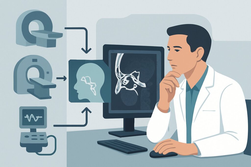
A 45-year-old patient presents with progressive, unilateral conductive hearing loss. On otoscopy, you identify a pearly white mass behind an intact tympanic membrane, classic for a cholesteatoma. More concerning, the patient reports recent-onset facial weakness on the same side and episodes of vertigo. You suspect the lesion has extended beyond the middle ear, and you need a detailed map for surgical planning. This clinical crossroads—evaluating a known middle ear mass for deeper invasion—is where precise imaging becomes critical. This article details the ACR-guided workflow for this exact scenario, explaining why specific studies are chosen to ensure a safe and effective surgical approach. For this presentation, the American College of Radiology (ACR) rates MRI head and internal auditory canal without and with IV contrast as Usually Appropriate.
Who Fits This Clinical Scenario?
This imaging workflow is designed for a specific patient population: those with an acquired conductive hearing loss where a middle ear mass, most often a cholesteatoma or neoplasm, has already been identified or is strongly suspected based on clinical examination. The critical factor that triggers this specific pathway is the additional suspicion of intracranial or inner ear extension.
Inclusion criteria for this workflow:
- Acquired Conductive Hearing Loss: The hearing deficit is not congenital.
- Known or Suspected Middle Ear Mass: Otoscopy or prior imaging suggests a cholesteatoma, paraganglioma, or other neoplasm.
- Suspicion of Extension: The patient exhibits signs or symptoms suggesting the lesion has breached the confines of the middle ear or mastoid. This includes facial nerve palsy, vertigo, sensorineural hearing loss component, or signs of intracranial involvement like headache or seizure.
- Goal is Surgical Planning: The primary purpose of the imaging is to define the full extent of the disease to plan a definitive surgical intervention.
This guidance does not apply if:
- The patient has conductive hearing loss without a clinically evident mass (see Acquired conductive hearing loss in absence of clinically evident mass).
- The hearing loss is purely sensorineural without a middle ear mass (see Acquired sensorineural hearing loss).
- The primary symptom is episodic vertigo without focal neurologic deficits, suggesting a peripheral cause (see Episodic vertigo with or without associated hearing loss).
Correctly identifying your patient’s scenario is the first step to ordering the most effective and appropriate imaging study.
What Diagnoses Are You Working Up in This Scenario?
When a middle ear mass shows signs of extension, the differential diagnosis narrows to aggressive or invasive pathologies. The imaging choice is tailored to differentiate these possibilities and, most importantly, to define their anatomic reach for the surgeon.
Cholesteatoma with Complications This is the most common diagnosis in this scenario. A cholesteatoma is not a tumor but an expanding sac of keratinizing squamous epithelium. Its danger lies in its ability to produce enzymes that erode bone. When it extends, it can destroy the ossicles, erode into the inner ear causing a labyrinthine fistula (leading to vertigo and sensorineural hearing loss), dehisce the facial nerve canal (causing palsy), or breach the tegmen tympani to extend intracranially, potentially causing meningitis, epidural abscess, or sigmoid sinus thrombosis. Imaging is crucial to identify these life-threatening complications before surgery.
Paraganglioma (Glomus Tumor) These are highly vascular neuroendocrine tumors that can arise in the middle ear (glomus tympanicum) or from the jugular bulb below (glomus jugulare). They often present with pulsatile tinnitus and conductive hearing loss. Their propensity for extensive local invasion and high vascularity makes preoperative characterization essential. Imaging must define their size, extension, and relationship to the carotid artery and jugular vein, which is vital for surgical planning and considering preoperative embolization.
Facial Nerve Schwannoma While less common, a schwannoma arising from the facial nerve can present as a middle ear mass. It can cause conductive hearing loss by encasing or displacing the ossicles. As it grows along the nerve, it can extend from the internal auditory canal through the temporal bone and into the parotid gland. Delineating its full course is paramount for planning a nerve-sparing surgery, if possible.
Malignant Neoplasms Rarely, a middle ear mass can be a primary malignancy like squamous cell carcinoma or adenocarcinoma. These are aggressive and require wide surgical margins. Imaging is used for staging, assessing for bone destruction, perineural invasion, and dural involvement, which radically alters the surgical plan and necessitates an oncology consultation.
Why Is MRI the Recommended Study for Suspected Extension?
While both MRI and CT play roles in evaluating the temporal bone, the specific question of soft tissue extension into the intracranial space or inner ear is best answered by magnetic resonance imaging. The ACR designates MRI head and internal auditory canal without and with IV contrast as Usually Appropriate for its superior ability to characterize soft tissues and identify enhancement patterns indicative of inflammation, tumor, or perineural spread.
The key advantage of MRI is its unparalleled contrast resolution. It can directly visualize:
- Intracranial Extension: MRI clearly shows dural enhancement, brain parenchymal involvement, or the formation of an epidural or brain abscess if the cholesteatoma has eroded through the tegmen tympani.
- Nerve Involvement: Post-contrast images are highly sensitive for abnormal enhancement of the facial or vestibulocochlear nerves, indicating perineural tumor spread or inflammation.
- Inner Ear Invasion: MRI can detect subtle fluid signal changes and enhancement within the cochlea and semicircular canals, confirming a labyrinthine fistula or tumor invasion.
- Differentiating Cholesteatoma from Inflammation: Specific sequences, like diffusion-weighted imaging (DWI), are highly sensitive and specific for cholesteatoma, which typically shows restricted diffusion, helping to distinguish it from surrounding inflammatory tissue or fluid.
The Complementary Role of CT
It is critical to note that CT temporal bone without IV contrast is also rated Usually Appropriate. The two tests answer different questions and are often used together for comprehensive surgical planning. While MRI excels at soft tissue, high-resolution CT is superior for evaluating fine bony anatomy. CT precisely delineates:
- Ossicular chain erosion or fixation.
- Bony defects in the facial nerve canal.
- Erosion of the scutum, a key sign of cholesteatoma.
- Dehiscence of the tegmen tympani or sigmoid plate.
- Integrity of the bony labyrinth.
For this scenario, MRI is the primary tool to assess for the suspected extension, but a non-contrast CT is almost always required to provide the detailed bony roadmap the surgeon needs.
Why Other Studies Are Rated Lower
- CT temporal bone with IV contrast (May be appropriate): For most cases of cholesteatoma, IV contrast adds little information to a non-contrast CT for bony assessment and provides significantly less soft tissue detail than an MRI. Its main role is in patients who cannot undergo MRI where a vascular lesion like a paraganglioma is suspected.
- CT head without or with IV contrast (Usually not appropriate): A standard head CT protocol lacks the thin-slice, high-resolution bone algorithm reconstructions necessary to evaluate the intricate structures of the temporal bone. It is the wrong test for this indication and will miss critical findings.
From a safety perspective, MRI is the preferred modality as it involves no ionizing radiation (0 mSv), compared to the moderate dose (☢☢☢ 1-10 mSv) from a temporal bone CT.
What’s Next After MRI? Downstream Workflow
The imaging results directly dictate the next steps in management, transforming the surgical plan from a standard procedure to a complex, multidisciplinary intervention.
- If the MRI confirms cholesteatoma with intracranial extension: The case immediately requires a neurosurgical consultation. The surgical approach will be a combined effort between otolaryngology and neurosurgery to address both the mastoid disease and the intracranial component (e.g., craniotomy for dural repair or abscess drainage) in a single or staged procedure.
- If the MRI shows a highly vascular neoplasm (e.g., paraganglioma): The workflow shifts. An interventional radiology consult for preoperative embolization is often the next step to reduce intraoperative bleeding. The surgical approach is fundamentally different from that for a cholesteatoma.
- If the MRI is negative for extension and confirms disease is limited to the middle ear/mastoid: This is a reassuring result. The surgeon can proceed with a more standard surgical approach (e.g., tympanomastoidectomy) with higher confidence. The preoperative high-resolution CT will remain the primary guide for navigating the bony anatomy during the procedure.
- If the findings are indeterminate: In rare cases, distinguishing between a sterile effusion, inflammation, and a small, non-enhancing lesion can be challenging. This may prompt a discussion between the radiologist and surgeon about the need for follow-up imaging or proceeding with surgery with the understanding that the full pathology will be determined intraoperatively.
Pitfalls to Avoid (and When to Get Help)
Navigating this clinical scenario requires avoiding several common missteps that can compromise surgical planning and patient safety.
- Ordering a standard “CT Head”: This is a frequent error. A routine head CT lacks the resolution to visualize the ossicles, facial nerve canal, or subtle bony erosion and is considered Usually not appropriate. Always specify “CT Temporal Bone” with thin cuts.
- Forgetting the non-contrast CT: While MRI is key for suspected extension, relying on it alone leaves the surgeon without a detailed bony map. In most surgical planning cases, both studies are complementary and necessary.
- Misinterpreting post-operative changes: In a patient with prior surgery, differentiating recurrent cholesteatoma from granulation tissue or scar on imaging can be difficult. This requires close collaboration with an experienced neuroradiologist.
- Ignoring facial nerve symptoms: New-onset facial weakness in the setting of a middle ear mass is a red flag for nerve involvement and demands urgent, high-quality imaging with MRI to assess the entire course of the nerve.
If you see evidence of intracranial extension, dural enhancement, or a large vascular mass, immediate escalation to neurosurgery and/or interventional neuroradiology is warranted before any otologic procedure is planned.
Related ACR Topics and Tools
For a comprehensive understanding of imaging for hearing loss and vertigo, and to access tools that streamline the ordering process, please refer to the following resources.
- For breadth across all scenarios in Hearing Loss and/or Vertigo, see our parent guide: Hearing Loss and/or Vertigo: ACR Appropriateness Decoded.
- Imaging Appropriateness Selector: For direct access to ratings for thousands of clinical variants.
- Imaging Protocol Library: To review detailed imaging techniques for recommended studies.
- Radiation Dose Calculator: To help facilitate conversations with patients about cumulative radiation exposure from medical imaging.
Frequently Asked Questions
Why are both MRI and CT rated ‘Usually Appropriate’ for this scenario?
They answer different but equally important questions for surgical planning. MRI is superior for evaluating soft tissue, which is essential for confirming suspected intracranial or inner ear extension, nerve involvement, and differentiating tumor from inflammation. High-resolution non-contrast CT is superior for delineating fine bony anatomy, such as ossicular erosion and the integrity of the facial nerve canal. Most surgeons will require both studies for a complete preoperative assessment.
If I can only order one study initially, which one should it be?
If the primary clinical question is the presence and extent of suspected intracranial or inner ear extension, the MRI without and with contrast is the more critical initial study. It directly addresses the most dangerous potential complications. The CT can be obtained subsequently for bony detail once the soft tissue extent is known.
Does the MRI need contrast if we are just looking for a cholesteatoma?
Yes. While non-contrast sequences like DWI are excellent for identifying the cholesteatoma itself, IV contrast is crucial for assessing complications and evaluating the differential. Contrast enhancement highlights dural inflammation, perineural spread of a tumor, abscess walls, and the vascularity of neoplasms like paragangliomas, all of which are critical for surgical planning in this scenario.
What if the patient has a contraindication to MRI, like a non-compatible pacemaker?
In cases where MRI is contraindicated, a CT temporal bone with and without IV contrast becomes the primary imaging modality. While less sensitive for subtle dural or nerve involvement, it can still identify gross intracranial extension, significant bone erosion, and will highlight highly vascular lesions. A CTA may also be considered to evaluate the relationship of a suspected paraganglioma to major vessels.
How does imaging change if this is for a post-operative patient with suspected recurrence?
In the post-operative setting, MRI with contrast, including a DWI sequence, is even more critical. Distinguishing recurrent cholesteatoma from post-surgical granulation tissue or scar can be very difficult on CT. The restricted diffusion seen on DWI is highly specific for recurrent cholesteatoma, making it the modality of choice for this question.
Reviewed by Pouyan Golshani, MD, Interventional Radiologist — May 26, 2026