Which Imaging Study Is Best for Brain Tumor Screening in Adults with Genetic Risk?
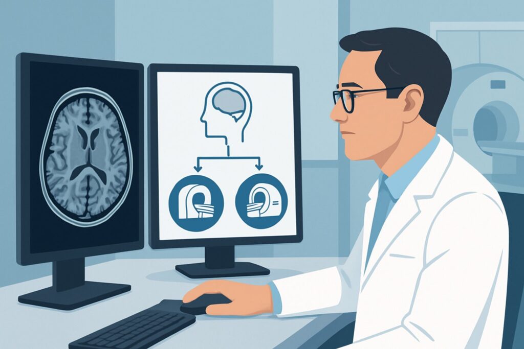
An asymptomatic 32-year-old patient with a confirmed diagnosis of neurofibromatosis type 2 (NF2) presents for their annual neurologic check-up. Given their high lifetime risk for developing vestibular schwannomas and meningiomas, the discussion turns to surveillance imaging. The central question for the ordering clinician is not if to screen, but how to screen effectively and safely over many years. This article provides a detailed clinical workflow for this specific scenario—primary brain tumor screening in an adult with known genetic risk factors. Based on the American College of Radiology (ACR) Appropriateness Criteria, an MRI head without and with IV contrast is rated Usually Appropriate for this indication.
Who Fits This Clinical Scenario for Brain Tumor Screening?
This guidance applies specifically to asymptomatic adults with a known genetic syndrome that predisposes them to the development of primary central nervous system (CNS) tumors. The goal is surveillance—to detect tumors at an early, often more treatable, stage.
Inclusion criteria for this workflow:
- Patient: Adult (18 years or older).
- Presentation: Asymptomatic, undergoing screening.
- Risk Factor: Confirmed diagnosis of a phakomatosis or other hereditary cancer syndrome with a known increased risk of primary brain tumors. Examples include:
- Neurofibromatosis type 1 (NF1)
- Neurofibromatosis type 2 (NF2)
- Tuberous Sclerosis Complex (TSC)
- Von Hippel-Lindau (VHL) disease
- Li-Fraumeni syndrome
- Gorlin syndrome (Nevoid Basal Cell Carcinoma Syndrome)
This workflow does NOT apply to patients who:
- Have new neurologic symptoms: A patient presenting with new-onset seizures, focal weakness, or persistent headaches requires a diagnostic workup, not a screening protocol.
- Have a known extracranial malignancy: An individual with lung or breast cancer being evaluated for brain metastases falls under the “Secondary or metastatic brain tumor screening” scenario.
- Have a previously identified brain lesion: If a lesion was found on prior imaging, the appropriate workflow is “Pretreatment evaluation” or “Posttreatment surveillance,” which involve different imaging considerations.
What Diagnoses Are You Screening For in Patients with Genetic Risk?
Unlike a diagnostic workup for a symptomatic patient, screening in this context is a targeted search for specific tumors known to be associated with the patient’s underlying genetic condition. The imaging study must be optimized to detect these “target lesions” with high sensitivity.
In a patient with Neurofibromatosis type 2 (NF2), the primary targets are bilateral vestibular schwannomas. Screening also aims to detect other potential tumors like multiple meningiomas, spinal schwannomas, and ependymomas. Early detection of vestibular schwannomas is critical to preserving hearing.
For individuals with Neurofibromatosis type 1 (NF1), the most common CNS tumor is the optic pathway glioma. While often found in childhood, they can be monitored into adulthood. Other potential findings include astrocytomas and the characteristic, non-neoplastic “unidentified bright objects” (UBOs) on T2-weighted MRI.
In Von Hippel-Lindau (VHL) disease, the key target is the hemangioblastoma, a highly vascular tumor that can occur in the cerebellum, brainstem, or spinal cord. Screening is essential as these lesions can cause significant morbidity from hemorrhage or mass effect.
For Tuberous Sclerosis Complex (TSC), screening focuses on identifying subependymal giant cell astrocytomas (SEGAs). These tumors typically arise near the foramen of Monro and can cause obstructive hydrocephalus, a neurologic emergency.
Why Is MRI Head Without and With IV Contrast the Recommended Screening Study?
The ACR panel designates MRI head without and with IV contrast as Usually Appropriate for this scenario because it provides the highest diagnostic yield with no ionizing radiation, a critical factor for patients requiring lifelong surveillance.
The rationale is multi-faceted:
- Superior Soft Tissue Contrast: MRI is unmatched in its ability to differentiate between gray matter, white matter, cerebrospinal fluid, and subtle parenchymal abnormalities. This is essential for detecting small, non-calcified tumors like low-grade gliomas or early schwannomas that would be invisible on other modalities.
- Necessity of IV Contrast: Many of the target lesions in these syndromes—including meningiomas, schwannomas, and hemangioblastomas—are highly vascular and demonstrate avid enhancement after the administration of a gadolinium-based contrast agent. The post-contrast sequences make these lesions conspicuous against the background brain parenchyma, dramatically increasing detection sensitivity. The pre-contrast sequences are still vital for identifying hemorrhage, calcification, or intrinsic T1 signal characteristics.
- Zero Ionizing Radiation: Since these patients often begin screening at a young age and require repeated scans over their lifetime, avoiding the cumulative radiation dose from CT is a primary safety consideration. MRI carries a radiation relative level of O (0 mSv).
Why are other studies rated lower for this screening purpose?
- CT head with IV contrast is rated Usually Not Appropriate. While faster and more accessible, its poor soft tissue resolution is a major limitation for detecting the subtle, non-calcified tumors common in these syndromes. Furthermore, the use of ionizing radiation (RRL ☢☢☢ 1-10 mSv) is undesirable for a repetitive screening examination.
- MRI head without IV contrast is rated May be appropriate. This can be a reasonable alternative for patients with severe contraindications to gadolinium (e.g., severe chronic kidney disease or known anaphylactic reaction). However, it is a compromise. It significantly reduces the sensitivity for detecting small, enhancing tumors like meningiomas or schwannomas, which could delay diagnosis.
When ordering, it is helpful to provide the clinical context (e.g., “Screening MRI for patient with known NF2”) to the reading radiologist. This ensures the protocol is tailored to look for the most likely pathologies.
What’s Next After the MRI? Downstream Workflow for Screening Results
The results of the screening MRI dictate the subsequent clinical pathway, which can range from continued routine surveillance to urgent specialist referral.
- If the study is negative: For a patient with a normal screening MRI, the next step is to schedule the next surveillance scan according to established guidelines for their specific condition. The frequency varies by syndrome and patient age but typically ranges from every one to three years.
- If the study is positive for a new lesion: The discovery of a tumor consistent with the patient’s syndrome (e.g., a new vestibular schwannoma in an NF2 patient) triggers a shift from a screening workflow to a diagnostic and management workflow. This involves:
- Referral to a multidisciplinary team, including a neurosurgeon and/or neuro-oncologist.
- Further characterization of the lesion, which may involve more advanced imaging. This next step aligns with a different ACR scenario, such as Adult. Suspected intra-axial brain tumor based on prior imaging. Pretreatment evaluation.
- Development of a management plan, which could be observation with short-interval follow-up imaging, surgery, or radiation therapy.
- If the study is indeterminate: If a finding is nonspecific, the initial step is often a discussion with the interpreting neuroradiologist. Depending on the level of suspicion, options include a short-interval follow-up MRI (e.g., in 3-6 months) to assess for stability or growth, or the use of advanced MRI sequences to better characterize the lesion.
Pitfalls to Avoid in Genetic-Risk Brain Tumor Screening
- Substituting CT for MRI: Do not use CT as the primary screening tool. Its lower sensitivity for key pathologies and its use of ionizing radiation make it an inappropriate choice for routine surveillance in this population.
- Omitting IV Contrast Without a Strong Contraindication: Unless the patient has a severe allergy to gadolinium or end-stage renal disease, a contrast-enhanced study should be performed. A non-contrast MRI is a significant compromise in sensitivity for this specific indication.
- Inconsistent Follow-up Intervals: Adhering to established, syndrome-specific screening guidelines is crucial. Allowing surveillance intervals to lapse can lead to delayed diagnosis of growing tumors at a point where intervention is less effective or carries higher risk.
- Ignoring the Spine: For certain syndromes, particularly NF2 and VHL, the risk of spinal tumors (e.g., ependymomas, hemangioblastomas) is also high. The ACR rates MRI complete spine without and with IV contrast as May be appropriate, and it should be considered as part of a comprehensive screening program based on the specific syndrome. If there is any concern for spinal pathology, escalate to a full neuro-axis imaging plan.
Related ACR Topics and Tools
This article focuses on a single, specific clinical scenario. For a broader view of imaging for all brain tumor presentations, or to explore the tools used to make these recommendations, the following resources are valuable.
- For breadth across all scenarios in Brain Tumors, see our parent guide: Brain Tumors: ACR Appropriateness Decoded.
- ACR Appropriateness Criteria Lookup — for adjacent scenarios
- Imaging Protocol Library — for technique on the recommended study
- Radiation Dose Calculator — for cumulative dose conversations
Frequently Asked Questions
At what age should screening for brain tumors begin in an adult with a known genetic syndrome?
The optimal age to begin screening varies significantly depending on the specific genetic syndrome. Guidelines are often established by consensus expert panels. For example, in NF2, baseline brain and spine MRI is often recommended in the early teenage years or at the time of diagnosis. For adults newly diagnosed with a predisposing condition, screening should begin promptly after diagnosis.
How often should screening MRI be performed if the initial scan is negative?
Surveillance frequency is syndrome-specific. For NF2, annual brain MRIs are common. For VHL, screening for CNS hemangioblastomas is often recommended every two years. For NF1 in adults, the need for routine screening is less established in the absence of symptoms. Clinicians should consult the most current guidelines for the specific condition.
Is a 3T MRI better than a 1.5T MRI for this type of screening?
While not strictly required, a 3 Tesla (3T) MRI can offer higher signal-to-noise ratio and improved spatial resolution compared to a 1.5T scanner. This may allow for the detection of smaller lesions and better characterization of tumor features. However, a high-quality, protocol-driven 1.5T MRI is still considered an excellent and acceptable tool for screening.
Should the entire spine be imaged during every screening session?
Not necessarily. The decision to image the spine depends on the specific genetic syndrome. For conditions with a high risk of spinal cord tumors, such as NF2 (ependymomas, schwannomas) and VHL (hemangioblastomas), periodic screening of the entire spine with a contrast-enhanced MRI is recommended. For other conditions like TSC, routine spinal imaging is not typically part of the standard screening protocol unless symptoms are present.
What if my patient has a contraindication to MRI, such as an incompatible pacemaker?
This is a challenging situation that requires a multidisciplinary discussion. If the MRI is absolutely contraindicated, a contrast-enhanced CT scan is the next best option, despite its limitations. The risks of missing a tumor must be weighed against the risks of the procedure. The patient and the device manufacturer’s guidelines should be consulted, as many newer cardiac devices are now MRI-conditional.
Reviewed by Pouyan Golshani, MD, Interventional Radiologist — May 30, 2026