Which Imaging Study Best Stages Liver Fibrosis in Chronic Liver Disease?
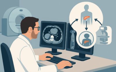
A 55-year-old patient with type 2 diabetes and metabolic syndrome presents for a follow-up visit. Routine labs reveal persistently elevated liver transaminases, and you suspect nonalcoholic fatty liver disease (NAFLD). The critical question is no longer just if they have liver disease, but how severe it is. You need to assess for the presence and degree of liver fibrosis, a key determinant of prognosis and management. This article details the ACR-guided imaging workflow for the initial diagnosis and staging of liver fibrosis in a patient with known or suspected chronic liver disease. For this specific clinical question, the ACR designates US shear wave elastography abdomen as a Usually Appropriate initial imaging study.
Who Fits This Clinical Scenario for Liver Fibrosis Staging?
This guidance applies to patients with known or suspected chronic liver disease from any etiology—including nonalcoholic fatty liver disease (NAFLD), alcohol-associated liver disease, or viral hepatitis (B or C)—where the primary clinical question is the non-invasive diagnosis and staging of hepatic fibrosis. The goal is to determine the severity of liver scarring, which ranges from none (F0) to cirrhosis (F4), as this information directly impacts management, prognosis, and the need for further surveillance.
This workflow is distinct from other related clinical scenarios. It does not apply to patients who are already known to have cirrhosis and require screening for liver cancer. That situation is covered by a different clinical pathway.
Specifically, this article is NOT for:
- Hepatocellular Carcinoma (HCC) Screening: Patients with established cirrhosis (F4 fibrosis) or high-risk chronic hepatitis B who need routine surveillance for liver cancer. That workflow focuses on detecting focal lesions, not staging background fibrosis.
- Post-Treatment Monitoring for HCC: Patients with a prior diagnosis of HCC who have undergone treatment (e.g., ablation, resection, TACE) and require follow-up imaging to assess for recurrence or treatment response.
The focus here is exclusively on the initial assessment of fibrosis in a patient not yet fully staged.
What Diagnoses Are You Working Up When Staging Liver Fibrosis?
When ordering imaging to stage liver fibrosis, you are not differentiating between causes of liver disease but rather determining the architectural severity of the disease. The result places the patient on a spectrum of risk, guiding subsequent management. The key thresholds you are assessing for are:
No or Mild Fibrosis (METAVIR Stage F0-F1): This is the most favorable outcome. A finding of minimal or no scarring suggests a lower risk of progression to cirrhosis and its complications. Management can focus on treating the underlying cause of liver disease (e.g., lifestyle modification for NAFLD, antiviral therapy for hepatitis C) to prevent fibrosis from developing or advancing.
Significant Fibrosis (METAVIR Stage F2): This is an important intermediate stage. Patients with significant fibrosis are at a higher risk of progressing to cirrhosis over time compared to those with mild fibrosis. Identifying this stage may prompt more aggressive management of the underlying condition, closer monitoring, and referral to a hepatologist to discuss potential therapies and strategies to halt progression.
Advanced Fibrosis or Cirrhosis (METAVIR Stage F3-F4): This is the most critical threshold to identify. A diagnosis of advanced fibrosis (F3) or established cirrhosis (F4) fundamentally changes the patient’s management plan. These patients are at substantial risk for decompensation (ascites, encephalopathy) and, most importantly, for developing hepatocellular carcinoma (HCC). Confirming F3-F4 fibrosis triggers the initiation of routine HCC surveillance (typically with ultrasound every six months) and endoscopic screening for esophageal varices.
Why Is Ultrasound Shear Wave Elastography a Recommended First Step for Fibrosis Staging?
The ACR panel rates both US shear wave elastography abdomen and MR elastography abdomen as Usually Appropriate for the initial staging of liver fibrosis. Both are excellent non-invasive tools that directly measure liver stiffness, a physical property that correlates strongly with the degree of histologic fibrosis. As the liver becomes more scarred, it becomes stiffer.
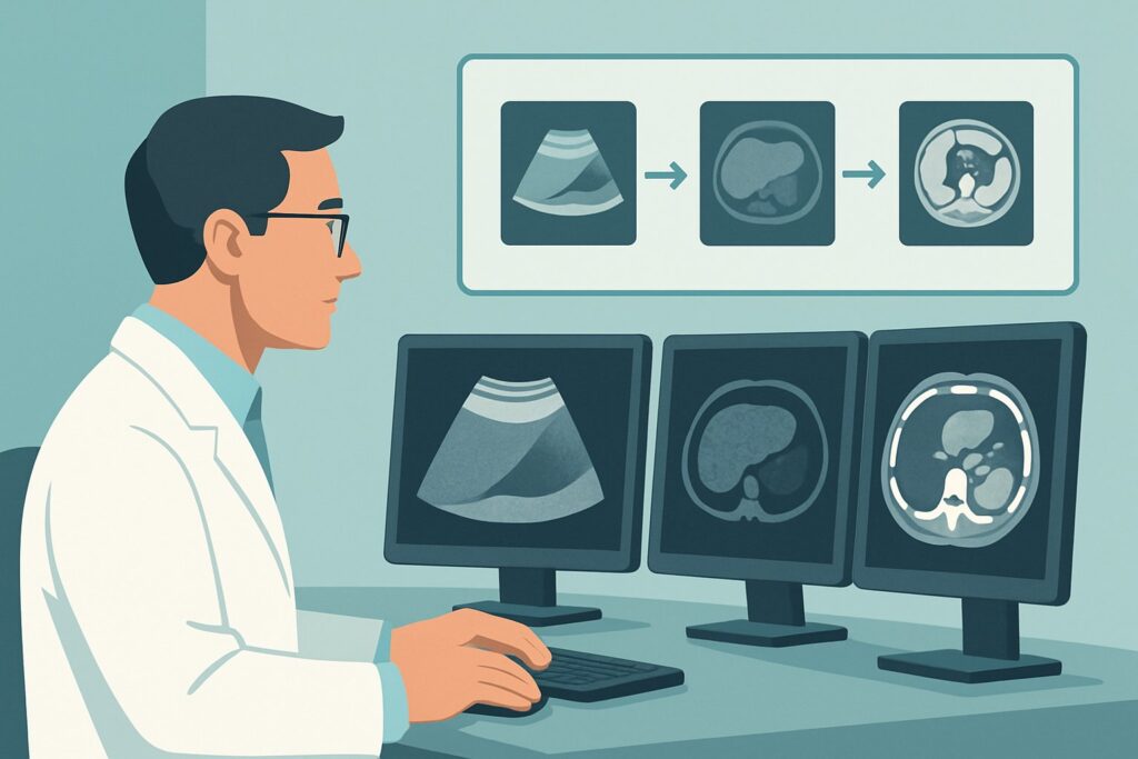
Ultrasound (US) shear wave elastography is often the practical first choice due to its wide availability, lower cost, and ease of performance at the point of care or in a standard imaging department. It uses focused acoustic pulses to generate microscopic shear waves within the liver tissue. The speed at which these waves travel is measured by the ultrasound system and converted into a quantitative stiffness value, typically reported in kilopascals (kPa). This technique provides a direct, numerical estimate of fibrosis without radiation (0 mSv) or intravenous contrast.
Comparing the top-rated studies to other options reveals why elastography is preferred:
- MR Elastography: Also rated Usually Appropriate, MR elastography is considered the most accurate non-invasive test for staging fibrosis. It uses a small, passive driver placed on the patient’s abdomen to generate shear waves, which are then imaged with an MRI. While highly accurate, especially in patients with obesity where US can be technically challenging, it is more expensive and less widely available than its ultrasound counterpart. It serves as an excellent primary option if available or as a problem-solving tool when US elastography is inconclusive.
- Standard US Abdomen: Rated as May be appropriate, a conventional ultrasound can only detect the late, morphologic signs of cirrhosis, such as a nodular liver surface, caudate lobe hypertrophy, or signs of portal hypertension like splenomegaly and ascites. It is insensitive for detecting or staging mild to moderate fibrosis (F1-F3) and cannot reliably rule out advanced disease.
- CT Abdomen with IV Contrast: Rated as May be appropriate, CT shares the same limitations as standard ultrasound for fibrosis staging—it primarily identifies late-stage, morphologic changes of cirrhosis. Furthermore, it exposes the patient to significant ionizing radiation (☢☢☢☢ 10-30 mSv) and requires IV contrast, making it unsuitable for the primary purpose of staging fibrosis.
For these reasons, a non-invasive stiffness measurement with either US or MR elastography is the clear, guideline-supported first step.
What’s Next After Elastography? Interpreting Results and Downstream Workflow
The quantitative result from an elastography study directly informs the next steps in clinical management. The stiffness cutoffs can vary slightly based on the etiology of the liver disease (e.g., NAFLD vs. Hepatitis C) and the specific elastography technique used, so results should always be interpreted in clinical context and with reference to established thresholds.
The downstream decision tree generally follows this pattern:
- If the result indicates low stiffness (ruling out advanced fibrosis, e.g., F0-F1): This result provides reassurance. The patient has a low risk of near-term liver-related complications. The focus should remain on managing the underlying cause of their chronic liver disease to prevent future progression. Repeat non-invasive testing can be performed in 2-3 years to monitor for any change.
- If the result indicates high stiffness (confirming advanced fibrosis or cirrhosis, e.g., F3-F4): This is a critical finding that triggers a new phase of management. The patient should be referred to a specialist (Gastroenterology/Hepatology) if not already established. They must be enrolled in a hepatocellular carcinoma (HCC) surveillance program, which typically involves a standard abdominal ultrasound every six months. An upper endoscopy (EGD) is also warranted to screen for esophageal varices.
- If the result is indeterminate or intermediate: This “gray zone” requires further assessment. The result may be discordant with other clinical data or non-invasive blood-based fibrosis scores (like FIB-4 or APRI). In these cases, options include repeating the US elastography, proceeding to the more robust MR elastography for clarification, or specialist consultation to consider the role of a liver biopsy, which remains the gold standard but is reserved for cases where the diagnosis remains uncertain and will change management.
Common Pitfalls in Non-Invasive Fibrosis Staging (and When to Escalate)
While powerful, non-invasive fibrosis assessment has several potential pitfalls that clinicians should be aware of to ensure accurate interpretation.
- Confounding Inflammation: Acute hepatic inflammation, such as in acute viral hepatitis or alcoholic hepatitis, can cause liver swelling and falsely elevate stiffness measurements. Elastography is most reliable in patients with stable, chronic liver disease.
- Technical Limitations: US elastography can be technically difficult or yield unreliable results in patients with severe obesity, narrow intercostal spaces, or significant ascites. The report should always be reviewed for a quality metric (e.g., IQR/median).
- Ignoring the Standard Ultrasound Findings: While elastography provides a quantitative score, the grayscale B-mode images from the standard ultrasound portion of the exam are still vital for detecting focal lesions, steatosis, or other abnormalities.
- Over-reliance on a Single Value: The stiffness value should be integrated with clinical data and serologic fibrosis scores (e.g., FIB-4) for a more robust assessment.
If an elastography study is technically inadequate or the results are highly discordant with the overall clinical picture, escalate by consulting a specialist or considering an alternative imaging modality like MR elastography.
Related ACR Topics and Tools
For a comprehensive overview of all clinical scenarios related to imaging in chronic liver disease, from screening to post-treatment surveillance, please see our parent topic hub article. For additional tools to help guide your imaging decisions, explore the resources below.
- For breadth across all scenarios in Chronic Liver Disease, see our parent guide: Chronic Liver Disease: ACR Appropriateness Decoded.
- Imaging Appropriateness Selector — for adjacent scenarios
- Imaging Protocol Library — for technique on the recommended study
- Radiation Dose Calculator — for cumulative dose conversations
Frequently Asked Questions
Is MR elastography better than US shear wave elastography for staging liver fibrosis?
Both are rated ‘Usually Appropriate’ by the ACR and are excellent non-invasive tests. MR elastography (MRE) is generally considered slightly more accurate, especially in patients with obesity or other factors that make ultrasound technically difficult. However, US shear wave elastography is more widely available, less expensive, and serves as an excellent and practical first-line imaging test for most patients.
My patient has ascites. Can I still order US elastography?
The presence of significant ascites is a major technical limitation for US elastography. The fluid can interfere with the propagation of shear waves, leading to unreliable or failed measurements. In patients with more than a small amount of ascites, MR elastography is a better choice if the goal is to assess underlying liver stiffness.
What is the difference between this scenario (fibrosis staging) and HCC screening?
This scenario focuses on answering the question: ‘What is the degree of liver scarring?’ It is a diagnostic and staging step. Once a patient is found to have advanced fibrosis or cirrhosis (METAVIR stage F3-F4), they enter a different clinical pathway: hepatocellular carcinoma (HCC) screening and surveillance. The HCC screening workflow uses imaging (typically standard ultrasound) to look for new focal liver lesions, not to re-stage the background fibrosis.
Can I use a standard abdominal ultrasound to rule out cirrhosis?
No. A standard abdominal ultrasound is rated ‘May be appropriate’ but is not a reliable tool for ruling out advanced fibrosis or cirrhosis. It can only detect the late-stage, morphologic signs of cirrhosis, such as a nodular liver contour or splenomegaly. The liver can appear normal on a standard ultrasound even in the presence of significant or advanced fibrosis. Elastography is required for sensitive detection.
Do elevated liver enzymes (transaminitis) affect the elastography result?
Yes, significant active liver inflammation can cause liver edema and falsely elevate stiffness measurements, making the liver appear more fibrotic than it actually is. For this reason, elastography is most accurate when liver enzymes are relatively stable and not acutely flared. If a patient has very high transaminases (e.g., >5 times the upper limit of normal), it may be prudent to wait for them to decrease before performing elastography for fibrosis staging.
Reviewed by Pouyan Golshani, MD, Interventional Radiologist — May 26, 2026