

Which Imaging Study Is Best for Acute Vision Loss from a Suspected Retinal Vascular Event?
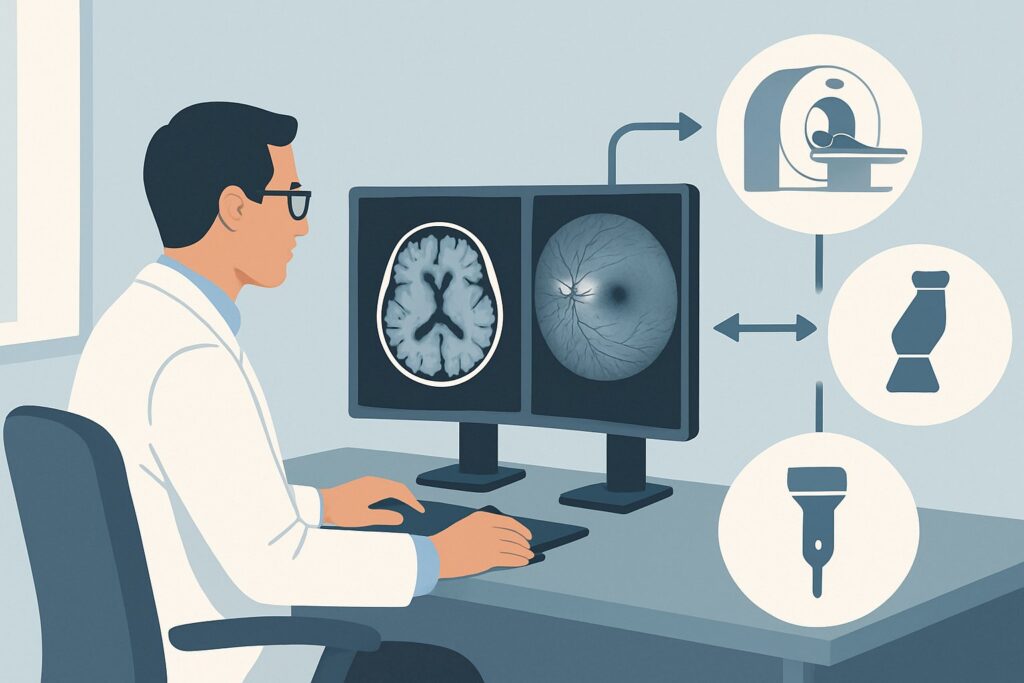
A 68-year-old male presents to the emergency department with sudden, painless, and profound vision loss in his left eye that started two hours ago. On fundoscopy, you note retinal pallor and a prominent cherry-red spot at the macula, classic for a central retinal artery occlusion (CRAO). This is a stroke equivalent, an ocular emergency demanding an immediate workup to identify the embolic source and prevent further ischemic events. The critical decision is which initial imaging study provides the most diagnostic yield for the underlying cause. This article details the clinical workflow for this specific scenario, where the American College of Radiology (ACR) Appropriateness Criteria rate MRA of the head and neck with IV contrast as Usually Appropriate.
Who Fits This Clinical Scenario?
This guidance applies to adult patients presenting with acute, non-traumatic, monocular vision loss where the clinical suspicion points toward a structural abnormality within the retina, most commonly from a vascular cause. The key features are a sudden onset (over minutes to hours) and findings on history or physical exam suggestive of retinal ischemia, such as a Central Retinal Artery Occlusion (CRAO) or Branch Retinal Artery Occlusion (BRAO).
This workflow is specific and should not be applied to patients with different presentations, even if they involve vision loss. Key exclusions include:
- Post-traumatic vision loss: If the patient has a history of recent trauma, the workup shifts to evaluating for orbital injury, a distinct clinical scenario.
- Signs of infection or inflammation: Patients with eye pain, redness, proptosis, or systemic signs of inflammation (e.g., fever, elevated ESR/CRP) fall under the “infection or inflammatory disorder suspected” category, which has a different imaging pathway.
- Bitemporal hemianopia: This specific visual field defect strongly suggests a lesion at the optic chiasm (e.g., a pituitary macroadenoma) and follows the “sellar or parasellar mass suspected” workflow.
- Gradual or chronic vision loss: This guidance is for acute events. Progressive vision loss over weeks to months suggests a different pathophysiology, such as a compressive lesion or a degenerative process.
What Diagnoses Are You Working Up in This Scenario?
When a patient presents with features of a retinal artery occlusion, the imaging workup is not for the eye itself but for the source of the presumed embolus. The differential diagnosis guides the choice of imaging to cover the most likely and most dangerous etiologies.
The most common cause is atherosclerotic disease of the carotid arteries. An unstable plaque in the ipsilateral internal carotid artery can break off, travel distally, and lodge in the central retinal artery. Identifying significant stenosis (>50-70%) is critical, as it may warrant urgent intervention like carotid endarterectomy or stenting to prevent a major hemispheric stroke.
Another primary consideration is a cardioembolic source. Atrial fibrillation is a major culprit, leading to clot formation in the left atrial appendage that can embolize to the cerebral or retinal circulation. Other cardiac sources include valvular disease, patent foramen ovale, or ventricular thrombus after a myocardial infarction. While neurovascular imaging won’t diagnose these, it is essential for ruling out a concurrent proximal vessel source.
Less common but highly consequential is large-vessel vasculitis, particularly Giant Cell Arteritis (GCA). This is a crucial diagnosis to consider in patients over 50, especially if they report associated symptoms like new-onset headache, jaw claudication, or scalp tenderness. While MRA can sometimes show vessel wall enhancement suggestive of vasculitis, the diagnosis often relies on clinical findings, inflammatory markers, and temporal artery biopsy.
Why Is MRA Head and Neck with IV Contrast the Recommended Study?
For an adult with acute vision loss from a suspected retinal vascular event, the ACR designates MRA head and neck with IV contrast as Usually Appropriate. This study provides a comprehensive, non-invasive evaluation of the entire arterial tree from the aortic arch to the intracranial vessels, directly addressing the most common and treatable cause: carotid artery disease.
The rationale for this recommendation includes:
- High Sensitivity for Stenosis: Contrast-enhanced MRA is highly sensitive and specific for detecting significant stenosis in the carotid bifurcation, the most common location for culprit plaques. It can accurately visualize the vessel lumen and characterize plaque morphology.
- Comprehensive Evaluation: The study covers both the extracranial (neck) and intracranial vessels, allowing for the detection of tandem lesions or alternative sources of ischemia like vertebral artery disease or intracranial atherosclerosis.
- No Ionizing Radiation: Unlike CTA, MRA does not use ionizing radiation (0 mSv). This is a significant advantage, particularly in patients who may require serial imaging.
Why are alternative studies rated lower for this initial workup?
- CTA head with IV contrast: While CTA is an excellent modality for vascular imaging, the ACR rates it Usually not appropriate as the initial study in this specific context. It provides similar diagnostic information to MRA but exposes the patient to ionizing radiation (☢☢☢ 1-10 mSv) and requires iodinated contrast, which carries a higher risk of contrast-induced nephropathy than gadolinium-based agents in patients with normal renal function.
- MRI orbits without IV contrast: This study is rated Usually not appropriate because it is designed to evaluate the optic nerves, extraocular muscles, and soft tissues of the orbit. While it can detect optic nerve ischemia, it does not visualize the large vessels of the head and neck, which are the primary target of the embolic workup. It answers the wrong clinical question for this scenario.
The goal is to rapidly and safely identify a treatable source of emboli. MRA of the head and neck with contrast achieves this with high accuracy and no radiation dose.
What’s Next After MRA Head and Neck with IV Contrast? Downstream Workflow
The results of the MRA will guide the subsequent management and workup. The pathway diverges based on whether a clear source is identified.
- If the MRA is positive for a significant stenosis: A finding of high-grade stenosis (typically >70%, or >50% if symptomatic) in the ipsilateral carotid artery is an actionable result. This should trigger an immediate consultation with vascular surgery or neurointerventional radiology to discuss urgent revascularization via carotid endarterectomy or stenting. The patient should also be started on aggressive medical therapy, including high-dose statins and antiplatelet agents.
- If the MRA is negative: A negative or non-obstructive MRA effectively rules out significant large-vessel atherosclerosis as the cause. The focus of the workup must then shift to a cardioembolic source. The next steps include obtaining an electrocardiogram (ECG), telemetry or Holter monitoring to look for atrial fibrillation, and a transthoracic or transesophageal echocardiogram to evaluate for valvular disease, intracardiac thrombus, or a patent foramen ovale.
- If the MRA is indeterminate or suggests vasculitis: In rare cases, the MRA findings may be unclear or show diffuse vessel wall thickening and enhancement suggestive of vasculitis (like GCA). This should prompt a rheumatology consultation, measurement of inflammatory markers (ESR and CRP), and consideration of a temporal artery biopsy. If clinical suspicion for GCA is high, empiric high-dose corticosteroids should be started immediately to prevent vision loss in the contralateral eye.
Pitfalls to Avoid (and When to Get Help)
Navigating the workup for acute retinal ischemia requires timely and precise decision-making. Several common pitfalls can compromise patient outcomes.
- Delaying the workup: CRAO is a “stroke of the eye.” The workup for an embolic source should be initiated with the same urgency as a cerebral stroke.
- Ordering a non-vascular study: Ordering a routine MRI of the brain or orbits without vessel imaging will miss the primary pathology (e.g., carotid stenosis) and delay the correct diagnosis.
- Stopping the workup after a negative MRA: A negative MRA does not mean the workup is over. It simply shifts the focus to a probable cardiac source, which must be diligently investigated.
- Forgetting Giant Cell Arteritis: In any patient over 50 with acute vision loss, GCA must be on the differential. Failing to ask about associated symptoms (headache, jaw claudication) and check inflammatory markers can lead to a missed diagnosis and irreversible bilateral blindness.
If the MRA identifies a critical stenosis or dissection, escalate immediately to the on-call stroke neurology and vascular surgery/neurointerventional radiology services.
Related ACR Topics and Tools
This article focuses on a single, specific clinical scenario. For a comprehensive overview of imaging for all types of vision loss, consult our parent guide. For detailed technical parameters or to explore other scenarios, the following resources are essential.
- For breadth across all scenarios in Vision Loss, see our parent guide: Vision Loss: ACR Appropriateness Decoded.
- ACR Appropriateness Criteria Lookup: For direct access to the guidelines for other clinical presentations.
- Imaging Protocol Library: For detailed technical specifications of the recommended MRA study.
- Radiation Dose Calculator: For discussing cumulative radiation exposure with patients when considering alternative studies like CTA.
Frequently Asked Questions
Why is MRA preferred over CTA if both can see the carotid arteries?
MRA is preferred as the initial study primarily because it does not involve ionizing radiation. While CTA is an excellent alternative and may be used if MRA is contraindicated (e.g., incompatible pacemaker, severe claustrophobia), the ACR guidance prioritizes MRA to avoid radiation exposure when diagnostic performance is comparable for the primary clinical question of identifying carotid stenosis.
Does the patient need a brain MRI at the same time as the MRA head and neck?
Yes, typically the order should include an MRI of the brain (specifically with a diffusion-weighted imaging or DWI sequence) along with the MRA of the head and neck. The DWI sequence is extremely sensitive for detecting acute ischemic stroke. Patients with CRAO have a high concurrent risk of silent cerebral infarcts, and identifying these is important for risk stratification and management.
What if my patient has renal failure and cannot receive gadolinium contrast?
In patients with severe renal impairment (e.g., eGFR < 30 mL/min/1.73m²) or on dialysis, gadolinium-based contrast agents are often contraindicated due to the risk of nephrogenic systemic fibrosis. In this situation, a non-contrast MRA of the neck can be performed, or the imaging modality can be switched to a CTA with IV contrast (if the risk-benefit of iodinated contrast is favorable) or a carotid duplex ultrasound.
Is a carotid duplex ultrasound sufficient for the initial workup?
Carotid duplex ultrasound is a good, non-invasive screening tool for carotid bifurcation disease. However, it does not evaluate the aortic arch or the intracranial vessels. MRA provides a more comprehensive evaluation of the entire vascular tree relevant to an embolic stroke workup, which is why it is recommended as the primary modality in the acute setting.
If the MRA is negative, is there any role for further neuroimaging?
If the MRA of the head and neck is negative and the cardiac workup is also unrevealing, the patient is considered to have embolic stroke of undetermined source (ESUS). Further imaging is typically not warranted unless new symptoms develop or a rare cause like vasculitis is suspected, which might prompt a dedicated vessel wall imaging MRI sequence.
Reviewed by Pouyan Golshani, MD, Interventional Radiologist — May 30, 2026


