Which Imaging Study Is Best for an Adult with New Onset Psychosis?
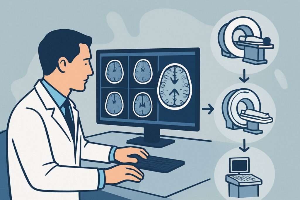
A 45-year-old patient with no prior psychiatric history is brought to the emergency department by family due to a week of escalating paranoid delusions and auditory hallucinations. The initial medical workup, including labs and a toxicology screen, is unrevealing. His neurologic exam is non-focal. You are now faced with a critical decision: does this patient need brain imaging to rule out an organic cause for this first psychotic break, and if so, which study provides the most diagnostic value? This article provides a deep-dive workflow for this specific scenario, guiding you through the differential diagnosis, imaging rationale, and downstream steps. Based on the American College of Radiology (ACR) Appropriateness Criteria, MRI of the head without and with IV contrast is rated May be appropriate as the initial imaging study.
Who Fits This Clinical Scenario?
This guidance applies specifically to an adult patient experiencing a first episode of psychosis. Psychosis is defined by the presence of delusions, hallucinations, or disorganized thought and speech. The key inclusion criteria for this workflow are a truly “new onset” presentation in an adult with no established primary psychiatric disorder (like schizophrenia or bipolar disorder) and a non-focal neurologic examination. The initial clinical and laboratory evaluation should not point toward an obvious toxic-metabolic cause, such as substance intoxication, withdrawal, or severe electrolyte imbalance.
It is crucial to distinguish this scenario from similar but distinct clinical presentations that follow different diagnostic pathways:
- Patients with focal neurologic deficits: If the patient with altered mental status also has a new focal finding like hemiparesis, aphasia, or a cranial nerve palsy, they fit the scenario of Altered mental status with suspected intracranial pathology, which has different imaging recommendations.
- Patients with a clear toxic-metabolic cause: If initial labs reveal severe hypoglycemia, uremia, or hyponatremia, the presentation fits the Altered mental status with suspected medical illness variant. Imaging may be deferred pending correction of the underlying abnormality.
- Patients with known psychiatric illness: This guidance does not apply to a patient with a known diagnosis of schizophrenia who presents with an exacerbation of their chronic symptoms.
What Diagnoses Are You Working Up in This Scenario?
While most cases of new onset psychosis in adults are ultimately attributed to a primary psychiatric disorder, imaging is performed to exclude underlying structural or inflammatory brain pathology. The goal is not to “see” the psychosis but to rule out organic mimics that can present identically. The differential diagnosis for this presentation is broad, and imaging is targeted at identifying these consequential causes.
Autoimmune Encephalitis: A critical and increasingly recognized cause, conditions like anti-N-methyl-D-aspartate (NMDA) receptor encephalitis can present with prominent psychiatric symptoms. MRI may reveal T2/FLAIR hyperintensities, often in the medial temporal lobes, though the scan can also be normal. This diagnosis is a key reason to consider imaging early.
Central Nervous System (CNS) Neoplasm: A primary brain tumor (like a glioma) or a metastatic lesion, particularly in the frontal or temporal lobes, can disrupt neural circuits and manifest as personality changes, disorganized thought, or hallucinations. These are often occult on neurologic exam until they become large.
Infectious Etiologies: Viral encephalitides, most classically Herpes Simplex Virus (HSV) encephalitis, can present with acute behavioral and psychotic features. Prompt diagnosis is vital as effective antiviral treatment exists. Other infections, such as neurosyphilis or HIV-associated neurocognitive disorders, can also be on the differential.
Demyelinating Disease: Multiple sclerosis (MS) can, in some cases, present with psychiatric symptoms as an initial manifestation. Imaging is the cornerstone of diagnosing demyelinating plaques.
Vascular Disease: While less common, a strategically located stroke or findings of CNS vasculitis could theoretically present with acute psychosis. This is a less frequent cause but remains an important consideration in patients with vascular risk factors.
Why Is MRI of the Head Without and With Contrast a Key Option for New Onset Psychosis?
In the setting of new onset psychosis, the ACR rates MRI head without and with IV contrast as May be appropriate. This rating reflects the clinical reality that while many patients will not have an organic cause, for those who do, MRI provides the most comprehensive evaluation. Its superior soft-tissue contrast is essential for detecting the subtle abnormalities associated with the key differential diagnoses.
The non-contrast portion of the MRI, particularly T2-weighted and Fluid-Attenuated Inversion Recovery (FLAIR) sequences, is highly sensitive for detecting edema and inflammation seen in encephalitis, the non-enhancing plaques of multiple sclerosis, and cytotoxic edema from an acute stroke. However, the addition of intravenous (IV) contrast is crucial. Contrast enhancement highlights areas of blood-brain barrier breakdown, which is characteristic of brain tumors, active demyelinating lesions, and infectious processes like abscesses or certain forms of encephalitis.
Let’s compare this to other imaging options for this specific scenario:
- CT head without IV contrast: Also rated May be appropriate, this study is often more readily available in an emergency setting. It is excellent for ruling out large masses, hemorrhage, or hydrocephalus. However, it has very low sensitivity for the most critical differential diagnoses in this scenario, including autoimmune encephalitis, early stroke, and demyelination. It exposes the patient to ionizing radiation (☢☢☢ 1-10 mSv) for a much lower diagnostic yield than MRI.
- MRI head without IV contrast: While still useful and rated May be appropriate, omitting contrast means you may miss enhancing tumors, active inflammatory plaques in MS, or infectious lesions. This can lead to a false-negative study and a delayed diagnosis for a treatable condition.
Given that the primary goal of imaging is to search for these specific organic causes, the combination of non-contrast and contrast-enhanced MRI sequences offers the highest probability of detecting them in a single, radiation-free (O 0 mSv) examination.
What Is the Downstream Workflow After an MRI for New Onset Psychosis?
The results of the MRI will dictate the next steps in the patient’s evaluation and management. The workflow branches significantly based on whether the study is positive, negative, or indeterminate.
If the MRI is positive: The findings guide the subsequent workup.
- Findings suggestive of encephalitis (e.g., temporal lobe T2/FLAIR hyperintensity): The immediate next step is a lumbar puncture to obtain cerebrospinal fluid (CSF) for analysis, including cell counts, protein, glucose, viral PCR (especially for HSV), and autoimmune/paraneoplastic antibody panels. A neurology consultation is essential.
- Findings of a mass lesion: An urgent neurosurgery consultation is required for consideration of biopsy or resection. Further systemic imaging may be needed to search for a primary cancer if metastasis is suspected.
- Findings of demyelination: This would prompt a neurology consultation for further workup for multiple sclerosis, which may include a spinal cord MRI and CSF analysis for oligoclonal bands.
If the MRI is negative: A normal MRI is a very common and reassuring result. It effectively rules out most major structural, inflammatory, and infectious causes. At this point, the diagnostic focus shifts more strongly toward a primary psychiatric disorder. The next step is a formal psychiatric consultation to confirm the diagnosis and initiate appropriate antipsychotic medication and therapy. Further neurologic workup, such as an electroencephalogram (EEG) to rule out non-convulsive seizures, may still be considered depending on the complete clinical picture.
Pitfalls to Avoid (and When to Get Help)
Navigating the workup for new onset psychosis requires careful consideration to avoid common diagnostic errors.
- Anchoring on a psychiatric diagnosis too early: The most significant pitfall is dismissing the presentation as “just psychiatric” without adequately considering organic mimics, especially in patients with atypical features (e.g., older age at onset, concurrent cognitive deficits).
- Ordering an incomplete study: Requesting an MRI without contrast to save time or avoid a gadolinium injection can miss critical diagnoses like tumors or active inflammation, defeating the purpose of the scan.
- Misinterpreting non-specific findings: Small, non-specific white matter hyperintensities are common in adults and are not, by themselves, indicative of a cause for psychosis. Avoid over-attributing symptoms to incidental findings.
- Ignoring the neurologic exam: A subtle focal deficit, even if minor, should shift the diagnostic algorithm toward a primary neurologic cause and heighten the urgency of imaging.
If the clinical picture is confusing, the MRI is equivocal, or the patient fails to respond to initial psychiatric treatment, it is crucial to escalate care by obtaining a neurology consultation.
Related ACR Topics and Tools
For a comprehensive overview of imaging for all related scenarios, including coma and delirium, please see our parent topic hub article. For further exploration of imaging techniques and safety, the following GigHz resources are available.
- Parent Topic Hub: For breadth across all scenarios in Altered Mental Status, Coma, Delirium, and Psychosis, see our parent guide: Altered Mental Status, Coma, Delirium, and Psychosis: ACR Appropriateness Decoded.
- ACR Criteria Lookup: To review guidelines for other, slightly different clinical presentations, use the ACR Appropriateness Criteria Lookup.
- Imaging Protocols: For detailed technical parameters of various MRI and CT studies, consult the Imaging Protocol Library.
- Dose Information: To discuss radiation exposure from CT scans with patients, the Radiation Dose Calculator can be a helpful tool.
Frequently Asked Questions
Is imaging always necessary for a first psychotic episode?
Not always, which is why the ACR rates it as ‘May be appropriate’ rather than ‘Usually appropriate’. The decision to image depends on the clinical context. Imaging is more strongly indicated in patients with atypical features such as older age of onset (e.g., >40), a non-focal but abnormal neurologic exam, cognitive decline, or a rapid, fulminant course. For a young adult with a classic presentation and a normal exam, a period of observation and psychiatric treatment may be initiated first.
If an MRI is not immediately available, is a CT scan a reasonable first step?
Yes, a non-contrast head CT is a reasonable first step if MRI is unavailable or contraindicated, particularly in the emergency setting. It is also rated ‘May be appropriate’. Its main role is to quickly rule out large, emergent pathologies like a major hemorrhage or a large tumor. However, a negative CT scan does not exclude the key differential diagnoses (like encephalitis or small tumors) and should be followed by an MRI if clinical suspicion for an organic cause remains high.
What clinical information should I include on the imaging requisition?
Providing a clear, concise history is critical for the radiologist. Instead of just writing ‘altered mental status,’ specify ‘New onset psychosis in a 45-year-old with no prior psychiatric history. Please rule out organic causes such as encephalitis, neoplasm, or demyelination.’ This level of detail allows the radiologist to tailor the imaging protocol and focus their interpretation on the most relevant potential findings.
Should I order an EEG in addition to an MRI?
An electroencephalogram (EEG) can be a valuable complementary test. It is particularly useful for evaluating for non-convulsive seizures or subclinical seizure activity, which can sometimes present as psychosis or acute confusion. If the MRI is negative and the clinical picture remains unclear, an EEG is often a logical next step in the neurologic workup.
Does a normal MRI completely rule out an organic cause for psychosis?
No. A normal MRI is very reassuring and rules out most structural, inflammatory, and infectious causes. However, some conditions, particularly early-stage autoimmune encephalitis (like anti-NMDAR), can present with a normal MRI. Additionally, toxic-metabolic encephalopathies and certain systemic autoimmune diseases can cause psychosis without producing specific changes on a brain MRI. The diagnosis remains a clinical one, integrating imaging, laboratory, and CSF findings.
Reviewed by Pouyan Golshani, MD, Interventional Radiologist — May 29, 2026