Which Imaging Study Is Best for Initial Workup of Suspected Gastric Adenocarcinoma?
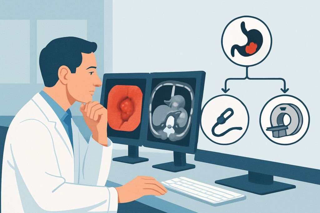
An older adult presents to your clinic with several months of progressive epigastric pain, unintentional weight loss, and early satiety. An initial esophagogastroduodenoscopy (EGD) with biopsy is pending, but your clinical suspicion for gastric adenocarcinoma is high. You need to order the right initial imaging study to assess for local disease and potential metastasis, which will be critical for preliminary staging and treatment planning. This decision point—choosing the first cross-sectional imaging study in a patient with suspected gastric cancer—is a common and consequential one. According to the American College of Radiology (ACR) Appropriateness Criteria, the recommended initial study for this scenario is `CT abdomen and pelvis with IV contrast`, which is rated as “Usually Appropriate.”
Who Fits This Clinical Scenario?
This guidance is for an adult patient with a new clinical suspicion of gastric adenocarcinoma who has not yet undergone definitive staging imaging. The typical presentation includes symptoms like persistent dyspepsia, dysphagia, unintentional weight loss, anorexia, or signs of gastrointestinal bleeding such as melena or iron deficiency anemia. This workflow applies before a full staging workup has been completed and is intended as the initial cross-sectional imaging step, often performed concurrently with or immediately following endoscopic evaluation.
This article does not apply to patients in slightly different clinical situations, which have their own distinct imaging pathways:
- Patients with a confirmed biopsy and who now require definitive locoregional and distant metastatic staging. While the imaging may be similar, the clinical question has advanced from suspicion to confirmation. This falls under the ACR variant for staging.
- Patients who have completed neoadjuvant or definitive therapy and require post-treatment evaluation to assess response.
- Asymptomatic patients undergoing long-term surveillance after successful treatment for gastric cancer.
Correctly identifying your patient’s clinical context ensures the most appropriate and resource-effective imaging is selected from the outset.
What Diagnoses Are You Working Up in This Scenario?
When ordering initial imaging for suspected gastric cancer, the primary goal is to identify and characterize a potential primary tumor and search for signs of metastatic disease. However, several other conditions can mimic this presentation clinically and radiologically.
Gastric Adenocarcinoma: This is the most common gastric malignancy and the primary target of the workup. Imaging aims to detect a focal or diffuse thickening of the gastric wall, an ulcerated mass, or linitis plastica (a rigid, shrunken stomach). Critically, imaging also seeks to identify regional lymphadenopathy, liver metastases, and peritoneal carcinomatosis, which profoundly impact staging and management.
Gastric Lymphoma: While less common than adenocarcinoma, primary gastric lymphoma can present with similar symptoms. On imaging, it often appears as marked, bulky, and uniform thickening of the gastric wall, sometimes with associated bulky lymphadenopathy. Unlike adenocarcinoma, it is less likely to cause gastric outlet obstruction for its size.
Gastrointestinal Stromal Tumor (GIST): These are mesenchymal tumors that typically arise from the stomach wall. On CT, a GIST often appears as a well-circumscribed, exophytic (outward-growing) mass that enhances avidly with contrast. They can be large and may show signs of central necrosis or hemorrhage.
Benign Peptic Ulcer Disease: Severe or complicated peptic ulcer disease can cause significant inflammation and gastric wall thickening that can be difficult to distinguish from malignancy on imaging alone. Associated findings like a clear ulcer crater without a discrete mass and the absence of metastatic disease can suggest a benign process, but endoscopic biopsy remains essential for confirmation.
Why Is CT Abdomen and Pelvis with IV Contrast the Recommended Study?
For the initial evaluation of suspected gastric adenocarcinoma, `CT abdomen and pelvis with IV contrast` is rated “Usually Appropriate” and serves as the workhorse imaging modality. Its primary value lies in its ability to provide a comprehensive assessment of both the primary tumor and potential sites of distant spread in a single, rapid acquisition.
Intravenous contrast is essential. It enhances the gastric wall, allowing for better visualization of tumor infiltration and differentiation from normal tissue. Furthermore, contrast enhancement is critical for detecting and characterizing liver metastases, identifying abnormal lymph nodes, and assessing for vascular involvement. The study provides excellent anatomic detail of the entire abdomen and pelvis, screening for common metastatic sites like the liver, peritoneum, distant lymph nodes, and ovaries (Krukenberg tumors).
While CT is the primary recommendation, other studies are rated for specific circumstances:
- FDG-PET/CT skull base to mid-thigh is also rated “Usually Appropriate.” It is highly sensitive for detecting metastatic disease, particularly in unexpected locations. However, it is often reserved for definitive staging after a tissue diagnosis is confirmed, rather than as the initial imaging test, due to higher cost, radiation dose (☢☢☢☢ 10-30 mSv), and potential for false positives from inflammatory conditions.
- MRI abdomen without and with IV contrast is rated “May be appropriate.” MRI offers excellent soft tissue contrast and can be superior for characterizing liver lesions that are indeterminate on CT. It is often used as a problem-solving tool for equivocal liver findings or in patients with a severe contraindication to iodinated CT contrast.
- Endoscopic ultrasound (US abdomen endoscopic) is rated “May be appropriate (Disagreement).” EUS is the most accurate modality for determining the depth of tumor invasion (T stage) and assessing perigastric lymph nodes (N stage). However, it is an invasive procedure and cannot evaluate for distant metastases, making it a complementary tool for locoregional staging rather than a primary, all-encompassing initial study.
The recommended CT carries a moderate radiation dose (adult RRL=☢☢☢ 1-10 mSv), a consideration that is generally outweighed by the critical diagnostic information gained in the setting of a suspected life-threatening malignancy. Once you’ve decided on this study, our protocol guide covers the technique, contrast, and reading principles: CT Chest/Abdomen/Pelvis with IV Contrast.
What’s Next After CT Abdomen and Pelvis with IV Contrast? Downstream Workflow
The results of the initial CT scan will guide the subsequent diagnostic and management pathway. The workflow typically branches based on the findings.
If the CT is positive for a suspicious gastric mass:
- No distant metastases seen: The patient should proceed with EGD and biopsy if not already done. An endoscopic ultrasound (EUS) is often the next step to refine the local T and N stage, which is crucial for determining if the patient is a candidate for neoadjuvant therapy versus upfront surgery. A staging laparoscopy may also be performed to rule out small-volume peritoneal disease not visible on CT.
- Distant metastases are identified (e.g., liver lesions, peritoneal carcinomatosis): The diagnosis is now metastatic (Stage IV) disease. The priority shifts to obtaining a tissue biopsy, either from the primary tumor via EGD or from a metastatic site (e.g., CT-guided liver biopsy), to confirm histology and molecular markers (like HER2, PD-L1) that guide systemic chemotherapy.
If the CT is negative or equivocal:
- If clinical suspicion remains high despite a negative CT, the focus returns to high-quality endoscopy. A CT scan can miss small or flat (linitis plastica) gastric cancers. EGD with multiple biopsies is the gold standard for diagnosis.
- If the CT shows an indeterminate finding, such as a non-specific liver lesion, a problem-solving study like a contrast-enhanced MRI of the abdomen may be warranted.
The initial CT scan is a pivotal decision point that triages patients into pathways for curative-intent local therapy or palliative-intent systemic therapy.
Pitfalls to Avoid (and When to Get Help)
Several common pitfalls can compromise the diagnostic value of the initial imaging workup for suspected gastric cancer:
- Ordering a non-contrast CT: A CT of the abdomen and pelvis without IV contrast is “Usually Not Appropriate.” It severely limits the evaluation of the gastric wall, liver parenchyma, and lymph nodes, potentially missing the primary tumor and metastatic disease.
- Inadequate gastric distention: A collapsed stomach can mimic or obscure wall thickening. While not always feasible in an emergent setting, having the patient drink water immediately before the scan can help distend the stomach for better evaluation.
- Overlooking subtle signs of peritoneal disease: Small peritoneal nodules or omental caking can be subtle. Careful review of the peritoneal surfaces and omentum is critical, as their presence indicates advanced disease.
- Not checking renal function: Always assess the patient’s renal function (e.g., eGFR) before ordering a study with IV contrast to ensure it is safe to administer.
If the imaging findings are complex, equivocal, or discordant with the clinical picture, a discussion with the reading radiologist or presentation at a multidisciplinary tumor board is the most appropriate next step.
Related ACR Topics and Tools
Navigating imaging decisions requires access to reliable, scenario-specific guidance. For a comprehensive overview of all clinical variants related to this condition, from staging to post-treatment follow-up, please consult our parent guide. For tools to help with ordering, protocoling, and discussing studies with patients, see the resources below.
- For breadth across all scenarios in Staging and Follow-up of Gastric Cancer, see our parent guide: Staging and Follow-up of Gastric Cancer: ACR Appropriateness Decoded.
- Explore other clinical scenarios with the ACR Appropriateness Criteria Lookup.
- Review technical details for this and other studies in the Imaging Protocol Library.
- For patient conversations about radiation exposure, use the Radiation Dose Calculator.
Frequently Asked Questions
Why not start with a PET/CT if it’s also rated ‘Usually Appropriate’?
While FDG-PET/CT is excellent for detecting distant metastases, it is generally reserved for definitive staging after a tissue diagnosis is confirmed. A standard contrast-enhanced CT is faster, less expensive, more widely available, and provides superior anatomical detail of the primary tumor and adjacent structures. It is the standard initial cross-sectional study, with PET/CT often used to clarify the extent of disease before major therapeutic decisions.
What if my patient has a severe allergy to iodinated contrast or poor renal function?
In cases of a true contraindication to iodinated contrast, `MRI abdomen without and with IV contrast` (using a gadolinium-based agent) becomes a strong alternative and is rated ‘May be appropriate.’ MRI provides excellent soft-tissue characterization and is highly sensitive for detecting liver metastases. A non-contrast CT is not a suitable substitute as it is rated ‘Usually Not Appropriate’ for this indication.
Is endoscopic ultrasound (EUS) a better first test than CT?
No, EUS is not a better *first* test for the overall initial workup. EUS is the most accurate tool for local T-staging (depth of tumor invasion) and N-staging (regional nodes), but it cannot evaluate for distant metastases in the liver, lungs, or peritoneum. Therefore, CT is the preferred initial study for a global assessment. EUS is a complementary tool used for local staging in patients who appear to have non-metastatic disease on CT.
Does the initial CT need to include the chest?
The ACR variant specifically lists `CT abdomen and pelvis`. While the lungs are a potential site of metastasis, they are less common than liver or peritoneal spread at initial presentation. Many institutional protocols for cancer staging will include the chest to establish a clean baseline. A dedicated `CT chest with IV contrast` is rated ‘Usually Not Appropriate’ as a standalone initial test for this scenario, but including the chest as part of a comprehensive CT Chest/Abdomen/Pelvis is a common and reasonable practice for staging.
Can a CT scan reliably differentiate gastric adenocarcinoma from gastric lymphoma?
While there are imaging features that can suggest one diagnosis over the other—for example, lymphoma often causes marked, bulky wall thickening without causing obstruction—there is significant overlap. A definitive diagnosis cannot be made on imaging alone. Endoscopic biopsy is always required to establish the histopathological diagnosis and guide appropriate treatment, which differs significantly between adenocarcinoma and lymphoma.
Reviewed by Pouyan Golshani, MD, Interventional Radiologist — May 30, 2026