Which Imaging Study Is Best for Suspected Dialysis Access Steal Syndrome?
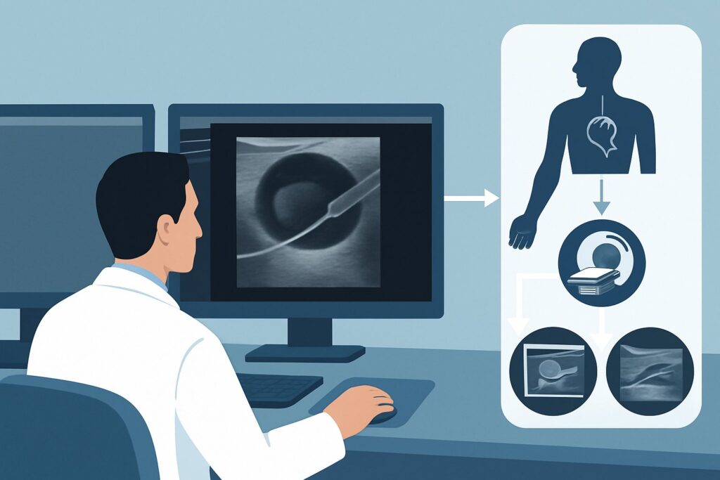
A 68-year-old man with end-stage renal disease and a mature left brachiocephalic arteriovenous fistula (AVF) presents to the clinic. For the past month, he’s noticed his left hand is persistently cold, with pain in his fingers, especially during dialysis. His nephrologist also notes worsening shortness of breath and peripheral edema, raising concern for high-output cardiac failure. You suspect dialysis access-associated steal syndrome (DASS) or a high-flow state, and you need to choose the right initial imaging study to confirm the diagnosis and plan a potential intervention. This article details the clinical workflow for this specific scenario, guiding you to the most appropriate imaging choice. According to the American College of Radiology (ACR) Appropriateness Criteria, Fluoroscopy fistulography hemodialysis access area of interest is rated Usually Appropriate for this presentation.
Who Fits This Clinical Scenario?
This guidance applies to patients with a mature arteriovenous fistula (AVF) or arteriovenous graft (AVG) who present with symptoms suggestive of either arterial insufficiency distal to the access or high-output cardiac failure. The key is the nature of the symptoms, which point toward a hemodynamic imbalance caused by the access itself.
Inclusion criteria for this workflow:
- An established, functioning AVF or AVG for hemodialysis.
- Symptoms of distal ischemia: new-onset pain at rest, coldness, pallor, paresthesias, or diminished pulses in the hand or foot distal to the access.
- Symptoms of high-output cardiac failure: unexplained or worsening dyspnea, tachycardia, cardiomegaly, or peripheral edema in a patient with a high-flow access.
This workflow is NOT for:
- Suspected Thrombosis: A patient with a suddenly absent thrill or pulse in the access. This represents acute occlusion and follows a different diagnostic pathway.
- Failure to Mature: A newly created fistula that has not developed adequate flow for dialysis within 2-3 months. This is a problem of inflow or outflow stenosis preventing maturation, not a hemodynamic steal from a mature access.
- General Dysfunction: A patient with decreased dialysis adequacy, high venous pressures, or prolonged bleeding times without specific ischemic or cardiac symptoms. While stenosis is likely, the urgency and differential are different.
What Diagnoses Are You Working Up in This Scenario?
When a patient presents with ischemic or cardiac symptoms related to their dialysis access, the imaging workup is focused on confirming a hemodynamic disturbance and identifying its anatomic cause. The differential diagnosis is narrow but includes critical, treatable conditions.
Dialysis Access-Associated Steal Syndrome (DASS) is the primary diagnosis to consider. In DASS, the low-resistance AVF or AVG shunts a large volume of arterial blood, “stealing” it from the distal extremity’s vascular bed. This leads to hypoperfusion and ischemic symptoms, which can range from mild coolness to limb-threatening gangrene. The imaging goal is to demonstrate retrograde flow in the artery distal to the anastomosis and quantify the severity of the steal.
High-Output Cardiac Failure is another major concern, particularly with large, proximal fistulas (e.g., brachiocephalic or thigh access). The massive blood volume shunted through the access significantly increases venous return (preload) to the heart. Over time, the sustained high-cardiac-output state can lead to ventricular hypertrophy, dilation, and eventual heart failure. Imaging aims to measure the access flow volume to confirm if it exceeds the threshold that typically precipitates cardiac compromise (often cited as >2.0 L/min).
Significant Access Stenosis can be a primary cause or a contributor to the patient’s symptoms. A juxta-anastomotic stenosis, for example, can alter pressure gradients and paradoxically worsen steal. A central venous stenosis can increase backpressure throughout the circuit, causing venous hypertension, severe limb edema, and symptoms that can mimic or overlap with steal syndrome. Identifying and locating any stenoses is a key objective of the workup.
Why Is Fluoroscopy Fistulography the Recommended Study for This Presentation?
The ACR Appropriateness Criteria rate both Duplex Doppler Ultrasound and Fluoroscopy Fistulography as Usually Appropriate for suspected steal syndrome. While ultrasound is an excellent non-invasive first step, fistulography is often the definitive study because it is both diagnostic and therapeutic.
Fluoroscopy fistulography, also known as a fistulogram, is the gold standard for evaluating the entire hemodialysis access circuit. It involves direct cannulation of the access and injection of iodinated contrast under live X-ray (fluoroscopy). Its primary advantages in this scenario are:
- Dynamic Flow Assessment: It provides a real-time map of blood flow, directly visualizing the location of the anastomosis, the degree of shunting, and whether there is retrograde flow in the distal artery—the hallmark of steal syndrome.
- Comprehensive Anatomy: It visualizes the entire circuit from the arterial inflow, through the fistula or graft, and all the way through the central venous outflow to the right atrium. This is crucial for identifying central venous stenosis, which can be missed by ultrasound.
- Therapeutic Capability: This is the key differentiator. If a significant stenosis is identified as the cause of the hemodynamic imbalance, it can be treated immediately with angioplasty or stenting during the same procedure. This “see-and-treat” capability streamlines patient care and avoids the delay of a second procedure.
The primary trade-off is the use of ionizing radiation (Relative Radiation Level ☢☢☢, 1-10 mSv) and iodinated contrast, which requires caution in patients with residual renal function or contrast allergies.
Alternative Studies and Their Ratings:
- US Duplex Doppler hemodialysis access: Also rated Usually Appropriate, this non-invasive study uses sound waves to visualize the access and measure blood flow velocities and volumes. It is excellent for screening, can diagnose steal by showing flow reversal in the distal artery, and can quantify access flow to diagnose a high-output state. It involves no radiation (RRL O, 0 mSv). However, it is highly operator-dependent and can be limited by patient body habitus or in visualizing the central veins. It is often used as the initial test, with fistulography reserved for confirmation and intervention.
- CTA extremity area of interest with IV contrast: Rated May be appropriate. Computed Tomography Angiography provides superb anatomic detail of the vasculature. However, it offers a static picture and lacks the dynamic, real-time flow information of fistulography or Doppler. It also requires a significant IV contrast load and has a variable radiation dose. It is best reserved as a problem-solving tool, such as for pre-operative planning of a complex revision.
Once you’ve decided on fistulography or another interventional procedure, our protocol guide covers the technique, contrast, and reading principles: IR Central Venous Access (PICC, Tunneled, Port).
What’s Next After Imaging? Downstream Workflow
The results of the imaging study directly guide the subsequent therapeutic steps, which are almost always managed by an interventional radiologist or vascular surgeon. The goal is to alleviate the patient’s symptoms while preserving the dialysis access whenever possible.
If the study is positive for significant steal or high-output state: The patient will be scheduled for a flow-reduction procedure. The fistulogram provides the roadmap for this intervention. Common options include:
- Surgical Banding: A minimally invasive procedure to place a suture or band around the fistula to narrow its diameter, thereby increasing resistance and reducing overall flow.
- Distal Revascularization-Interval Ligation (DRIL): A more complex surgical bypass created to restore arterial flow to the ischemic hand.
- Revision of the Anastomosis: The connection between the artery and vein may be surgically revised to reduce its size.
If the study shows a treatable stenosis causing the symptoms: The most common downstream step is percutaneous transluminal angioplasty (PTA), with or without stent placement. This can often be performed during the initial diagnostic fistulogram. Correcting a critical stenosis can normalize flow dynamics and resolve the steal or high-flow physiology.
If the study is negative: A normal fistulogram and Doppler ultrasound in a symptomatic patient suggest the cause is not related to the dialysis access. The workup should then pivot to other potential causes of extremity ischemia (e.g., peripheral artery disease, embolism) or cardiac failure (e.g., primary cardiomyopathy, coronary artery disease).
Pitfalls to Avoid (and When to Get Help)
Navigating the workup for suspected steal syndrome requires careful clinical correlation and an understanding of the imaging modalities’ limitations. Here are common pitfalls to avoid:
- Dismissing Mild Symptoms: Early ischemic symptoms like cold intolerance or mild pain can progress to severe, limb-threatening ischemia. A low threshold for imaging is warranted.
- Relying Solely on Physical Exam: While a diminished distal pulse is a classic sign, many patients with significant steal have a palpable radial pulse at rest. Dynamic assessment with imaging is necessary.
- Incomplete Imaging: Failing to visualize the entire outflow tract to the right atrium can miss a central venous stenosis, which can be the root cause of the patient’s symptoms. This is a key advantage of fistulography over ultrasound.
- Attributing All Symptoms to Steal: Remember to consider other causes. A patient with diabetes and an AVF may have ischemic symptoms from underlying peripheral artery disease, not steal.
If a patient presents with acute, severe ischemic symptoms (the “6 Ps”: pain, pallor, pulselessness, paresthesia, paralysis, poikilothermia), this is a vascular emergency. Escalate immediately to vascular surgery or interventional radiology for emergent evaluation.
Related ACR Topics and Tools
For a comprehensive overview of all clinical scenarios related to dialysis access imaging, please refer to our parent topic hub article. For tools to help with ordering, protocoling, and discussing studies with patients, see the resources below.
- For breadth across all scenarios in Dialysis Fistula Malfunction, see our parent guide: Dialysis Fistula Malfunction: ACR Appropriateness Decoded.
- ACR Appropriateness Criteria Lookup — for adjacent scenarios
- Imaging Protocol Library — for technique on the recommended study
- Radiation Dose Calculator — for cumulative dose conversations
Frequently Asked Questions
Is Duplex Ultrasound or a Fistulogram the better first test for suspected steal syndrome?
Both are rated ‘Usually Appropriate’ by the ACR. Duplex Ultrasound is an excellent non-invasive first step to screen for steal syndrome by measuring flow volumes and detecting flow reversal. However, a Fluoroscopy Fistulogram is often required for definitive diagnosis, especially to evaluate the central veins, and it allows for immediate intervention (like angioplasty) in the same session. The choice often depends on local practice and the severity of symptoms.
What is the difference between steal syndrome and high-output cardiac failure from a fistula?
Steal syndrome is a problem of distal arterial insufficiency, where the fistula shunts too much blood away from the hand or foot, causing ischemic symptoms. High-output cardiac failure is a systemic problem where the total flow volume through the fistula is so large that it overwhelms the heart’s ability to pump, leading to symptoms of heart failure. While they have different clinical manifestations, both are caused by excessive fistula flow and are evaluated with similar imaging techniques.
Can a patient have a palpable distal pulse and still have steal syndrome?
Yes. The presence of a palpable radial or pedal pulse does not rule out steal syndrome. The ischemia is often exertional or becomes most apparent during dialysis when systemic blood pressure may be lower. The diagnosis relies on demonstrating retrograde arterial flow on imaging, not just the absence of a pulse.
How much radiation is involved in a fistulogram?
According to the ACR, a fistulogram has a relative radiation level of ☢☢☢, which corresponds to an effective dose of 1-10 mSv. This is comparable to the amount of natural background radiation a person receives over several years. While this is a moderate dose, the diagnostic and therapeutic benefits in a symptomatic patient generally far outweigh the risk.
If the fistulogram is normal, what should I do next?
If a comprehensive fistulogram and a high-quality duplex ultrasound are both normal, the patient’s symptoms are unlikely to be caused by the dialysis access. The clinical workup should then focus on other potential causes, such as intrinsic peripheral artery disease, embolic phenomena, diabetic neuropathy, or primary cardiac dysfunction.
Reviewed by Pouyan Golshani, MD, Interventional Radiologist — May 30, 2026