What Is the Best Imaging for Routine Surveillance After Primary Liver Cancer Treatment?

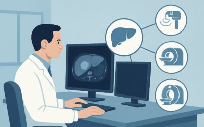
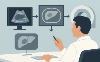
A 68-year-old man with a history of hepatocellular carcinoma (HCC) in the setting of nonalcoholic steatohepatitis (NASH) cirrhosis is in your clinic for a six-month follow-up. He underwent successful radiofrequency ablation of a 2.5 cm lesion and has been feeling well, with stable liver function tests and a normal alpha-fetoprotein level. It is time for his scheduled surveillance imaging to assess for tumor recurrence. You need to decide between MRI, CT, and ultrasound, considering the need for repeated scans over the coming years. This article details the evidence-based workflow for this specific clinical decision. For routine surveillance in a treated adult with primary liver cancer, the American College of Radiology (ACR) rates `MRI abdomen without and with IV contrast` as Usually Appropriate.
Who Fits This Clinical Scenario for Liver Cancer Surveillance?
This guidance applies specifically to adult patients with a confirmed diagnosis of primary liver cancer, most commonly hepatocellular carcinoma (HCC), who have already undergone treatment with curative or locoregional intent. This includes patients post-resection, ablation (RFA or microwave), transarterial chemoembolization (TACE), radioembolization (Y-90), or stereotactic body radiation therapy (SBRT). The key context is routine surveillance—asymptomatic, scheduled follow-up imaging intended to detect recurrence at an early, treatable stage.
This workflow is distinct from other, similar-sounding clinical situations. This article does not apply to:
- Initial screening for liver cancer: This applies to high-risk patients (e.g., with cirrhosis) who have not yet been diagnosed with cancer. That scenario follows a different imaging pathway, often starting with ultrasound.
- Initial staging of a new diagnosis: For a newly discovered and untreated liver tumor, the imaging goals are to characterize the primary lesion, determine the extent of disease, and plan initial therapy. This is covered in the Staging and Follow-up of Primary Liver Cancer parent topic.
- Immediate post-treatment evaluation: Imaging performed shortly after a procedure (e.g., 1-3 months post-ablation) is intended to assess treatment response and confirm technical success, a different clinical question than long-term surveillance for new disease.
Correctly identifying your patient’s situation as routine surveillance is the critical first step to ordering the most appropriate imaging study.
What Diagnoses Are You Working Up in This Scenario?
In routine surveillance after liver cancer treatment, the primary goal is the early detection of tumor recurrence. The liver, especially if cirrhotic, remains a high-risk environment for new tumor development. The differential considerations you are evaluating with imaging include several patterns of recurrence.
The most critical distinction is between expected post-treatment changes and viable, recurrent cancer. After therapies like ablation or embolization, the treated area will form a scar, which can have complex imaging features. The radiologist’s task, and the purpose of high-quality imaging, is to differentiate benign, evolving scar tissue from enhancing, active tumor.
You are specifically looking for:
- Local Recurrence: The reappearance of viable tumor at the edge of the treatment cavity. This is a common failure pattern and requires precise imaging to detect small nodules of enhancement adjacent to the scar.
- New Intrahepatic Tumors: The development of entirely new HCC lesions elsewhere in the liver. In a cirrhotic liver (a “field defect”), the risk for new primary tumors remains high even after successful treatment of the initial lesion.
- Vascular Invasion: New or progressive tumor growth into branches of the portal vein or hepatic veins. This is a significant negative prognostic indicator that drastically alters management options.
- Extrahepatic Metastases: While less common, primary liver cancer can spread to regional lymph nodes, the lungs, adrenal glands, and bones. Abdominal cross-sectional imaging provides a first look for nodal and adrenal involvement.
Why MRI abdomen without and with IV contrast Is the Recommended Study for This Presentation
The ACR designates `MRI abdomen without and with IV contrast` as Usually Appropriate for routine surveillance of treated primary liver cancer. This recommendation is driven by MRI’s superior soft-tissue contrast resolution, which is essential for solving the central diagnostic challenge in this scenario: differentiating post-treatment scar from recurrent tumor.
MRI provides detailed information on tissue characteristics that CT cannot. Key sequences like diffusion-weighted imaging (DWI) can show restricted diffusion in viable tumor cells, helping to distinguish them from necrotic or fibrotic post-treatment tissue. Furthermore, dynamic contrast-enhanced imaging after administration of a gadolinium-based contrast agent allows for assessment of tumor vascularity. Recurrent HCC typically demonstrates avid arterial phase hyperenhancement followed by “washout” on later phases—a hallmark finding that MRI is highly sensitive in detecting. The use of hepatobiliary-specific contrast agents can further increase the conspicuuity of small lesions.
While MRI is the top-rated study, it’s important to understand the rationale for the ratings of alternatives:
- `CT abdomen with IV contrast multiphase` is also rated Usually Appropriate. It is an excellent alternative when MRI is contraindicated (e.g., incompatible implanted devices, severe claustrophobia) or unavailable. Multiphase CT is highly effective at demonstrating the characteristic arterial enhancement and washout of HCC. However, its primary drawback is the use of ionizing radiation (☢☢☢☢ 10-30 mSv). For patients requiring surveillance every 3-6 months for years, the cumulative radiation dose becomes a significant consideration.
- `US abdomen transabdominal` is rated Usually Not Appropriate for this specific scenario. While ultrasound is the primary tool for initial screening in patients with cirrhosis, it lacks the sensitivity needed for post-treatment surveillance. The liver architecture is often distorted by scarring, surgical changes, and regeneration, which creates a complex acoustic environment where small recurrent nodules can be easily missed.
Given that these patients will undergo many scans over their lifetime, the lack of ionizing radiation (0 mSv) with MRI is a powerful advantage, making it the preferred modality when available and not contraindicated. When ordering, be sure to specify a “liver protocol” and provide the clinical history of treated HCC to ensure the correct multiphase contrast sequences are performed.
What’s Next After MRI abdomen without and with IV contrast? Downstream Workflow
The results of the surveillance MRI will direct the next phase of management, which almost always involves a multidisciplinary discussion. The imaging findings are typically categorized using the Liver Imaging Reporting and Data System (LI-RADS), which standardizes the interpretation and reporting of observations in patients at risk for HCC.
Here is a typical post-imaging decision tree:
- If the study is negative (LI-RADS 1 or 2): This is the desired outcome. The patient continues with their established surveillance schedule (e.g., repeat imaging in 3-6 months, per institutional or society guidelines). No immediate action is needed.
- If the study is positive for definite recurrence (LI-RADS 5 or LR-TIV for tumor in vein): This finding triggers a re-evaluation by the multidisciplinary tumor board. The team (including hepatology, medical oncology, interventional radiology, radiation oncology, and surgery) will review the patient’s case to determine the best salvage therapy. Options may include repeat locoregional therapy, systemic therapy, or clinical trial enrollment.
- If the study is indeterminate (LI-RADS 3 or 4): These findings are equivocal and represent a common clinical challenge. The next step depends on the size and specific features of the observation, as well as the patient’s overall clinical status. The most common management is short-interval follow-up imaging, typically with MRI in 3 months, to assess for stability or progression. In some cases, if a definitive diagnosis would immediately change therapy and the lesion is safely accessible, a biopsy may be considered.
The goal of the surveillance workflow is to catch recurrence when it is small and asymptomatic, allowing for the timely application of effective salvage therapies.
Pitfalls to Avoid (and When to Get Help)
Navigating post-treatment surveillance for liver cancer requires careful attention to imaging protocol and clinical context. Avoiding common errors can prevent delayed diagnoses and unnecessary procedures.
Key pitfalls to watch for include:
- Using the wrong modality: Do not use standard abdominal ultrasound for post-treatment surveillance. Its sensitivity is unacceptably low in a liver with post-therapeutic changes. Stick with multiphase MRI or CT.
- Ordering an incomplete study: A non-contrast MRI or a single-phase CT is inadequate for this indication and rated Usually Not Appropriate. The dynamic, multiphasic assessment of contrast enhancement is essential for lesion characterization.
- Over-reliance on a single data point: Do not interpret a rising alpha-fetoprotein (AFP) level in isolation if imaging is negative. A rising AFP is a red flag for recurrence that may be extrahepatic. Consider imaging the chest or a PET/CT if suspicion remains high despite a negative liver MRI.
- Misinterpreting benign enhancement: Benign post-treatment inflammation and perfusion alterations can mimic tumor. Always ensure the interpreting radiologist has access to prior imaging studies for comparison, which is critical for accurate diagnosis.
If you encounter any new or equivocal findings suggestive of recurrence, the appropriate next step is to present the case at a multidisciplinary liver tumor board for consensus recommendations.
Related ACR Topics and Tools
For a comprehensive overview of imaging across all clinical situations involving primary liver cancer, from screening to staging and follow-up, please consult our parent guide. Additional GigHz tools can help you apply these guidelines in your daily practice.
- For breadth across all scenarios in Staging and Follow-up of Primary Liver Cancer, see our parent guide: Staging and Follow-up of Primary Liver Cancer: ACR Appropriateness Decoded.
- To look up appropriateness ratings for thousands of other clinical scenarios, visit the ACR Appropriateness Criteria Lookup tool.
- For detailed technical specifications on how to perform the recommended studies, see the Imaging Protocol Library.
- To discuss cumulative radiation exposure with your patients, use the Radiation Dose Calculator.
Frequently Asked Questions
How often should surveillance imaging be performed after treatment for primary liver cancer?
The optimal interval varies based on the specific tumor, treatment received, risk of recurrence, and societal guidelines (e.g., AASLD, NCCN). A typical schedule is every 3 to 6 months for the first 2-3 years post-treatment, after which the interval may be extended if the patient remains disease-free.
Is a multiphase CT scan an acceptable alternative to MRI for routine surveillance?
Yes. The ACR rates `CT abdomen with IV contrast multiphase` as ‘Usually Appropriate,’ the same rating as MRI. It is the preferred alternative if MRI is contraindicated or unavailable. The main trade-off is CT’s use of ionizing radiation, which can become a concern with the need for repeated scans over many years.
What if my patient has a contraindication to both MRI gadolinium-based contrast and CT iodinated contrast?
This is a challenging clinical situation that requires multidisciplinary discussion. Options are limited and suboptimal. A non-contrast MRI may provide some information, particularly with diffusion-weighted imaging, but it is rated ‘Usually Not Appropriate’ as it cannot assess vascularity. Contrast-enhanced ultrasound (CEUS) is another option in some centers but is also rated ‘Usually Not Appropriate’ by the ACR for this specific surveillance scenario. The decision must balance the risks of contrast agents against the risk of missing a treatable recurrence.
Does FDG-PET/CT have a role in routine surveillance for primary liver cancer?
For routine surveillance, `FDG-PET/CT skull base to mid-thigh` is rated ‘Usually Not Appropriate.’ While PET/CT is valuable for staging and assessing extrahepatic disease in certain situations, it has lower sensitivity for detecting small, well-differentiated intrahepatic HCC recurrences compared to multiphase MRI or CT. Its primary role is reserved for specific clinical questions, such as a rising tumor marker with negative conventional imaging, or before major procedures like transplant or resection to rule out distant metastases.
Why is ultrasound not recommended for surveillance after treatment if it’s used for initial screening?
The acoustic properties of the liver change dramatically after treatment. Therapies like ablation and embolization create scar tissue, inflammation, and architectural distortion. These changes make it very difficult for ultrasound to reliably detect small new or recurrent tumors, which may be obscured by shadowing or altered echotexture. The superior contrast and spatial resolution of MRI and CT are required to overcome these challenges.
Reviewed by Pouyan Golshani, MD, Interventional Radiologist — May 30, 2026