What Is the Best Imaging Study for an Indeterminate Liver Lesion on Ultrasound?
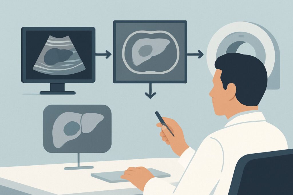
A 48-year-old male undergoes an abdominal ultrasound for vague, intermittent right upper quadrant discomfort. The scan is largely unremarkable, but the radiologist notes an incidental, well-circumscribed, 2.5 cm hyperechoic lesion in the right hepatic lobe, indeterminate on the non-contrast images. The patient has no history of cancer, normal liver function tests, and no signs of chronic liver disease. You are now faced with a common clinical question: what is the most appropriate next step to characterize this lesion and determine its significance? This scenario requires a methodical approach to avoid unnecessary procedures while ensuring a definitive diagnosis. According to the American College of Radiology (ACR) Appropriateness Criteria, for this specific presentation, an `US abdomen with IV contrast` is rated Usually Appropriate and is a primary recommended study for further evaluation.
Who Fits This Clinical Scenario for an Indeterminate Liver Lesion?
This diagnostic workflow is specifically for patients who meet a precise set of criteria. Applying this guidance outside of this context can lead to suboptimal imaging choices. This article applies to patients with:
- An indeterminate liver lesion greater than 1 cm discovered on an initial, non-contrast abdominal ultrasound.
- A normal underlying liver parenchyma, with no sonographic or clinical evidence of cirrhosis, steatosis, or other chronic liver disease.
- No known history or current suspicion of an extrahepatic malignancy. The patient should have no personal history of cancers that commonly metastasize to the liver (e.g., colon, breast, lung, pancreatic cancer) and no constitutional symptoms like unexplained weight loss to suggest an occult primary tumor.
It is crucial to distinguish this presentation from similar but distinct clinical scenarios that follow different diagnostic pathways. This guidance does not apply if:
- The patient has known chronic liver disease or cirrhosis. In these patients, any new lesion is suspicious for hepatocellular carcinoma (HCC), and the workup follows specific guidelines like the Liver Imaging Reporting and Data System (LI-RADS).
- The patient has a known extrahepatic malignancy. Here, the primary concern is metastatic disease, and the imaging choice (often multiphase CT or MRI) is tailored to that suspicion.
- The lesion was initially found on CT or MRI. If a lesion is indeterminate on these higher-level modalities, repeating an ultrasound is not the logical next step.
What Diagnoses Are You Working Up in This Scenario?
In a healthy patient with a normal liver, an incidentally discovered lesion is overwhelmingly likely to be benign. The goal of the imaging workup is to confidently diagnose a benign entity, thereby avoiding unnecessary follow-up or invasive procedures, while remaining vigilant for the rare possibility of malignancy.
Cavernous Hemangioma
This is the most common benign solid tumor of the liver. Hemangiomas are vascular malformations composed of blood-filled spaces. On imaging, they have a highly characteristic enhancement pattern that allows for a definitive, non-invasive diagnosis. They are typically asymptomatic and require no treatment or follow-up unless very large or causing symptoms.
Focal Nodular Hyperplasia (FNH)
FNH is the second most common benign liver lesion. It is thought to be a hyperplastic response of hepatocytes to a congenital vascular anomaly. Like hemangiomas, FNH has a classic imaging appearance, often featuring a central scar and avid arterial enhancement, which usually allows for a confident diagnosis without biopsy.
Hepatocellular Adenoma
This is a less common benign tumor but is clinically significant due to its potential for hemorrhage and, in rare cases, malignant transformation into HCC. Adenomas are most frequently seen in young and middle-aged women with a history of oral contraceptive use. Distinguishing an adenoma from FNH is a key goal of the workup, as adenomas may require cessation of oral contraceptives, surveillance, or even resection depending on their size and subtype.
Primary or Metastatic Malignancy
While unlikely in this specific clinical scenario, an indeterminate lesion could represent a previously unknown malignancy. This could be a primary hepatocellular carcinoma (very rare in a non-cirrhotic liver) or a solitary metastasis from an undiscovered primary tumor. The enhancement characteristics on dynamic, contrast-enhanced imaging are critical for differentiating these from benign lesions.
Why Is Contrast-Enhanced Ultrasound the Recommended Study for This Presentation?
When an indeterminate liver lesion is found on a standard ultrasound, the primary question is related to its vascularity and enhancement pattern. Contrast-Enhanced Ultrasound (CEUS) directly addresses this by using microbubble contrast agents to visualize blood flow within the lesion in real-time. The ACR rates `US abdomen with IV contrast` as Usually Appropriate for this scenario.
The key advantage of CEUS is its ability to provide a dynamic, continuous assessment of all vascular phases (arterial, portal venous, and late phases) without ionizing radiation (0 mSv). This allows for precise characterization of enhancement patterns that are pathognomonic for certain lesions. For example, the classic peripheral, discontinuous, nodular enhancement that fills in centripetally over time is diagnostic of a cavernous hemangioma. The rapid, uniform arterial enhancement and presence of a central scar can be highly suggestive of FNH.
Two other studies are also rated Usually Appropriate but have different trade-offs:
- MRI abdomen without and with IV contrast: This is often considered the gold standard for liver lesion characterization due to its superior soft tissue resolution. It is an excellent alternative if CEUS is unavailable or if the results of CEUS are equivocal. Like CEUS, it uses no ionizing radiation.
- CT abdomen with IV contrast multiphase: This study is also highly effective at characterizing lesions based on their enhancement across different phases. However, it delivers a significant dose of ionizing radiation (☢☢☢☢ 10-30 mSv), making it a less desirable first-line option than CEUS or MRI in a patient without a high pre-test probability of malignancy.
Conversely, some studies are rated lower for this specific purpose. For instance, an Image-guided biopsy is rated Usually not appropriate at this stage. Biopsy is an invasive procedure with risks, including bleeding and tumor seeding. It is reserved for cases where non-invasive imaging fails to provide a confident diagnosis or when malignancy is strongly suspected. Similarly, a CT abdomen without IV contrast is Usually not appropriate because it provides little to no additional information about vascularity, which is the key to characterizing the lesion.
What’s Next After Contrast-Enhanced Ultrasound? Downstream Workflow
The results of the contrast-enhanced ultrasound will dictate the next steps in the patient’s management. The goal is to reach a definitive conclusion and exit the diagnostic pathway whenever possible.
Result: Classic Benign Features
If the CEUS demonstrates a classic enhancement pattern for a benign lesion, such as a hemangioma or FNH, the workup is complete. The diagnosis is considered confirmed. No further imaging, follow-up, or treatment is typically required for these lesions. The referring physician can provide the patient with reassurance.
Result: Indeterminate or Atypical Features
If the enhancement pattern on CEUS is not classic for a specific benign entity, the lesion remains indeterminate. The next logical step is to proceed to the other highly-rated, non-radiation modality: `MRI abdomen without and with IV contrast`. The superior soft-tissue contrast and the use of hepatobiliary-specific contrast agents with MRI can often clarify the diagnosis, particularly in differentiating FNH from hepatocellular adenoma.
Result: Features Suspicious for Malignancy
If the CEUS shows features concerning for malignancy, such as rapid “washout” of contrast in the portal venous or late phases, the clinical pathway changes significantly. The patient should be referred to a specialist (hepatologist or GI oncologist). This will typically trigger a more comprehensive workup, which may include an MRI for further characterization and staging, a search for a primary extrahepatic tumor (e.g., colonoscopy, chest CT), and potentially an image-guided biopsy to obtain a tissue diagnosis.
Pitfalls to Avoid (and When to Get Help)
Navigating the workup of an incidental liver lesion requires careful attention to detail to avoid common errors.
- Ordering a Premature Biopsy: The most significant pitfall is proceeding to biopsy before exhausting non-invasive characterization. Biopsying a cavernous hemangioma, for example, carries a risk of significant hemorrhage.
- Using the Wrong Imaging Protocol: Ordering a single-phase contrast CT or MRI is insufficient. Characterization depends on observing the lesion’s behavior across multiphase imaging (arterial, portal venous, delayed phases). Be specific in your order.
- Ignoring Clinical Context: Failing to consider the patient’s history, such as oral contraceptive use in a woman, can lead to under-appreciation of the risk of a hepatocellular adenoma, which has different management implications than FNH.
- Accepting an “Indeterminate” Report Without Action: If a study like CEUS is indeterminate, the workup is not over. The next step (typically MRI) should be pursued to achieve diagnostic certainty.
If a lesion remains indeterminate after both CEUS and a high-quality multiphase MRI, or if it demonstrates growth on any subsequent imaging, it is time to escalate. This warrants a referral to a hepatologist or a center with expertise in liver imaging and pathology for multidisciplinary review and potential biopsy.
Related ACR Topics and Tools
For further reading on related scenarios and access to decision-support tools, the following resources are available. For breadth across all scenarios in Liver Lesion-Initial Characterization, see our parent guide: Liver Lesion-Initial Characterization: ACR Appropriateness Decoded.
- Imaging Appropriateness Selector — for adjacent scenarios
- Imaging Protocol Library — for technique on the recommended study
- Radiation Dose Calculator — for cumulative dose conversations
Frequently Asked Questions
Why is Contrast-Enhanced Ultrasound (CEUS) recommended over a multiphase CT scan?
For this specific scenario—an indeterminate lesion in a healthy patient with a normal liver—both CEUS and multiphase CT are rated ‘Usually Appropriate’ by the ACR. However, CEUS is often preferred as the initial step because it provides excellent real-time vascular information without any ionizing radiation. A multiphase CT scan delivers a significant radiation dose (10-30 mSv), which is best avoided if a non-radiation alternative can answer the clinical question.
If my institution doesn’t offer Contrast-Enhanced Ultrasound, what is the best alternative?
If CEUS is not available, the other ‘Usually Appropriate’ option is an MRI of the abdomen without and with IV contrast. MRI offers superb soft tissue contrast, does not use ionizing radiation, and is highly effective for characterizing liver lesions. It is an excellent and widely used alternative to CEUS.
What if the lesion is smaller than 1 cm?
Liver lesions less than 1 cm are often too small to characterize definitively with any imaging modality and are almost always benign cysts or hemangiomas. For a sub-centimeter lesion in this low-risk patient population, the general recommendation is often no further workup or follow-up, though this can depend on institutional guidelines and specific patient factors.
Does a ‘hyperechoic’ appearance on the initial ultrasound mean it’s likely a hemangioma?
A uniformly hyperechoic (bright) appearance on ultrasound is a classic feature of a cavernous hemangioma, the most common benign liver lesion. While this finding increases the likelihood of a hemangioma, it is not specific enough to be considered diagnostic on its own. Other lesions can appear hyperechoic, so further characterization with a dynamic contrast-enhanced study (like CEUS or MRI) is still necessary to confirm the diagnosis.
If the lesion is confirmed to be a benign hemangioma or FNH, does it need to be monitored with follow-up scans?
Once a liver lesion is confidently characterized as a typical cavernous hemangioma or focal nodular hyperplasia (FNH) via high-quality imaging, no further follow-up imaging is generally recommended. These are benign entities with no malignant potential, and routine surveillance is not necessary unless the patient develops new, attributable symptoms or the lesion is exceptionally large.
Reviewed by Pouyan Golshani, MD, Interventional Radiologist — May 26, 2026