Incidental Liver Lesion >1cm in Chronic Liver Disease: Which Imaging Study Is Best?
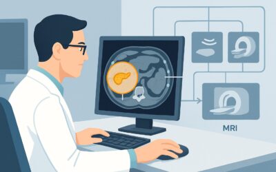
A hepatologist reviews the chart of a new patient with cirrhosis secondary to nonalcoholic steatohepatitis (NASH). An abdominal ultrasound, ordered by the patient’s primary care physician for unrelated reasons, incidentally noted a new 1.8 cm solid-appearing lesion in the right hepatic lobe. The report is non-specific, labeling it “indeterminate.” The immediate clinical question is how to definitively characterize this lesion in a high-risk liver. Is this early hepatocellular carcinoma? A dysplastic nodule? The next imaging study must provide a clear answer without unnecessary risk or delay. According to the American College of Radiology (ACR) Appropriateness Criteria, for this specific presentation, a US abdomen with IV contrast is rated Usually appropriate and is one of the primary recommended examinations.
Who Fits This Clinical Scenario for an Incidental Liver Lesion?
This clinical workflow is designed for a specific patient population: individuals with known chronic liver disease who are found to have an incidental liver lesion greater than 1 cm on a non-definitive initial imaging study.
Inclusion criteria for this guidance:
- The patient has a confirmed diagnosis of chronic liver disease, most commonly cirrhosis from any etiology (e.g., viral hepatitis, alcohol-related liver disease, NASH).
- A liver lesion measuring more than 1 cm has been discovered incidentally.
- The initial imaging was an ultrasound (US), a noncontrast Computed Tomography (CT), a single-phase contrast CT, or a noncontrast Magnetic Resonance Imaging (MRI). These studies are sufficient to detect a lesion but insufficient to characterize it.
Exclusion criteria (patients who fit a different workflow):
- Patients with a normal liver: An incidental lesion in a patient without underlying chronic liver disease has a different differential diagnosis and workup.
- Patients with a known extrahepatic malignancy: In this case, the primary concern is metastatic disease, which follows a separate ACR guideline.
- Lesions smaller than 1 cm: Sub-centimeter lesions in cirrhotic livers are often managed with surveillance rather than immediate characterization, representing a distinct clinical pathway.
- Lesions with definitive benign features on initial imaging: A lesion clearly identified as a simple cyst on ultrasound, for example, does not require further workup.
What Diagnoses Are You Working Up in a Patient with Chronic Liver Disease?
In the setting of a cirrhotic liver, a new, solid, indeterminate lesion raises immediate concern for malignancy. The imaging workup is primarily focused on confirming or excluding diagnoses that require urgent management, guided by the Liver Imaging Reporting and Data System (LI-RADS).
The most critical diagnosis to exclude is Hepatocellular Carcinoma (HCC). This is the most common primary liver cancer, and cirrhosis is its single greatest risk factor. The entire surveillance and diagnostic strategy for liver lesions in this population is built around the early detection of HCC, as treatment options and prognosis are highly dependent on the stage at diagnosis.
A high-grade dysplastic nodule is another key consideration. These are pre-malignant lesions that exist on the continuum of hepatocarcinogenesis. While not yet cancer, they have a high rate of progression to HCC and require close follow-up or treatment. Distinguishing them from well-differentiated HCC is a primary goal of advanced imaging.
Benign nodules, such as regenerative nodules, are extremely common in a cirrhotic liver. These represent the liver’s response to chronic injury and are not malignant. However, on non-contrast imaging, they can be indistinguishable from early HCC, making definitive characterization with dynamic contrast-enhanced imaging essential.
While less common than HCC, Intrahepatic Cholangiocarcinoma (ICC) is the second most common primary liver cancer and can also arise in patients with chronic liver disease. It has different imaging features and a different treatment pathway than HCC, making an accurate diagnosis crucial.
Why Is Contrast-Enhanced Ultrasound the Recommended Study for This Presentation?
For an indeterminate liver lesion over 1 cm in a patient with chronic liver disease, the ACR identifies three imaging modalities as Usually appropriate: US abdomen with IV contrast (CEUS), MRI abdomen without and with IV contrast, and CT abdomen with IV contrast multiphase. Among these, CEUS offers a powerful combination of diagnostic accuracy and safety.
The rationale for recommending CEUS rests on its unique ability to assess tumor vascularity in real-time. CEUS uses microbubble contrast agents that remain entirely within the blood vessels, unlike CT or MRI contrast agents which leak into the interstitial space. This property allows for a pure and dynamic visualization of blood flow through the lesion. The hallmark of HCC is intense arterial phase hyperenhancement (APHE) followed by “washout” of contrast in the portal venous or later phases. CEUS provides exceptional temporal resolution to capture these fleeting vascular events, which are central to a LI-RADS 5 (definitely HCC) diagnosis.
Key advantages of CEUS in this scenario include:
- No Ionizing Radiation: The study uses ultrasound waves, carrying a radiation dose of 0 mSv. This is particularly valuable for patients who may require multiple follow-up scans.
- Excellent Safety Profile: The microbubble contrast agents are not nephrotoxic, making CEUS a safe option for patients with renal insufficiency, a common comorbidity in chronic liver disease.
- High Diagnostic Performance: For characterizing focal liver lesions, the sensitivity and specificity of CEUS are comparable to those of multiphase CT and MRI.
How do other studies compare?
- MRI abdomen without and with IV contrast is also rated Usually appropriate and is an excellent alternative or complementary study. It offers superior soft-tissue contrast and can provide additional diagnostic clues from sequences like diffusion-weighted imaging and hepatobiliary phase imaging. It is often preferred for lesions in locations that are difficult to visualize with ultrasound.
- CT abdomen with IV contrast multiphase is also Usually appropriate but exposes the patient to significant ionizing radiation (☢☢☢☢ 10-30 mSv). While it provides the necessary dynamic enhancement information, it is typically reserved for situations where CEUS or MRI are contraindicated or unavailable.
- Image-guided biopsy is rated May be appropriate. It is not a first-line diagnostic tool because of the risks of bleeding and tumor seeding along the needle tract. Non-invasive diagnosis via imaging is the standard of care for typical HCC. Biopsy is reserved for lesions with atypical imaging features where a tissue diagnosis is required to guide therapy.
What’s Next After Contrast-Enhanced Ultrasound? Downstream Workflow
The results of the contrast-enhanced ultrasound will guide the subsequent management steps, which should ideally be determined by a multidisciplinary liver tumor board.
If the study is diagnostic for HCC (LI-RADS 5):
A definitive, non-invasive diagnosis of hepatocellular carcinoma has been made. The next step is staging to determine the extent of the disease and plan treatment. This typically involves a multiphase CT of the chest, abdomen, and pelvis. The patient should be referred to a specialist (hepatologist, surgeon, or interventional radiologist) to discuss treatment options, which may include ablation, resection, trans-arterial chemoembolization (TACE), or evaluation for liver transplantation.
If the study is indeterminate (LI-RADS 3 or 4):
The lesion shows some suspicious features but does not meet the strict criteria for HCC. The standard next step is to perform the other high-rated imaging modality: a multiphase MRI abdomen without and with IV contrast. The additional information from MRI, particularly from hepatobiliary phase agents, can often clarify the diagnosis and upgrade the lesion to LI-RADS 5 or downgrade it to a more benign category.
If the study suggests a benign or probably benign entity (LI-RADS 1 or 2):
The lesion does not demonstrate features of malignancy. In this case, the patient can typically return to their standard HCC surveillance protocol, which usually involves a non-contrast ultrasound every six months.
If the study is technically inadequate or remains indeterminate after both CEUS and MRI:
In this rare situation, an image-guided biopsy becomes the necessary next step to obtain a tissue diagnosis.
Pitfalls to Avoid (and When to Get Help)
Navigating the workup of a liver lesion in a cirrhotic patient requires careful attention to imaging protocols and clinical context.
Common pitfalls include:
- Ordering a single-phase CT: A standard “CT with contrast” is often only a portal venous phase scan. This is insufficient for LI-RADS characterization, which requires non-contrast, late arterial, portal venous, and delayed phases. Always specify “multiphase liver protocol.”
- Accepting a non-diagnostic study: If the initial study (e.g., ultrasound) was limited by body habitus or lesion location, do not simply repeat it. Proceed directly to a higher-quality modality like multiphase CT or MRI.
- Prematurely biopsying a potentially resectable lesion: For a lesion with classic HCC features, biopsy is often unnecessary and carries a small but real risk of tumor seeding. Non-invasive diagnosis is preferred.
- Ignoring alpha-fetoprotein (AFP) levels: While not a primary diagnostic tool, a significantly elevated or rising AFP level in conjunction with an indeterminate lesion increases the suspicion for HCC and may lower the threshold for proceeding to biopsy or treatment.
If a lesion has atypical features, or if the diagnosis remains unclear after both CEUS and MRI, it is crucial to escalate the case to a multidisciplinary liver tumor board for consensus review.
Related ACR Topics and Tools
For a comprehensive overview of all clinical scenarios related to the initial characterization of a liver lesion, please consult the main topic guide. Further resources for selecting appropriate imaging and understanding protocols are also available.
- For breadth across all scenarios in Liver Lesion-Initial Characterization, see our parent guide: Liver Lesion-Initial Characterization: ACR Appropriateness Decoded.
- To explore adjacent clinical questions, use the ACR Appropriateness Criteria Lookup.
- For detailed procedural techniques, see the Imaging Protocol Library.
- To discuss cumulative radiation exposure with patients, use the Radiation Dose Calculator.
Frequently Asked Questions
Why is contrast-enhanced ultrasound (CEUS) recommended over a multiphase CT or MRI?
CEUS, multiphase CT, and multiphase MRI are all rated ‘Usually appropriate’ by the ACR for this scenario and have similar diagnostic performance. CEUS is often highlighted because it avoids ionizing radiation (0 mSv) and uses a contrast agent that is not harmful to the kidneys, which is a significant advantage in patients with chronic liver disease who may have concurrent renal dysfunction. However, the best choice may depend on local expertise, patient factors (like body habitus), and lesion location.
What if my institution does not offer contrast-enhanced ultrasound (CEUS)?
If CEUS is not available, a multiphase MRI of the abdomen without and with IV contrast is an excellent and equally appropriate alternative. It provides outstanding soft-tissue detail and diagnostic information. A multiphase CT of the abdomen is the next best choice if MRI is also unavailable or contraindicated.
Is a biopsy always necessary if the imaging is indeterminate?
Not always. For indeterminate lesions (e.g., LI-RADS 3 or 4), the standard approach is to perform a second type of high-quality imaging (e.g., MRI if the first study was CEUS). Biopsy is generally reserved for cases that remain indeterminate after a full non-invasive imaging workup, or for lesions with atypical features where a non-HCC malignancy like cholangiocarcinoma is suspected.
Does the size of the lesion (e.g., 1.5 cm vs 4 cm) change the initial imaging choice?
No, for any incidental lesion greater than 1 cm in a patient with chronic liver disease, the initial recommended diagnostic studies (CEUS, MRI, or CT) are the same. The size of the lesion becomes more critical for determining treatment options after a diagnosis of HCC is made, but it does not change the initial choice of imaging for characterization.
What is the role of PET/CT in this specific scenario?
For the initial characterization of a newly discovered liver lesion in a patient with cirrhosis, FDG-PET/CT is rated ‘Usually not appropriate’ by the ACR. While it is used for staging many cancers, its sensitivity for well-differentiated HCC is relatively low. The primary diagnostic tools rely on the specific vascular patterns of the lesion, which are best assessed with dynamic contrast-enhanced ultrasound, CT, or MRI.
Reviewed by Pouyan Golshani, MD, Interventional Radiologist — May 29, 2026