ACR Workflow: What Is the Right First Imaging Study for a Palpable Breast Mass in a Woman Aged 30-39?

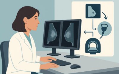
A 34-year-old woman presents to your clinic for her annual exam and mentions a new, firm, mobile lump she felt in her right breast last week. The clinical exam confirms a palpable, non-tender, 1.5 cm mass in the upper outer quadrant. She has no family history of breast cancer and no other symptoms. You now face the decision of which imaging study to order first to evaluate this finding. The patient is anxious, and you need a clear, evidence-based path forward to differentiate a benign finding from the less likely but critical possibility of malignancy. This article provides a focused workflow for this exact clinical question, grounded in the American College of Radiology (ACR) Appropriateness Criteria. For this specific scenario, a breast ultrasound is rated Usually Appropriate as the initial imaging examination.
Who Fits This Clinical Scenario for a Palpable Breast Mass?
This guidance is specifically tailored for the initial imaging workup of a palpable breast mass in an adult female patient between 30 and 39 years of age. The key inclusion criteria are a distinct, palpable lump discovered by either the patient or a clinician, with no prior imaging performed for this specific finding. This workflow applies whether the mass is focal and well-defined or a more general area of focal asymmetry or thickening.
It is crucial to distinguish this scenario from others that require a different diagnostic approach. This guidance does not apply to:
- Patients younger than 30: While the initial imaging is often the same (ultrasound), the pre-test probability of malignancy is lower, which can influence downstream management.
- Patients 40 years of age or older: For this group, diagnostic mammography is the cornerstone of the initial workup, often supplemented with ultrasound. The higher baseline risk of cancer in this demographic mandates a different primary imaging strategy.
- Patients with other suspicious signs: This workflow is for an isolated palpable mass. If the patient also presents with bloody nipple discharge, skin retraction, erythema, or axillary adenopathy, the workup is more urgent and may follow a different pathway.
- Pregnant or lactating patients: Hormonal changes significantly alter breast tissue, requiring specialized imaging considerations.
This article addresses only the initial diagnostic step for a new, isolated palpable mass in a woman in her thirties.
What Diagnoses Are You Working Up in a Woman Aged 30-39 with a Breast Lump?
When evaluating a palpable breast mass in this age group, the differential diagnosis is broad, with benign causes being far more common than malignant ones. However, the primary goal of imaging is to confidently exclude or identify cancer.
The most common benign solid mass in women in their 30s is a fibroadenoma. These are typically firm, rubbery, well-circumscribed, and mobile on physical exam. On ultrasound, they have a classic appearance that often allows for a confident, non-invasive diagnosis.
Breast cysts are another extremely common cause of palpable lumps. These fluid-filled sacs can appear suddenly and may be tender. Ultrasound is exceptionally effective at identifying simple cysts, which are definitively benign and require no further workup unless they are large and symptomatic, in which case aspiration can be considered for relief.
Fibrocystic changes represent a spectrum of benign histologic alterations in breast tissue that can cause lumpy, dense, and often tender breasts, particularly in relation to the menstrual cycle. While these changes are diffuse, they can sometimes present as a focal palpable area of thickening or a dominant lump, which requires imaging to rule out an underlying discrete mass.
While less common, breast cancer must be the primary consideration to exclude. The incidence of breast cancer begins to increase in this decade. Although the absolute risk is lower than in older women, a new, persistent, solid mass warrants a thorough evaluation to ensure a malignancy is not missed.
Other less frequent possibilities include fat necrosis (often related to prior trauma), galactoceles (in women who have recently lactated), and abscesses (typically associated with clinical signs of infection).
Why Is Ultrasound the Recommended First Study for a Palpable Mass in This Age Group?
For a woman aged 30 to 39 with a palpable breast mass, the ACR rates US breast as Usually Appropriate. This is the primary recommendation for the initial imaging evaluation. Diagnostic mammography and digital breast tomosynthesis are also rated Usually Appropriate but are typically used as adjuncts to ultrasound in this specific demographic, not as the first-line study.
The rationale for prioritizing ultrasound is threefold:
- Tissue Characterization: Ultrasound excels at the first critical step in evaluating a lump: determining if it is cystic or solid. A simple cyst identified on ultrasound is considered benign (BI-RADS 2), effectively ending the diagnostic workup. This capability is fundamental and cannot be achieved with mammography alone.
- Breast Density: Women in their 30s typically have denser breast tissue compared to older, postmenopausal women. This dense fibroglandular tissue can appear white on a mammogram, potentially obscuring an underlying mass (the “camouflage effect”). Ultrasound is not limited by breast density and can visualize masses within dense tissue with high sensitivity.
- Radiation Safety: Ultrasound uses no ionizing radiation (Relative Radiation Level: O 0 mSv). While the dose from a modern diagnostic mammogram is low (RRL: ☢☢ 0.1-1mSv), avoiding radiation exposure when a non-ionizing modality offers superior or equivalent diagnostic information is a key principle, especially in younger patients who may require cumulative imaging over their lifetime.
While diagnostic mammography is also Usually Appropriate, it is often performed secondarily if the ultrasound reveals a solid mass that is indeterminate or suspicious. In these cases, mammography provides complementary information about morphology, calcifications, and the surrounding breast architecture.
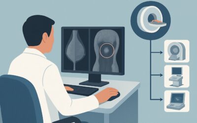
Conversely, MRI breast with and without IV contrast is rated Usually Not Appropriate for the initial workup of a palpable mass. While highly sensitive, MRI has lower specificity, which can lead to false positives and subsequent unnecessary biopsies. It is reserved for specific clinical situations, such as problem-solving after a full conventional workup, evaluating the extent of known disease, or for screening high-risk patients.
What Is the Downstream Workflow After the Initial Breast Ultrasound?
The results of the initial breast ultrasound will dictate the next steps, which are typically guided by the Breast Imaging Reporting and Data System (BI-RADS) assessment category.
- Negative or Benign Finding (BI-RADS 1 or 2): If the ultrasound shows no abnormality at the site of the palpable lump (BI-RADS 1) or identifies a definitively benign finding like a simple cyst or a classic intramammary lymph node (BI-RADS 2), the workup is complete. The patient can be reassured and returned to routine age-appropriate screening.
- Probably Benign Finding (BI-RADS 3): If the ultrasound identifies a solid mass with classic features of a fibroadenoma, a BI-RADS 3 assessment is often appropriate. The standard recommendation is for short-term imaging follow-up, typically with a repeat targeted ultrasound in 6 months, to ensure stability. Biopsy is an alternative if the patient has high anxiety or if the finding has atypical features.
- Suspicious Finding (BI-RADS 4 or 5): If the mass has features that are indeterminate, suspicious, or highly suggestive of malignancy (e.g., irregular shape, spiculated margins, posterior acoustic shadowing), a BI-RADS 4 or 5 assessment is made. This finding requires immediate action. The next step is an image-guided core needle biopsy, most commonly performed under ultrasound guidance, to obtain a tissue diagnosis. Diagnostic mammography is also typically performed at this stage to fully characterize the lesion and evaluate the rest of the breast for other findings.
- Incomplete Assessment (BI-RADS 0): Occasionally, the ultrasound may be inconclusive or require correlation with another imaging modality. This BI-RADS 0 assessment means more imaging is needed. The most common next step is to obtain a diagnostic mammogram for further characterization.
Pitfalls to Avoid (and When to Get Help)
Navigating the workup of a palpable breast mass in a younger woman requires careful attention to detail to avoid common errors.
A primary pitfall is inadequate clinical-radiologic correlation. If the ultrasound is reported as negative (BI-RADS 1) but a distinct, persistent mass is clearly palpable on clinical exam, do not stop the workup. The finding may be mammographically or sonographically occult. In this situation, escalation to a breast specialist or breast surgeon for consideration of further imaging (like MRI) or clinical follow-up is warranted.
Another error is ordering a screening mammogram instead of a diagnostic study. A screening study is for asymptomatic patients and may not include the specific views or radiologist oversight needed to evaluate a palpable problem. Always order a “diagnostic” mammogram and/or a “targeted” ultrasound, specifying the exact location of the palpable concern.
Finally, avoid false reassurance based on age. While cancer is less common in this age group, it is not rare. A persistent, solid mass found on imaging should not be dismissed without a definitive benign diagnosis or appropriate follow-up, regardless of the patient’s age.
Related ACR Topics and Tools
This article covers one specific scenario in depth. For a comprehensive overview of all patient presentations and age groups related to this topic, please consult our parent guide. Additional GigHz tools can help you apply these criteria in your daily practice.
- For breadth across all scenarios in Palpable Breast Masses, see our parent guide: Palpable Breast Masses: ACR Appropriateness Decoded.
- To look up other clinical scenarios, use the Imaging Appropriateness Selector.
- To understand the technical details of the recommended study, visit the Imaging Protocol Library.
- To discuss cumulative radiation exposure with your patients, reference the Radiation Dose Calculator.
Frequently Asked Questions
Why not just order a mammogram first for a woman in her 30s with a breast lump?
In women aged 30-39, breast tissue is often dense, which can hide a mass on a mammogram. Ultrasound is more sensitive for detecting masses in dense tissue and can definitively distinguish between a simple cyst (benign) and a solid mass, which is the most critical first step. Ultrasound also avoids ionizing radiation. A mammogram is often used as a secondary test if the ultrasound is suspicious or inconclusive.
If the ultrasound shows a ‘probably benign’ solid mass (BI-RADS 3), is a biopsy always necessary?
No. For a finding with classic benign features on ultrasound, such as a fibroadenoma, the standard of care is typically short-term imaging follow-up (e.g., a repeat ultrasound in 6 months) to ensure stability. This approach has a very high negative predictive value for cancer (<2%) and avoids unnecessary invasive procedures. Biopsy is an alternative for patient preference or if any atypical features are present.
What if the patient has a strong family history of breast cancer? Does that change the initial imaging choice?
For the initial workup of a new palpable lump, ultrasound remains the recommended first study even with a strong family history. However, the patient’s risk status is critical for downstream management. A suspicious finding may be biopsied more readily, and the patient should be evaluated for high-risk screening (which often includes annual breast MRI in addition to mammography), but this is separate from the diagnostic workup of the palpable problem itself.
The ultrasound report was negative (BI-RADS 1), but I can still feel a definite lump. What should I do?
This is a critical situation requiring clinical-radiologic correlation. Do not dismiss the patient’s or your own clinical finding. The mass may be sonographically occult. The appropriate next step is to refer the patient to a breast surgeon or breast imaging specialist for further evaluation. This may involve a repeat clinical exam, repeat imaging, or consideration of a problem-solving MRI.
Is it okay to just watch a palpable lump for a few months without imaging in a 35-year-old?
No, a new, persistent, palpable breast mass in any woman, including those in their 30s, should be evaluated with imaging. While many lumps are benign, delaying the diagnosis of a potential malignancy can negatively impact outcomes. The initial imaging with ultrasound is non-invasive, safe, and provides crucial diagnostic information to guide management.
Reviewed by Pouyan Golshani, MD, Interventional Radiologist — June 27, 2026