Ankle/Foot X-Ray — Dictation, Appropriateness, and Dose for Residents
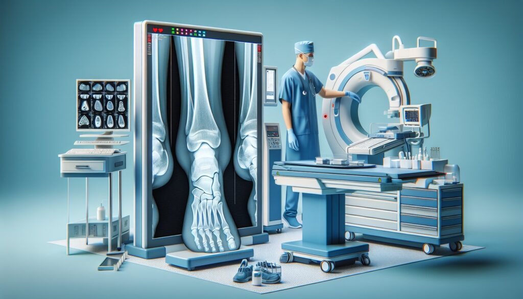
Stat from the ED. Ankle trauma, meets Ottawa rules. You pull up the images and see what looks like subtle widening of the medial clear space on the mortise view. Is it a true syndesmotic injury or just positioning? The attending is going to want a definitive read, and they’ll expect you to know the Weber classification if you call a fibular fracture. Getting these details right, fast, is the job.
When I was a junior resident, I kept a cheat sheet taped to my monitor for the key ankle and foot measurements—medial clear space, Boehler’s angle, the Lisfranc interval. It’s one of those high-volume, high-stakes studies where small details change management. This guide is that cheat sheet, built for your call shifts and daily readouts. We’ll cover a structured template, the key classifications attendings expect, and the ACR appropriateness criteria. For more high-yield content, check out the residents and fellows resource hub.
What an Ankle and Foot X-Ray Covers and What Attendings Look For
Plain radiographs are the first-line imaging for acute ankle and foot trauma, guided by the Ottawa Ankle and Foot Rules. The goal is to rapidly identify or exclude fractures, dislocations, and significant malalignment. While MRI is superior for ligamentous injury and stress fractures, the radiograph sets the stage for all subsequent management.
Your attending expects a systematic and complete report that addresses:
- Alignment: Is the ankle mortise congruent? Is there evidence of a Lisfranc injury?
- Bones: A thorough search for fractures, paying special attention to high-miss areas like the proximal fibula (Maisonneuve), the base of the fifth metatarsal (Jones vs. Pseudo-Jones), the navicular, and the calcaneus.
- Joints: Evaluation of all joint spaces for symmetry, widening, or degenerative changes. The medial clear space is critical.
- Soft Tissues: Note any significant swelling, effusions, or radiopaque foreign bodies.
- Classifications: Application of relevant classification systems like Weber for lateral malleolar fractures or Salter-Harris for pediatric physeal injuries.
Radiology Report Template for Ankle and Foot X-Ray
This template provides a solid foundation. You can adapt it for your institution’s specific macros in PowerScribe or other dictation software. The key is to be systematic so you don’t miss a subtle finding.
Technique
Ankle: AP, lateral, and mortise views of the [right/left] ankle were obtained.
Foot: AP, lateral, and oblique views of the [right/left] foot were obtained.
[As needed: Weight-bearing views were also obtained.]
Findings
Alignment: The ankle mortise is [congruent/widened]. The talar dome is normally positioned. No evidence of dislocation. The relationship between the first and second metatarsal bases is anatomic, without diastasis to suggest Lisfranc injury.
Bones:
- Tibia/Fibula: No acute fracture of the distal tibia or fibula. [Describe fracture location, orientation, displacement, and intra-articular extension. Apply Weber classification for lateral malleolar fractures.]
- Tarsal Bones: The talus, calcaneus, navicular, cuboid, and cuneiforms are intact. [Boehler’s angle is normal (20-40 degrees).]
- Metatarsals/Phalanges: No acute fracture or dislocation. [Pay special attention to the base of the fifth metatarsal to differentiate a Jones from a tuberosity avulsion fracture.]
- Osseous structures are otherwise unremarkable. Normal bone density for age.
Joints:
- Ankle Joint: The tibiotalar and tibiofibular joints are maintained. The medial clear space is normal [typically <4 mm]. No joint effusion.
- Other Joints: The subtalar, talonavicular, and calcaneocuboid joints are unremarkable. The tarsometatarsal (Lisfranc) joints are congruent.
Soft Tissues: Soft tissues are [unremarkable/demonstrate swelling centered at the ___].
Impression
1. [e.g., Acute, nondisplaced transverse fracture of the lateral malleolus at the level of the ankle mortise, consistent with a Weber B fracture.]
2. [e.g., No acute fracture or dislocation.]
3. [e.g., Widening of the medial clear space, concerning for deltoid ligament injury.]
Free Template Sources for Your Personal Library
Building your own template library is a rite of passage. If you’re looking for more examples or templates for other modalities, two great free repositories exist. These are excellent, non-commercial resources maintained by and for radiologists.
- RadReport.org: Curated by the RSNA, this is a comprehensive library of peer-reviewed templates covering nearly every study imaginable.
- Radiology Templates (AU): An Australian-maintained library with a clean interface and practical, clinically-focused templates.
The Next-Level Move: From Free-Form Dictation to Structured Report
The challenge on a busy shift isn’t just spotting the finding—it’s getting it into a clean, structured report that your attending and the clinical team can easily digest. This is where AI-powered tools can streamline your workflow. Instead of meticulously navigating a macro, you can dictate the positive findings in free form—”Weber B fracture of the left fibula with 2 mm of displacement and some widening of the medial clear space”—and let the software handle the rest.
Tools like GigHz Precision AI are designed for this. It takes your natural language dictation of positive findings and generates a complete, structured report using pre-loaded templates from the American College of Radiology (ACR) and Society of Interventional Radiology (SIR). This process helps ensure all key elements are included and formatted correctly, allowing you to focus on the images, not the clicks.
When Should You Order an Ankle or Foot X-Ray? ACR Appropriateness Criteria
The decision to order an ankle or foot radiograph is almost always guided by the Ottawa Ankle and Foot Rules. The American College of Radiology (ACR) has codified this into its Appropriateness Criteria, providing clear guidance for referring providers that is essential for radiologists to know.
For an adult or child over 5 with acute ankle trauma, the guidelines are clear. If the patient meets the Ottawa Ankle Rules (e.g., inability to bear weight or point tenderness over the malleoli), an ankle X-ray is Usually Appropriate (ACR rating 8/9). Conversely, if the Ottawa Ankle Rules are negative, imaging is also Usually Appropriate (ACR rating 8/9), though often deferred in favor of clinical follow-up. In cases where exclusionary criteria like a neurologic disorder prevent the use of the Ottawa rules, an X-ray is Usually Appropriate (ACR rating 9/9).
The logic is similar for acute foot trauma. If the Ottawa Foot Rules are positive (e.g., tenderness at the base of the fifth metatarsal or navicular), a foot X-ray is Usually Appropriate (ACR rating 8/9). If the rules are negative, it is also Usually Appropriate (ACR rating 7/9). When the clinical suspicion involves an area not covered by the Ottawa rules, such as the toes, an X-ray is Usually Appropriate (ACR rating 9/9).
If initial radiographs are negative but there is high clinical suspicion for a Lisfranc injury, occult fracture, or significant ligamentous damage, further imaging with CT or MRI may be appropriate.
How Much Radiation Does an Ankle or Foot X-Ray Deliver?
Patients and referring providers often ask about radiation dose. For an ankle or foot X-ray, the dose is extremely low. The estimated effective dose is between 0.005 and 0.05 mSv. To put this in perspective, this is significantly less than the average annual background radiation a person receives just from living on Earth (about 3 mSv per year).
The ACR designates any exam with a dose under 0.1 mSv as very low. This study falls well within that category, making it one of the lowest-dose imaging studies performed in radiology. Dose is minimized by collimating the beam strictly to the area of interest and using appropriate technical factors.
| Study | Typical Effective Dose (mSv) |
|---|---|
| Ankle/Foot X-Ray | 0.005 – 0.05 mSv |
| Chest X-Ray (PA/Lat) | ~0.1 mSv |
| Annual Background Radiation | ~3 mSv |
| CT Head | ~2 mSv |
Ankle and Foot X-Ray Imaging Protocol — Standard Views and Technique
A standard, high-quality radiographic series is essential for an accurate diagnosis. The protocol consists of specific views designed to evaluate the complex anatomy of the ankle and foot without superimposition of key structures. For the ankle, the mortise view is critical, while for the foot, the oblique view helps clarify the tarsal and metatarsal relationships.
The following table outlines the standard protocol. Weight-bearing views are optional but crucial when there is suspicion for a Lisfranc injury or subtle ligamentous instability, as they can unmask malalignment not visible on non-weight-bearing images.
| Region | View | Key Technical Parameters | Purpose |
|---|---|---|---|
| Ankle | AP | kVp: 55-65 | General overview of distal tibia, fibula, and talus. |
| Ankle | Lateral | kVp: 55-65 | Assesses for posterior malleolar fractures, calcaneal fractures, and joint effusions. |
| Ankle | Mortise | 15-20° internal rotation | True AP of the ankle joint, showing the tibiotalar and tibiofibular joints without overlap. Best view for assessing mortise symmetry. |
| Foot | AP (Dorsoplantar) | kVp: 55-65 | Evaluates metatarsals, phalanges, and tarsometatarsal joints. |
| Foot | Lateral | kVp: 55-65 | Assesses foot arches, calcaneus, and talonavicular joint. |
| Foot | Medial Oblique | kVp: 55-65 | Visualizes the cuboid, navicular, and anterior calcaneus. Separates the metatarsal bases. |
A common pitfall is inadequate imaging of the entire fibula in the setting of an ankle injury. If a medial malleolar fracture or deltoid ligament injury is seen with a mechanism of pronation-external rotation, a Maisonneuve fracture (proximal fibular fracture) must be excluded by obtaining radiographs of the entire tibia and fibula.
3+ months free for radiology residents and fellows
Look like a rockstar on your reports. With the GigHz Precision AI, you can dictate your positive findings in free form, and the AI will generate a complete, structured draft using the latest ACR and SIR templates. The appropriate clinical decision support, like classifications for complex fractures or liver lesions, fires automatically.
We’re offering extended free access to residents and fellows. All we ask in return is your feedback so we can keep improving the product for trainees.
To apply, just send us these three items:
- Your PGY year (e.g., PGY-2, PGY-4)
- Your training type (radiology residency or specific fellowship)
- Your training program / hospital name
The signup process is simple. No credit card, no long forms. Just reply to the application with the information above, and we’ll get you set up. You can apply for the residents free-access program here.
Free GigHz Tools That Pair With This Article
Three free tools that complement the material above:
- Imaging Appropriateness Selector — Type an indication or clinical scenario in plain language and get the imaging studies the ACR rates for it, with adult and pediatric radiation levels. Built directly from 297 ACR topics, 1,336 clinical variants, and 15,823 procedure ratings.
- GigHz Imaging Protocol Library — A searchable library of 131 imaging protocols with the physics specs surfaced and the matching ACR Appropriateness Criteria alongside. Plain-English narratives readable in 60 seconds, organized by modality.
- GigHz Radiation Dose Calculator — Pick the imaging studies a patient has had and see total dose in millisieverts (mSv) with comparisons to natural background radiation, transatlantic flights, and chest X-rays. Useful for shared decision-making.
Frequently Asked Questions
Is it HIPAA-compliant?
Yes. The platform is designed for de-identified workflows by default. You dictate findings, not patient data. It operates securely in the cloud and does not require integration with your hospital’s EMR or PACS to function, ensuring a clear separation from protected health information (PHI).
Do I need my IT department to set it up?
No. GigHz Precision AI is browser-based and requires no local software installation or special permissions from your IT department. It works on any modern computer, including the shared workstations in the reading room or your personal laptop or iPad at home.
Does it work with PowerScribe or other dictation systems?
Yes. It works alongside any existing dictation system. Most residents use it on a second monitor or an iPad. You read the study in your PACS, dictate your findings into the GigHz tool, and then copy the generated structured report into your hospital’s system. It complements, rather than replaces, your current workflow.
Can I use this on my phone or iPad?
Absolutely. The platform is fully responsive and works well on mobile devices, making it perfect for reviewing a template or generating a report structure from your iPad on call.
Can I customize the templates?
Yes. While the system comes pre-loaded with ACR and SIR standard templates, you can create, save, and modify your own templates to match your personal preferences or your institution’s specific formatting requirements.
What happens after I finish residency or fellowship?
The extended free access is specifically for trainees. After you graduate, you can transition to a standard plan for practicing radiologists. Your customized templates and settings will be saved and carry over with your account.
Last reviewed by Pouyan Golshani, MD — 2026-06-23.
Reviewed by Pouyan Golshani, MD, Interventional Radiologist — August 4, 2026